Chapter 14: The Brain and Cranial Nerves Overview
1/202
There's no tags or description
Looks like no tags are added yet.
Name | Mastery | Learn | Test | Matching | Spaced | Call with Kai |
|---|
No analytics yet
Send a link to your students to track their progress
203 Terms
What are the major subdivisions of the brain?
Forebrain, cerebellum, and brainstem.
What is the largest part of the forebrain?
The cerebrum, which accounts for 83% of brain volume.
What are gyri and sulci in the context of the brain?
Gyri are thick folds on the cerebrum's surface, while sulci are shallow grooves between the gyri.
What is the longitudinal cerebral fissure?
A deep groove that separates the cerebral hemispheres.
What connects the cerebral hemispheres?
The corpus callosum, a thick nerve bundle at the bottom of the longitudinal fissure.
Where is the cerebellum located?
In the posterior cranial fossa, separated from the cerebrum by the transverse cerebral fissure.
What is the second-largest part of the brain?
The cerebellum.
What are the components of the brainstem?
The midbrain, pons, and medulla oblongata.
What are the directional terms used to describe the brain's orientation?
Rostral (toward the forehead) and caudal (toward the spinal cord).
What is the significance of gray and white matter in the brain?
Gray matter contains neuronal cell bodies, while white matter consists of myelinated axons.
What is the embryonic development of the CNS related to?
It is related to adult brain anatomy.
What are the two main types of matter in the brain?
Gray matter and white matter.
What does gray matter contain?
Nerve cell bodies, dendrites, and synapses.
What is the cortex in relation to gray matter?
The cortex is the surface layer of gray matter in the cerebrum and cerebellum.
What are nuclei in the brain?
Deeper masses of gray matter surrounded by white matter.
What is white matter composed of?
Tracts, which are bundles of nerve fibers (axons).
Where is white matter located in relation to gray matter?
Deep to cortical gray matter in the brain.
What is the function of white matter?
To connect one part of the brain to another and to the spinal cord.
What are the meninges?
Three membranes surrounding the brain and spinal cord.
What is the primary function of the meninges?
To protect the brain and provide structural framework for its arteries and veins.
What are the three layers of the meninges, from outermost to innermost?
Dura mater, arachnoid mater, pia mater.
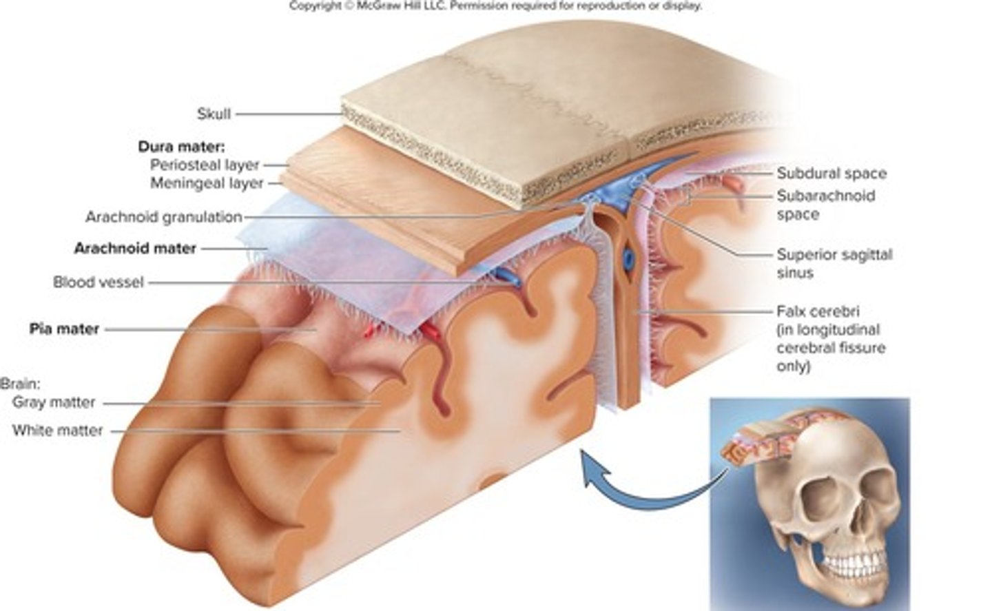
What are the two layers of the cranial dura mater?
Outer periosteal layer and inner meningeal layer.
What is the function of dural sinuses?
To collect blood circulating through the brain.
What is the superior sagittal sinus?
A dural sinus located just under the calvaria along the median line.
What does the falx cerebri do?
Separates the two cerebral hemispheres.
What is the role of the arachnoid mater?
A transparent membrane over the brain surface.
What is contained in the subarachnoid space?
Cerebrospinal fluid and blood vessels.
What is meningitis?
Inflammation of the meninges.
What are common signs of meningitis?
High fever, stiff neck, drowsiness, intense headache.
How is meningitis diagnosed?
By examining cerebrospinal fluid obtained by lumbar puncture (spinal tap).
What are the four ventricles of the brain?
Two lateral ventricles, third ventricle, and fourth ventricle.
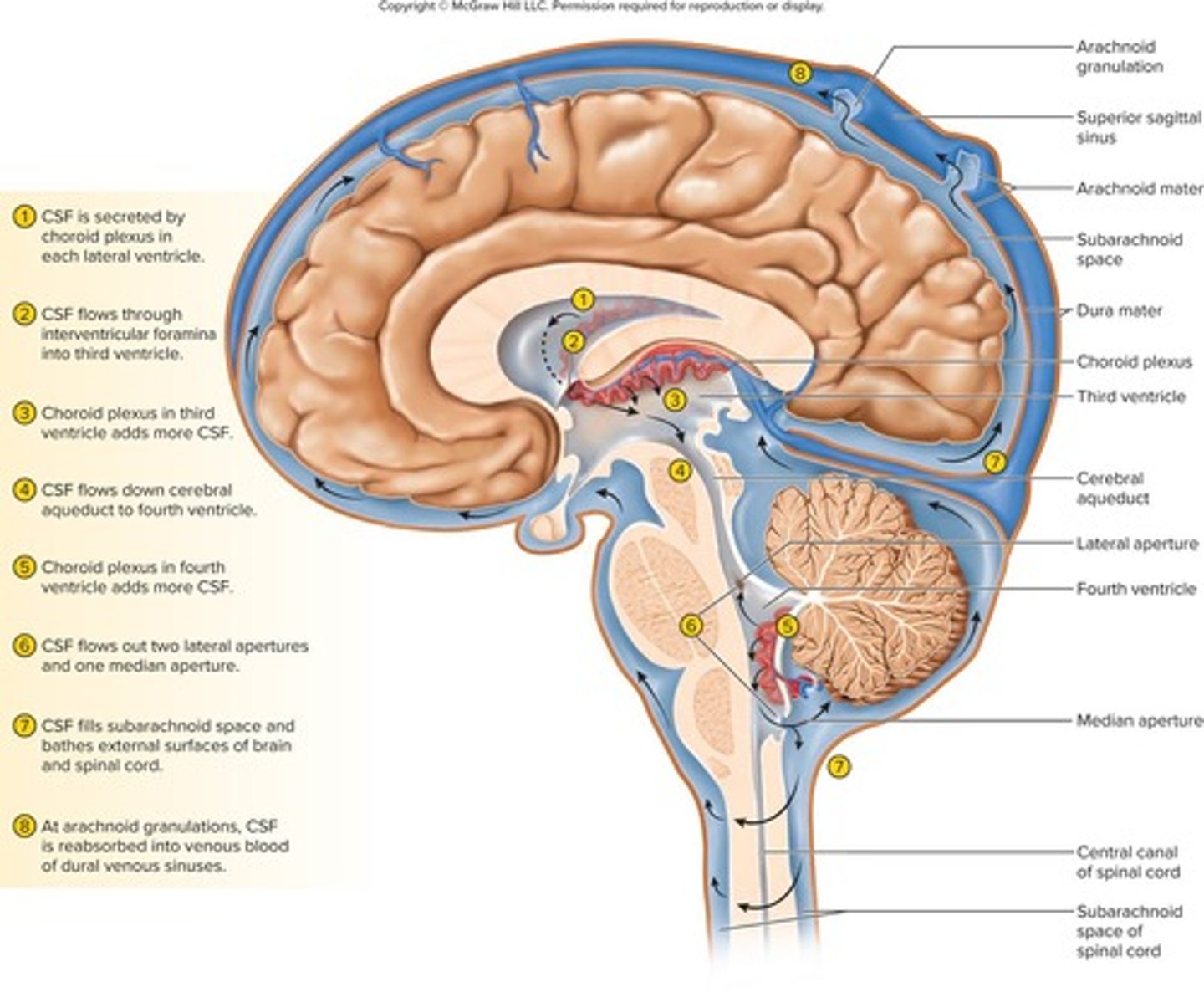
What connects the lateral ventricles to the third ventricle?
The interventricular foramen.
What connects the third ventricle to the fourth ventricle?
The cerebral aqueduct.
What is the central canal?
A tube that connects to the fourth ventricle and runs through the center of the spinal cord.
What is cerebrospinal fluid (CSF)?
A clear, colorless liquid that fills the ventricles, canals of the CNS, and bathes its external surface.
How is cerebrospinal fluid (CSF) produced?
CSF production begins with the filtration of blood plasma through capillaries of the brain.
What is the choroid plexus?
A spongy mass of blood capillaries located on the floor of each ventricle that produces cerebrospinal fluid.
What role do ependymal cells play in relation to cerebrospinal fluid?
Ependymal cells are neuroglia that line the ventricles and cover the choroid plexus, modifying the filtrate.
How does the composition of cerebrospinal fluid (CSF) differ from blood plasma?
CSF has more sodium and chloride, less potassium, calcium, glucose, and very little protein compared to plasma.
What drives the continuous flow of cerebrospinal fluid (CSF) through the CNS?
The flow is driven by its own pressure, the beating of ependymal cilia, and pulsations of the brain produced by each heartbeat.
What is the path of cerebrospinal fluid (CSF) through the ventricles?
CSF is secreted in the lateral ventricles, flows through intervertebral foramina into the third ventricle, down the cerebral aqueduct into the fourth ventricle, with the third and fourth ventricles adding more CSF along the way.
How does cerebrospinal fluid (CSF) escape into the subarachnoid space?
CSF escapes through three pores: the median aperture and two lateral apertures.
What are arachnoid granulations?
Cauliflower-shaped extensions of the arachnoid meninx that protrude through the dura mater into the superior sagittal sinus.
How is cerebrospinal fluid (CSF) reabsorbed?
CSF is reabsorbed by arachnoid granulations, which allow CSF to penetrate the walls of the villi and mix with blood in the sinus.
What are the three main functions of cerebrospinal fluid (CSF)?
Buoyancy, protection, and chemical stability.
How does CSF provide buoyancy to the brain?
It allows the brain to attain considerable size without being impaired by its own weight.
What protection does CSF offer to the brain?
It protects the brain from striking the cranium when the head is jolted.
What is the consequence of a 10-second interruption in blood supply to the brain?
Loss of consciousness.
What happens after a 1- to 2-minute interruption of blood supply to the brain?
Significant impairment of neural function.
What can occur after a 4-minute interruption of blood supply to the brain?
Irreversible brain damage.
What is the blood-brain barrier (BBB)?
A system that regulates what substances can enter the brain from the bloodstream.
What substances are highly permeable through the blood-brain barrier?
Water, glucose, and lipid-soluble substances such as oxygen, carbon dioxide, alcohol, caffeine, nicotine, and anesthetics.
What is the role of astrocytes in the formation of the blood-brain barrier?
They induce endothelial cells to form tight junctions that create the barrier.
What distinguishes the blood-CSF barrier from the blood-brain barrier?
The blood-CSF barrier is formed by tight junctions between ependymal cells, allowing exchange between brain tissue and CSF.
What is a stroke, also known as a cerebral vascular accident (CVA)?
A sudden death of brain tissue due to interruption of blood supply.
What are the two types of strokes and their causes?
Hemorrhagic stroke (rupture of a blood vessel) and ischemic stroke (obstruction by a clot or lipid deposit).
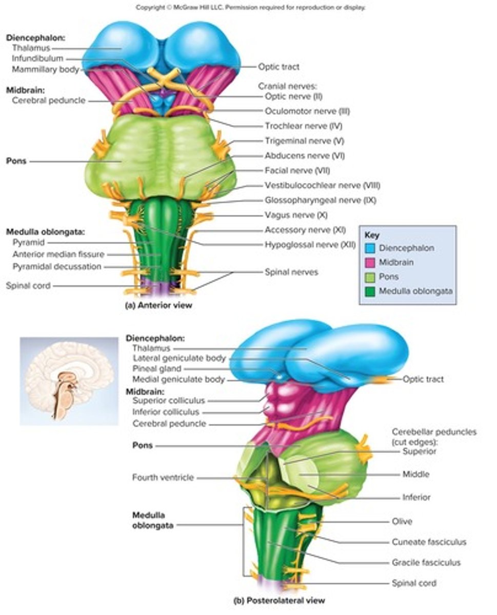
What is the medulla oblongata and where is it located?
An adult brain region that develops from the embryonic myelencephalon, beginning at the foramen magnum and extending about 3 cm rostrally.
What are the anatomical features of the medulla oblongata?
Pyramids on the anterior surface, resembling side-by-side baseball bats, separated by the anterior median fissure.
What is the significance of the blood-brain barrier being selectively permeable?
It allows necessary substances to pass while excluding harmful ones.
What is the consequence of the absence of tight junctions in ependymal cells elsewhere?
It allows for exchange between brain tissue and cerebrospinal fluid.
What percentage of blood does the brain receive relative to its body weight?
The brain receives 15% of the blood despite contributing only 2% of adult body weight.
What is the importance of a constant blood supply to neurons?
Neurons have a high demand for ATP, oxygen, and glucose, making a constant blood supply critical.
What is the typical age demographic for stroke occurrences?
Two-thirds of stroke patients are over 65 years old.
What is the role of the blood-CSF barrier?
It protects the brain at the choroid plexus and regulates the exchange of substances.
What happens to brain tissue during a stroke?
There is a sudden death of brain tissue due to blood supply interruption.
What is the anatomical location of the medulla oblongata in relation to the pons?
It ends at a groove just below the pons.
What are the two points of entry that must be guarded by the brain barrier system?
Blood capillaries throughout the brain tissue and capillaries of the choroid plexus.
What is the function of the inferior olivary nucleus in the medulla oblongata?
It serves as a relay center for signals to the cerebellum.
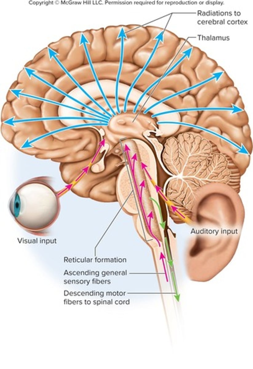
What does the reticular formation in the brainstem contain?
It contains the cardiac center, vasomotor center, and respiratory centers.
What is the pons and how long is it?
The pons is an adult brain region that develops from the embryonic metencephalon and measures 2.5 cm long.
What anatomical feature connects the pons to the cerebellum?
Cerebellar peduncles, which are thick stalks on the posterior pons.
What additional functions does the reticular formation in the pons serve?
It contains nuclei concerned with sleep, respiration, and posture.
What is the midbrain and what does it connect?
The midbrain is a brain region that develops from the embryonic mesencephalon and connects the hindbrain to the forebrain.
What is the role of the cerebral aqueduct in the midbrain?
It is a channel that connects the third and fourth ventricles.
What is the function of the periaqueductal gray substance in the midbrain?
It is involved in pain awareness.
What are the two parts of the tectum in the midbrain?
The two superior colliculi, which are involved in visual attention and tracking moving objects, and the two inferior colliculi, which relay signals from the inner ear to the thalamus.
What are the three parts of the cerebral peduncles in the midbrain?
Tegmentum, substantia nigra, and cerebral crus.
What is the reticular formation?
A loose web of gray matter that runs vertically through all levels of the brainstem and into the upper spinal cord.
What is the primary function of the cerebellum?
Motor coordination and locomotor ability.
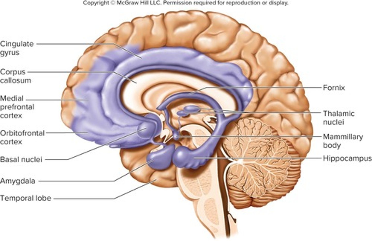
What are the two main parts of the forebrain?
The diencephalon and telencephalon.
What does the diencephalon enclose?
The third ventricle.
What are the three major components of the diencephalon?
Thalamus, hypothalamus, and epithalamus.
What is the location of the thalamus in the brain?
It is perched at the superior end of the brainstem beneath the cerebral hemispheres.
What is the interthalamic adhesion?
It is a structure that joins the right and left thalami medially.
How much of the diencephalon does the thalamus constitute?
About four-fifths of the diencephalon.
How many nuclei are composed in the thalamus?
At least 23 nuclei within five major groups: anterior, posterior, medial, lateral, and ventral.
What is the main function of the hypothalamus?
It regulates various autonomic functions, including temperature control, hunger, and thirst.
What is the role of the epithalamus?
It includes the pineal gland, which secretes melatonin and regulates sleep-wake cycles.
What is the significance of the basal nuclei?
They are involved in the regulation of voluntary motor control and procedural learning.
What is the limbic system responsible for?
It is involved in emotion, behavior, motivation, long-term memory, and olfaction.
What is the role of the thalamus in relation to the cerebral cortex?
The thalamus acts as a gateway to the cerebral cortex, processing nearly all input to the cerebrum and screening out most information.
How does the thalamus contribute to motor control?
The thalamus relays signals from the cerebellum to the cerebrum and provides feedback loops between the cerebral cortex and basal nuclei.
What functions are associated with the limbic system in relation to the thalamus?
The limbic system includes some of the anterior thalamic nuclei, which are involved in memory and emotion.
What anatomical structures define the hypothalamus?
The hypothalamus forms part of the walls and floor of the third ventricle, extending anteriorly to the optic chiasm and posteriorly to the mammillary bodies.
What is the function of the mammillary bodies in the hypothalamus?
Each mammillary body contains nuclei that relay signals from the limbic system to the thalamus.
How is the hypothalamus connected to the pituitary gland?
The hypothalamus attaches to the pituitary gland through a stalk-like structure called the infundibulum.
What are the major functions of the hypothalamus?
The hypothalamus is a major control center for the autonomic nervous system and endocrine system, regulating homeostasis across all body systems.
What role do hypothalamic nuclei play in hormone secretion?
They control the anterior pituitary, regulating growth, metabolism, reproduction, and stress responses, and produce hormones for labor contractions, lactation, and water conservation.
How does the hypothalamus regulate autonomic effects?
It serves as a major integrating center for the autonomic nervous system, influencing heart rate, blood pressure, and gastrointestinal functions.
What is the function of the hypothalamic thermostat?
It monitors body temperature and activates mechanisms to adjust temperature as necessary.