Topic 13 ankle and foot.
 Call Kai
 Learn
Learn Practice Test
 Spaced Repetition
 Match
Match Flashcards
 Knowt Play
1/36

There's no tags or description
Looks like no tags are added yet.
Name | Mastery | Learn | Test | Matching | Spaced | Call with Kai | Chat |
|---|
No analytics yet
Send a link to your students to track their progress
37 Terms

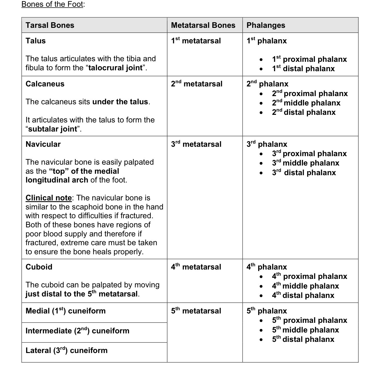

Name the bones labeled 1-7
Talus - articulates with the tibia and fibula to create “talocrural joint”
Calcaneus - sits under the talus, forms the subtalar joint
Navicular - easily palpated as the “top” of the medial longitudinal arch of the foot. Clinical note - the navicular bone is similar to the scaphoid bone in the respect to difficulties if fractured, this bone has poor blood supply so extreme meeasures have to be made to ensure it heals properly
Cuboid - the cuboid can be palpated by moving just distal to the 5th metatarsal
Medial (1st) cuneiform
Intermediate (2nd) cuneiform
Lateral (3rd) cuneiform


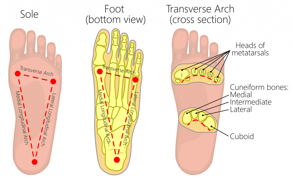
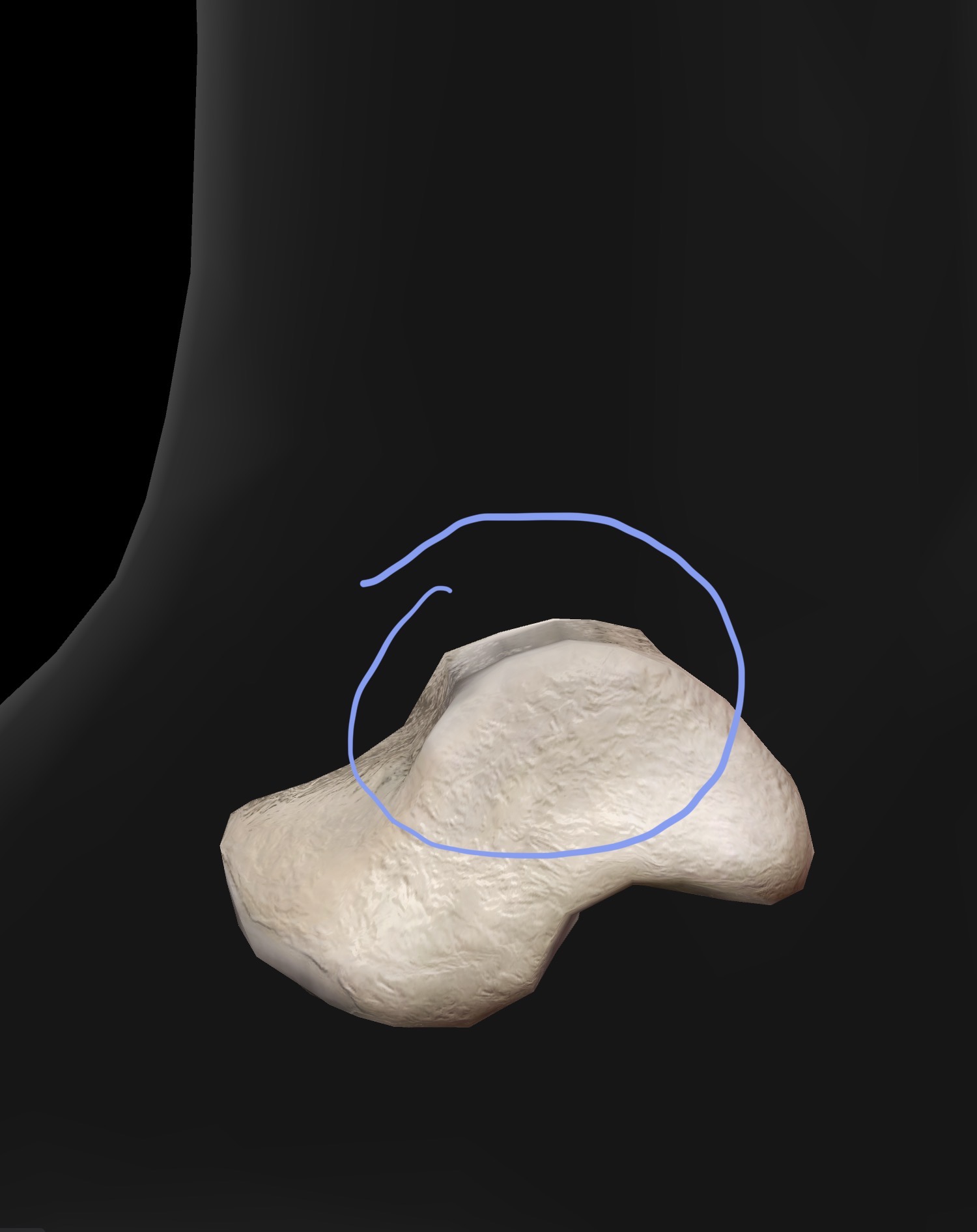
What bone is this? What are the bone landmarks?
Talar dome (trochlea) - rounded top of proximal aspect of the talus that articulates with the tibia and fibula to form the “talocrural“
The Talar dome can be palpated at the “top of the talus” best felt with the ankle (talocrural joint) in a plantar flexed position
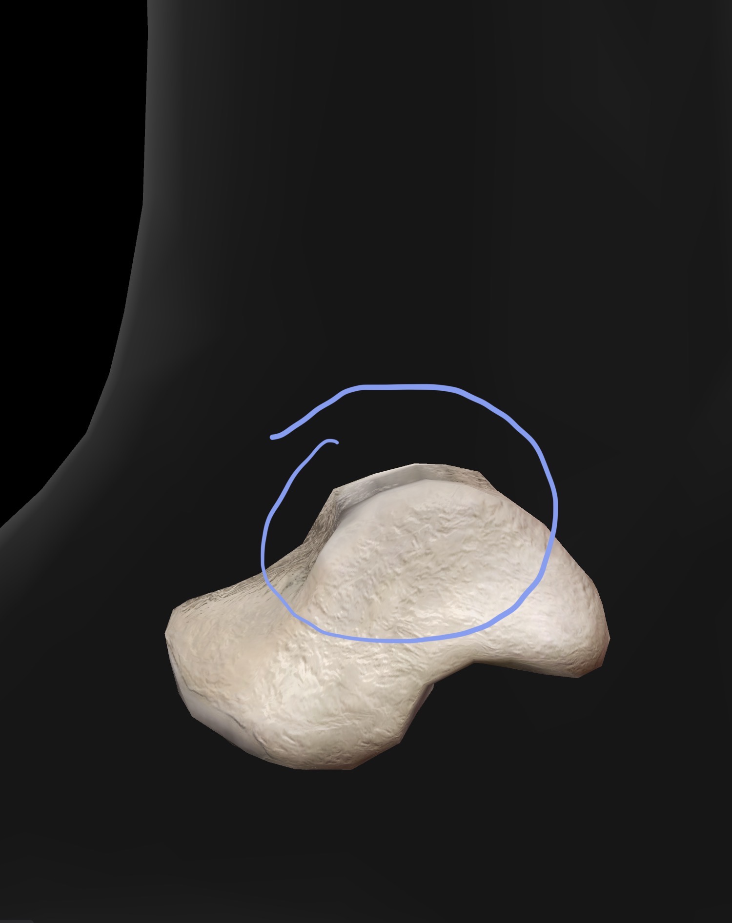

What bone is this? What are the bone landmarks?
calcaneus

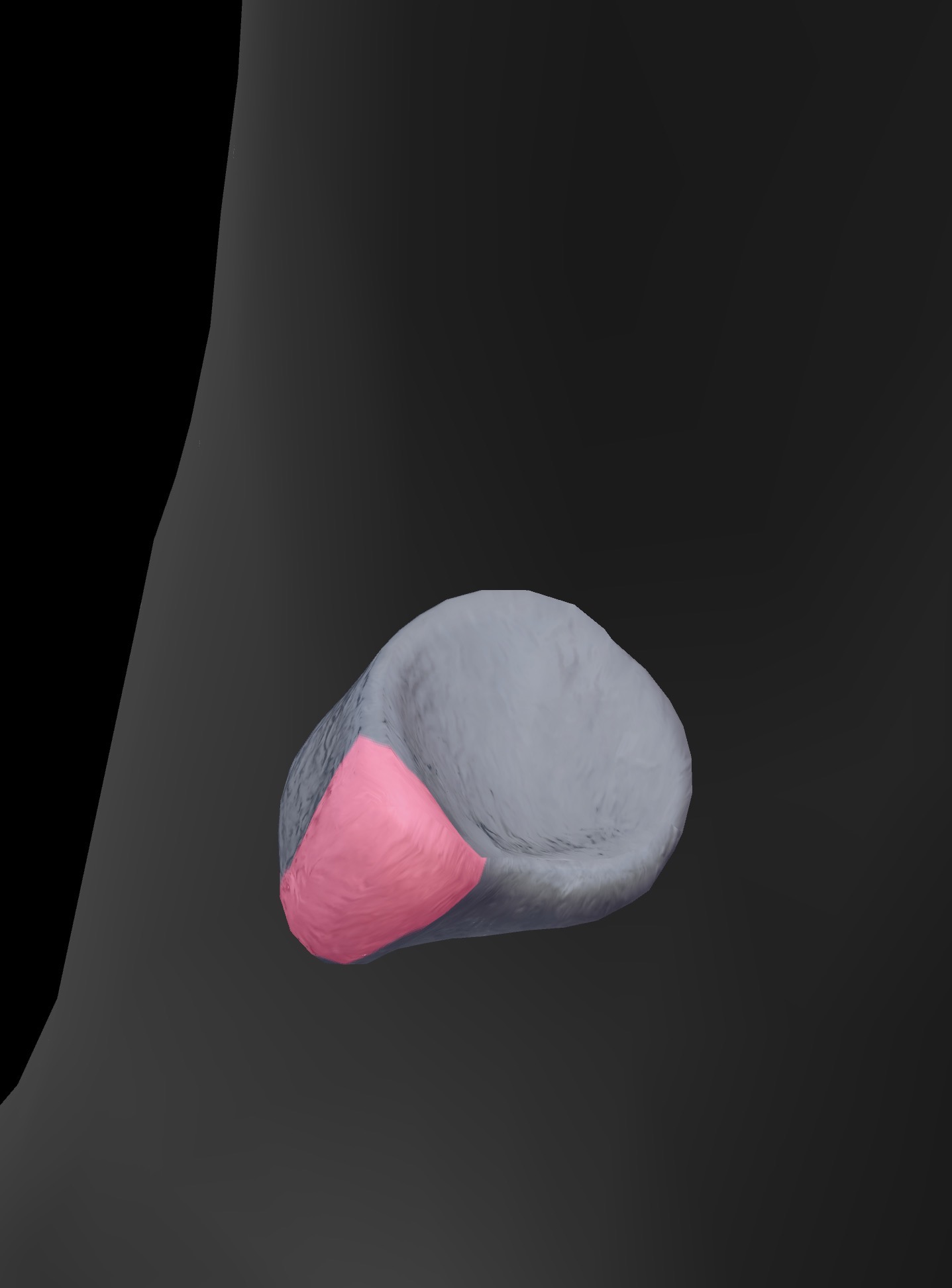
What bone is this? What are the bone landmarks?
Navicular - navicular tuberosity
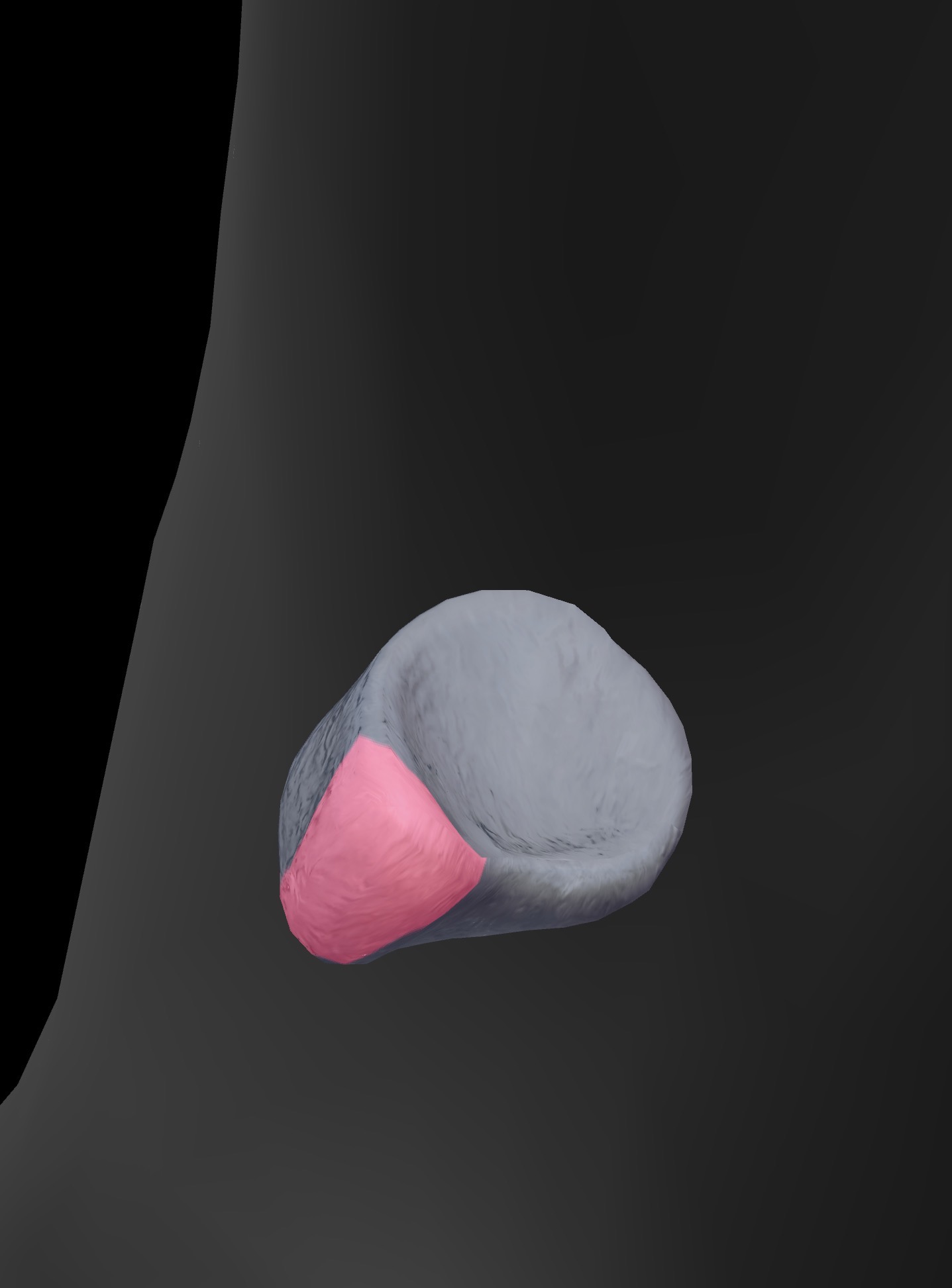
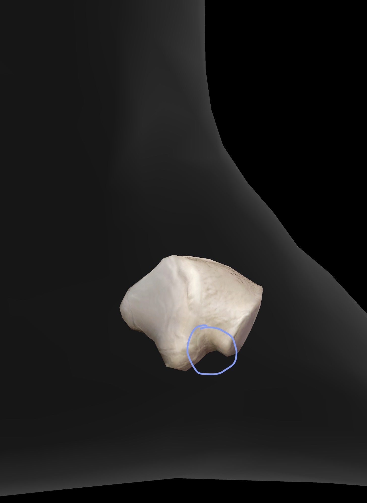
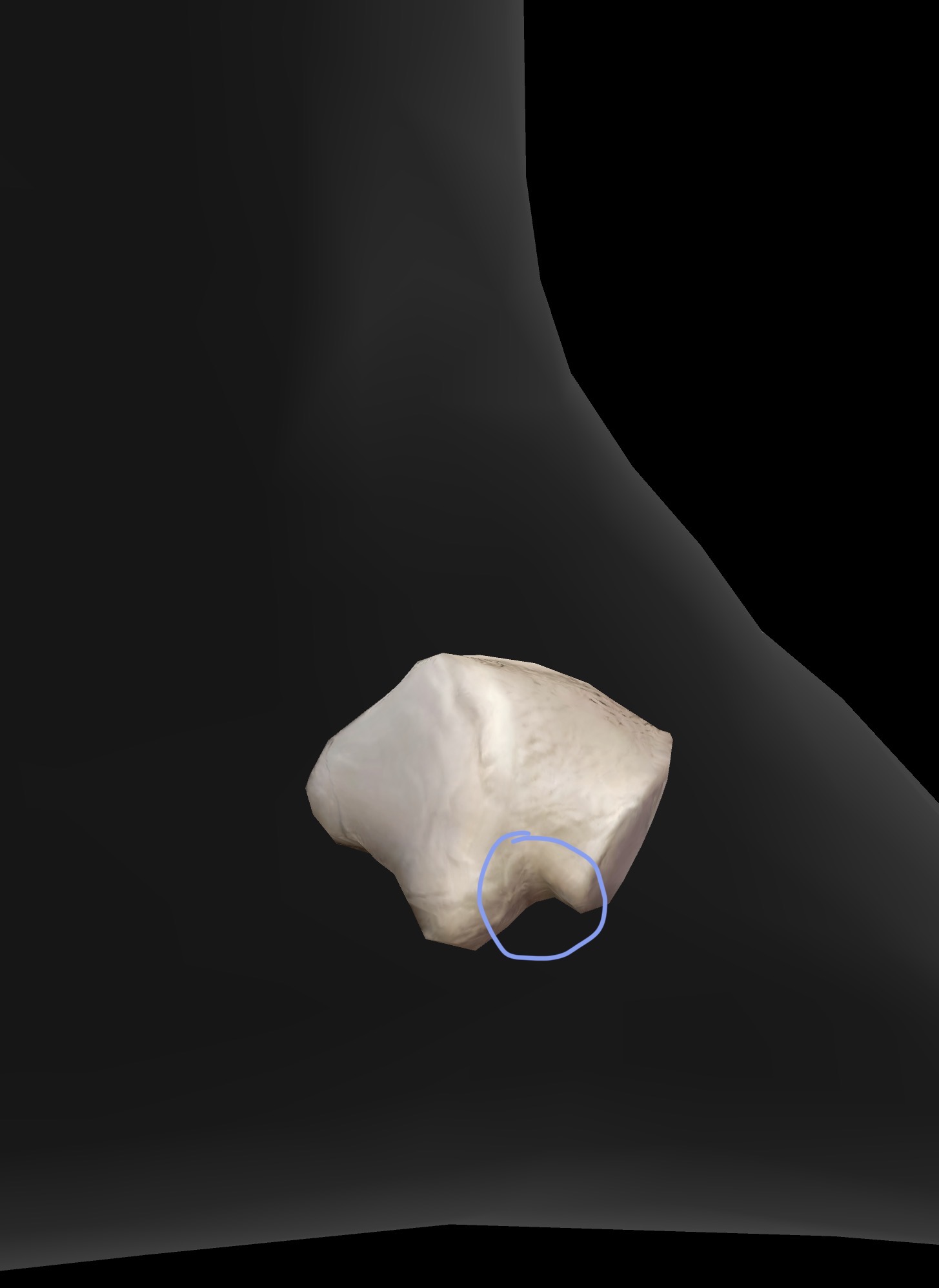
Cuboid - peroneal groove (tendon peroneus Longus passes through and attaches on the base of medial cuneiform and base of 1st metatarsal

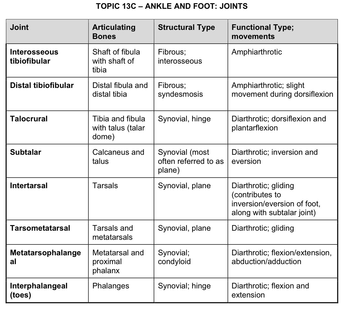
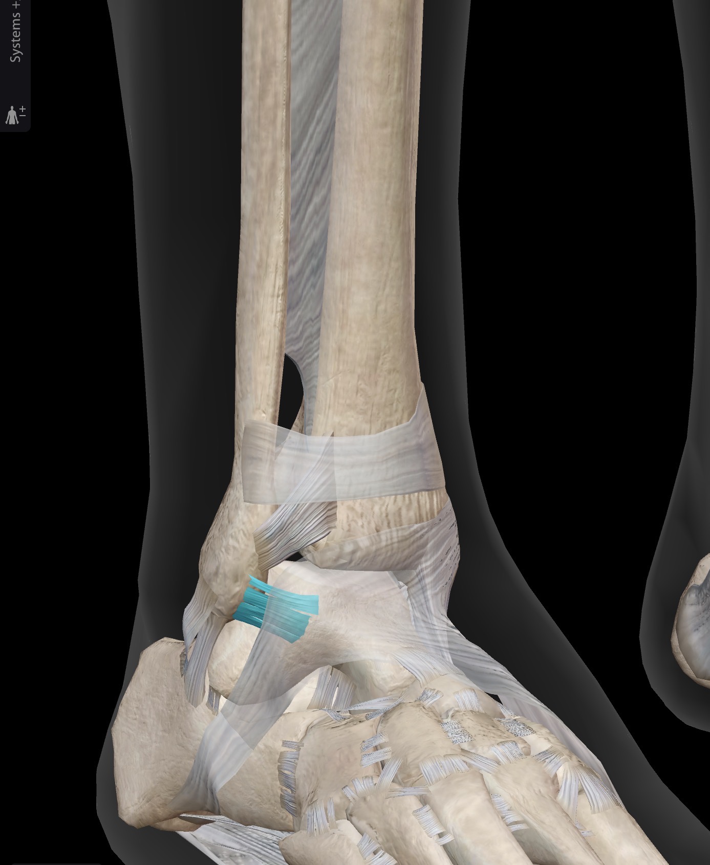
What ligament is this? What does it connect?
Anterior talofibular ligament (ATFL)
Lateral malleolus to lateral aspect of anterior talus
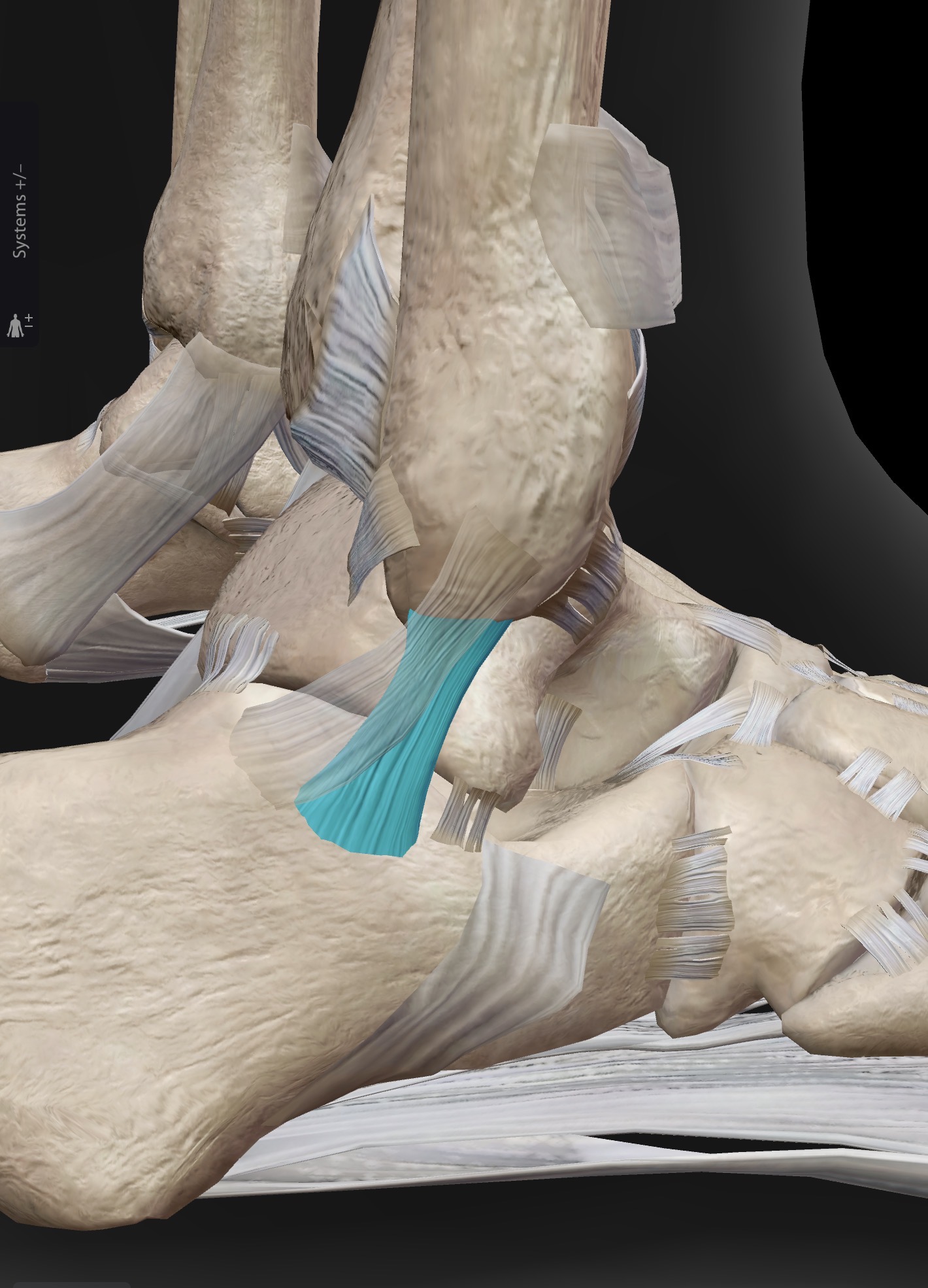
What ligament is this? What does it connect?
Calcaneofibular ligament
Lateral malleolus to lateral calcaneus
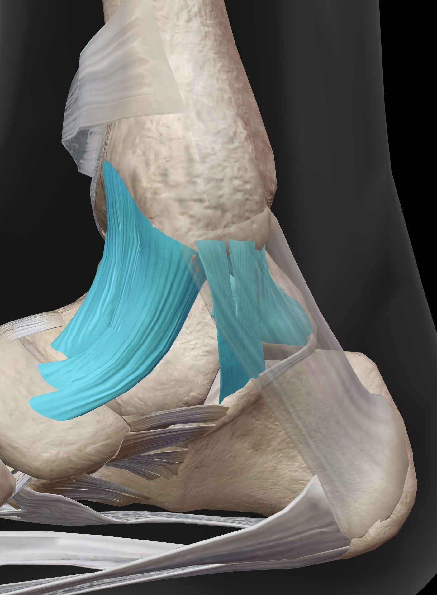
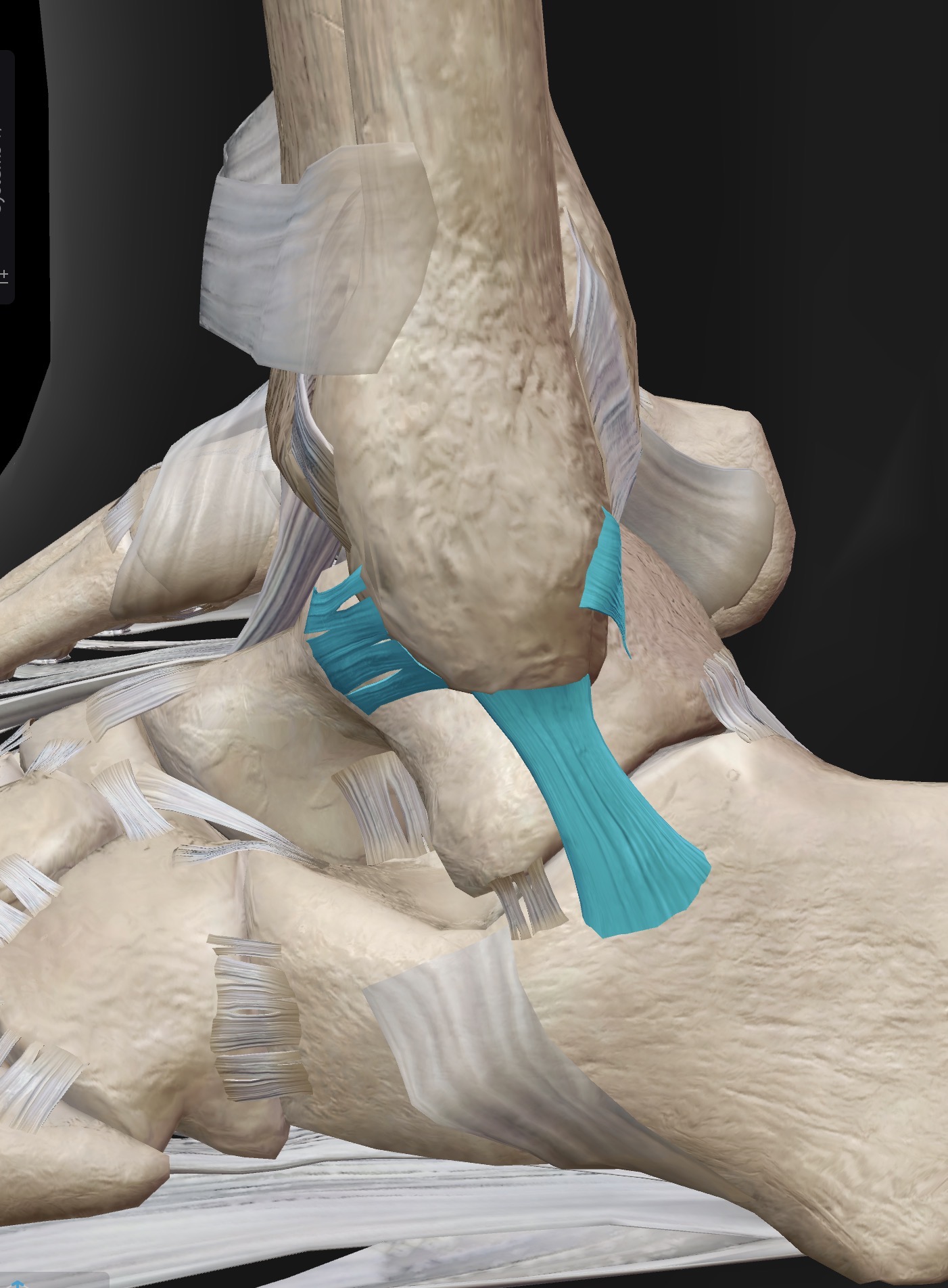
What ligament is this? What does it connect?
Medial ankle ligaments collectively known as “Deltoid ligaments”
Medial malleolus to medial aspect of talus, navicular and medial aspect of calcaneus
From left to right
Anterior tibiotalar, tibionavicular, tibiocalcaneal, posterior tibiotalar (behind flexor retinaculum)
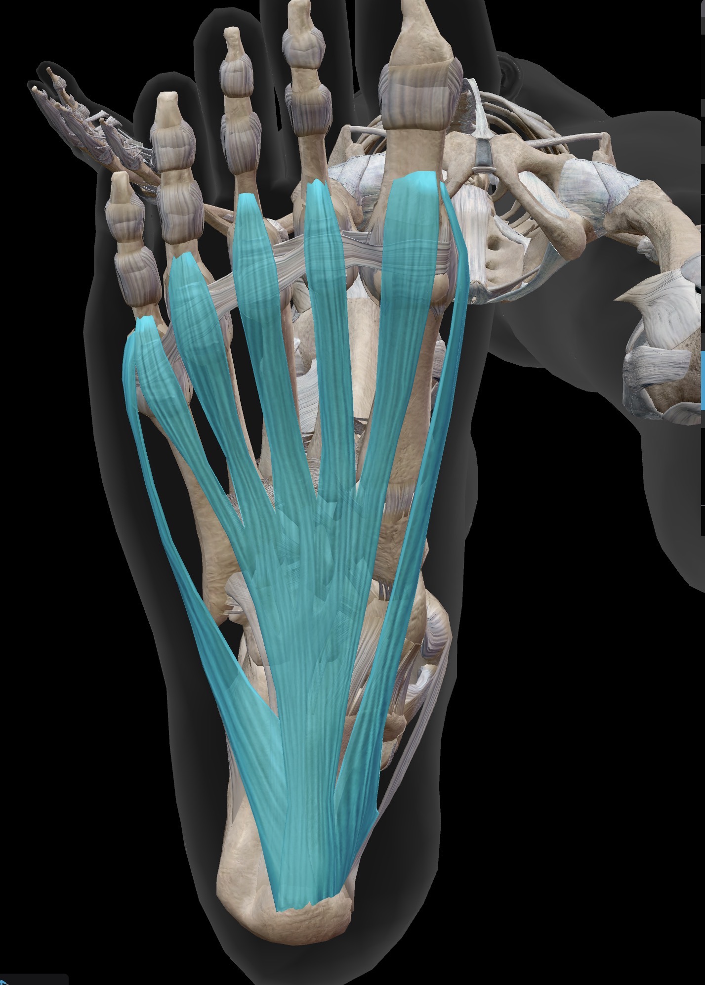
What ligament is this? What does it do?
Plantar fascia is a flat sheet of connective tissue that runs from the plantar aspect of the calcaneus (including the medial calcaneal process) to the bases of the proximal phalanges of the foot.
Clinical note: Inflammation of the plantar fascia often presents with pain on the bottom of the heel, at the medial calcaneal process (especially with the first few steps out of bed in the morning, and then later in the day with increased activity).
This condition is known as plantar fasciitis
What are the most common ankle sprains? What ligaments do they affect. What are the least common? What do they affect? What’s a high ankle sprains?
Inversion ankle sprains are the most common, affecting mostly the anterior tibiofibular ligament (ATFL) is the most commonly injured, followed by the calcaneofibular ligament
Less common are eversion ankle sprains which involve stretching the deltoid ligaments on the medial side of the ankle
Distal ends of the tibia and fibula are united by a strong syndesmosis formed by the interosseous membrane and ligaments at the distal tibiofibular joint
Injuries that produce a lateral twisting on a planted foot can result in stretching or tearing of the tibiofibular ligaments, producing a syndesmotic ankle sprain or “high ankle sprain”
What movements occur at the ankle and foot?
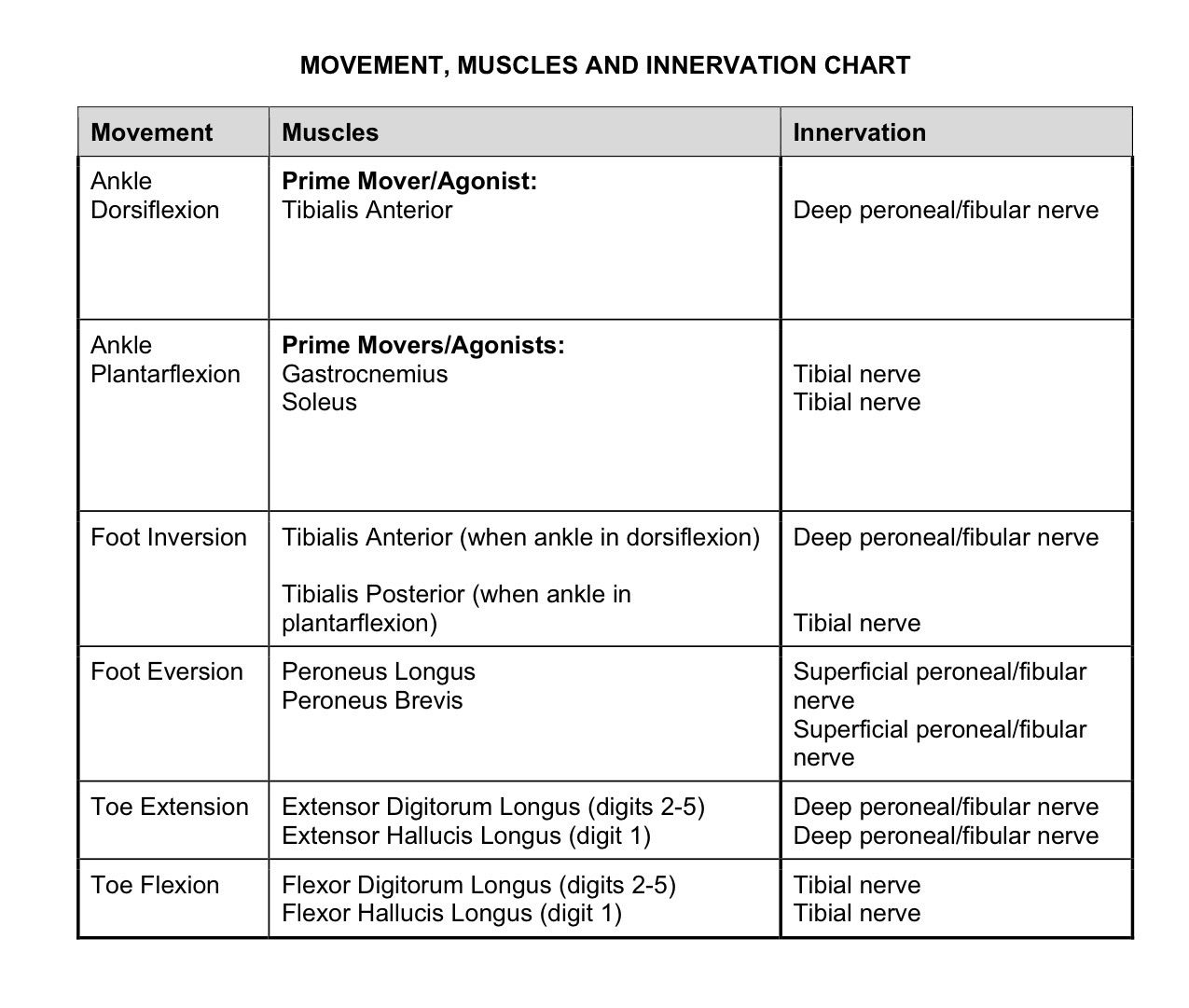
Plantarflexion/dorsiflexion of the foot at the talocrural joint (ankle joint)
Inversion/eversion of the foot at the subtalar and intertarsal joints
Flexion/extension of the toes at the interphalangeal joints
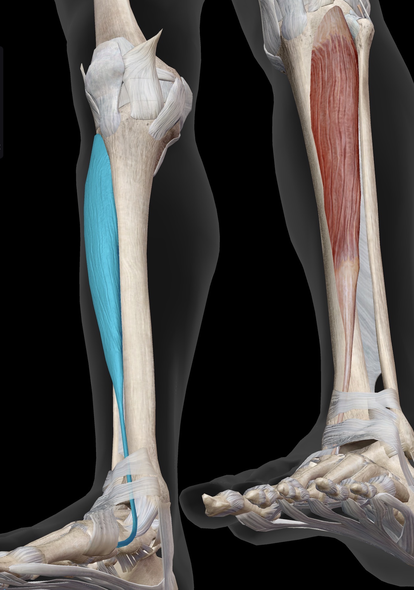
Name the muscle and corresponding letter ( O = origin, I = insertion, A = actions, IN = innervation)
O
I
A
IN
Tibialis anterior
O: Anterior surface of the tibia, on the lateral side
I: Plantar aspect of the 1st metatarsal and media cuneiform
A: Prime mover of dorsiflexion of ankle (talocrural joint). Also, one of the prime movers of inversion of the foot (at the subtalar and intertarsal joints)
Because the tibialis anterior tendon travels anterior to the medial malleolus at the ankle joint, if you invert your foot in dorsiflexion, you are mainly using tibialis anterior
IN: Deep peroneal (deep fibular) nerve
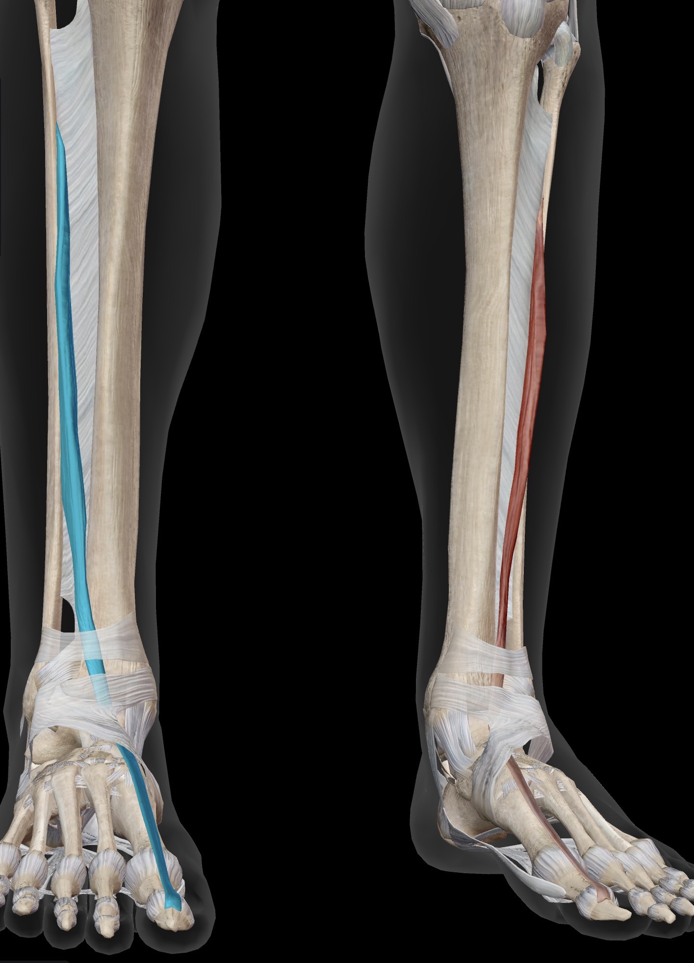
Name the muscle and corresponding letter ( O = origin, I = insertion, A = actions, IN = innervation)
O
I
A
IN
Extensor hallucis longus
O: Anterior fibula
I: Dorsal surface of distal phalanx of great (1st) toe
A: Prime mover of extension of the great toe. Synergistic of dorsiflexion of talocrural joint/ankle
IN: Deep peroneal (deep fibular) nerve
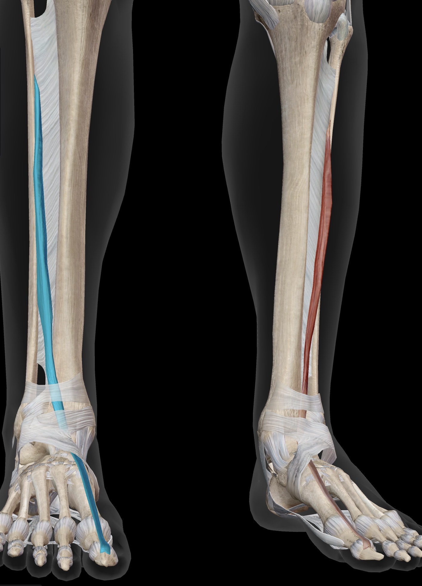
Name the muscle and corresponding letter ( O = origin, I = insertion, A = actions, IN = innervation)
O
I
A
IN
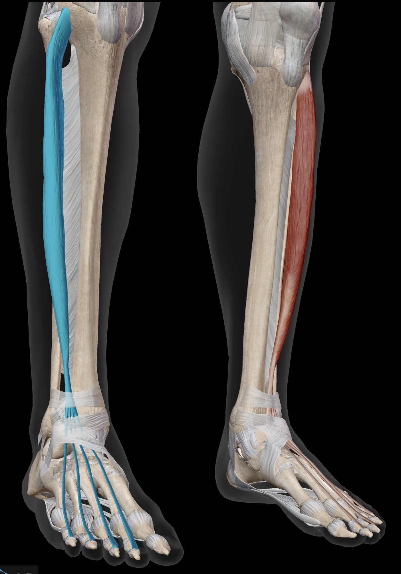
Extensor digitorum longus
O: Mainly anterior fibula
I: By four tendons into middle and distal phalanges of 2nd-5th toes
A: Prime mover of toe extension (digits 2-5). Synergistic of dorsiflexion of the talocrural joint/ankle
IN: Deep peroneal (Deep fibular) nerve
Name the muscle and corresponding letter ( O = origin, I = insertion, A = actions, IN = innervation)
O
I
A
IN
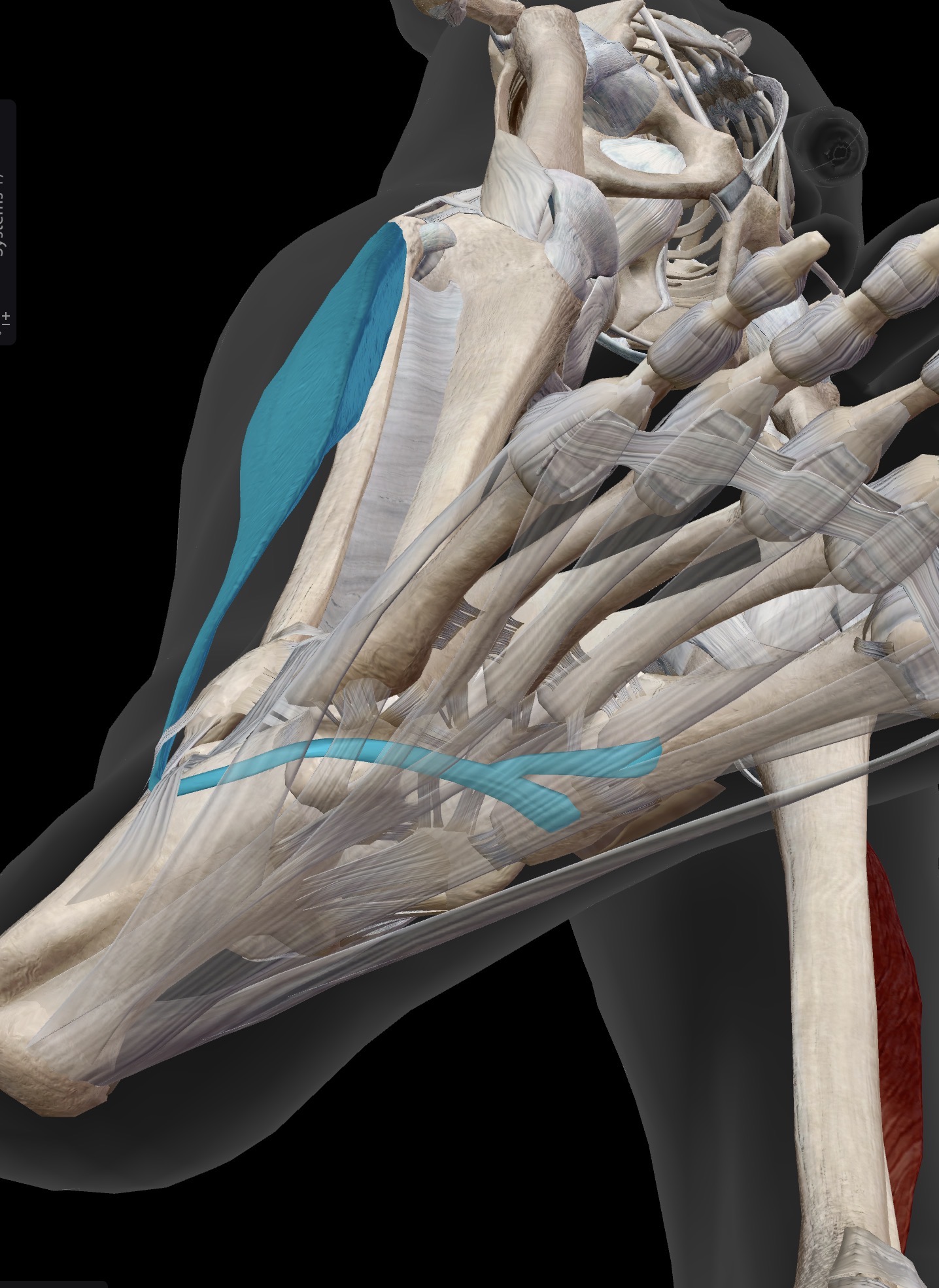
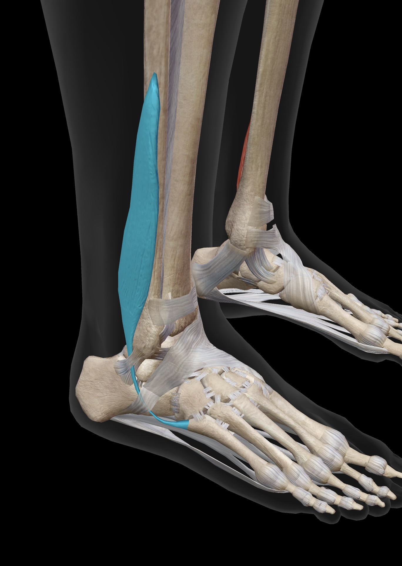
Peroneus longus (fibular is longus)
O: proximal lateral surface of fibula
I: Plantar aspect of the 1st metatarsal and medial cuneiform (beside insertion of tibialis anterior)
A: Prime mover of eversion of the subtalar and intertarsal joints (foot)
IN: Superficial peroneal (superficial fibular) nerve
Name the muscle and corresponding letter ( O = origin, I = insertion, A = actions, IN = innervation)
O
I
A
IN
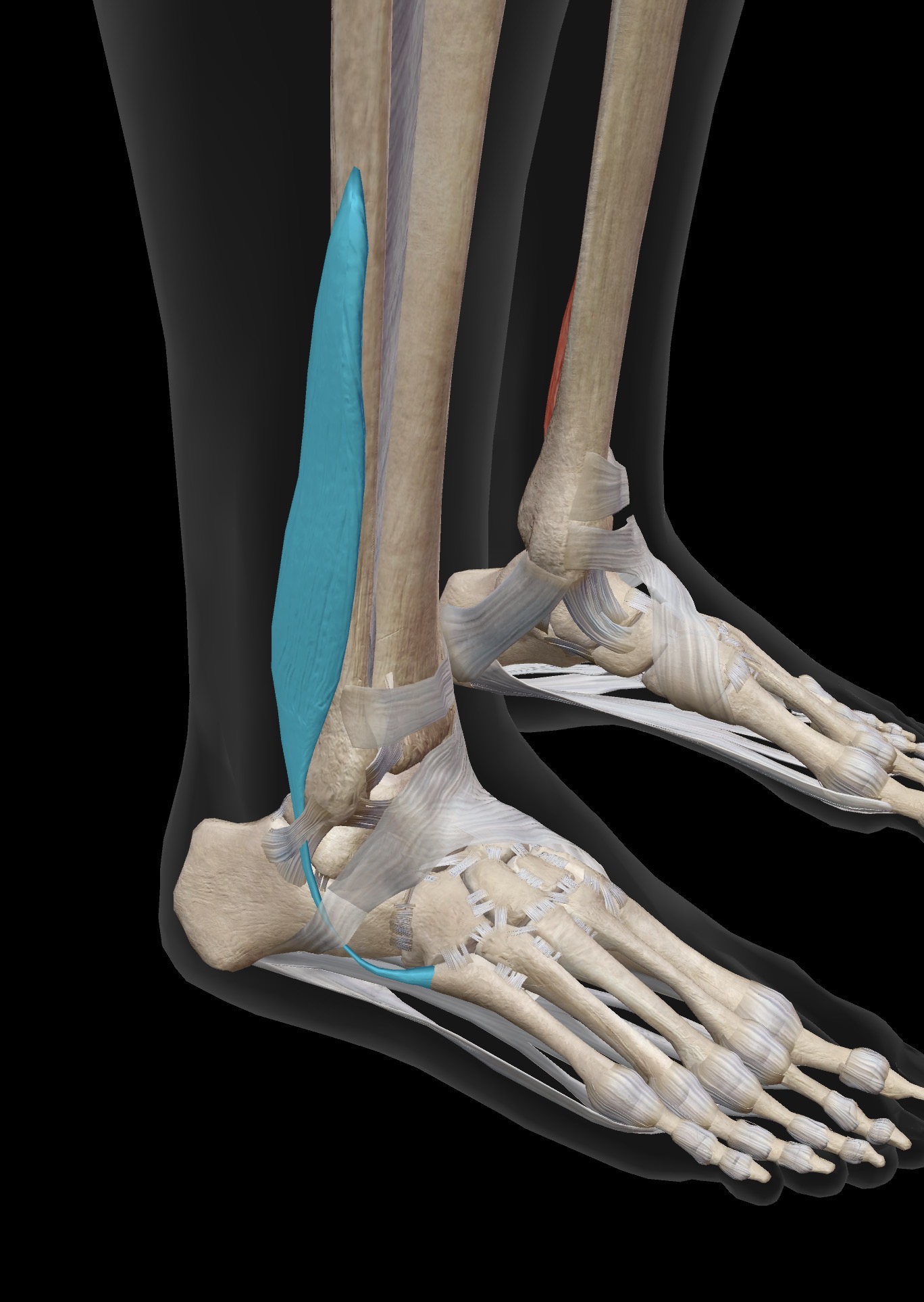
Peroneus Brevis (fibularis brevis)
O: Distal lateral surface of fibula
I: Base of the 5th metatarsal
A: Other important muscle for eversion of the subtalar and intertarsal joints (foot)
IN: Superficial peroneal (superficial fibular) nerve
Name the muscle and corresponding letter ( O = origin, I = insertion, A = actions, IN = innervation)
O
I
A
IN
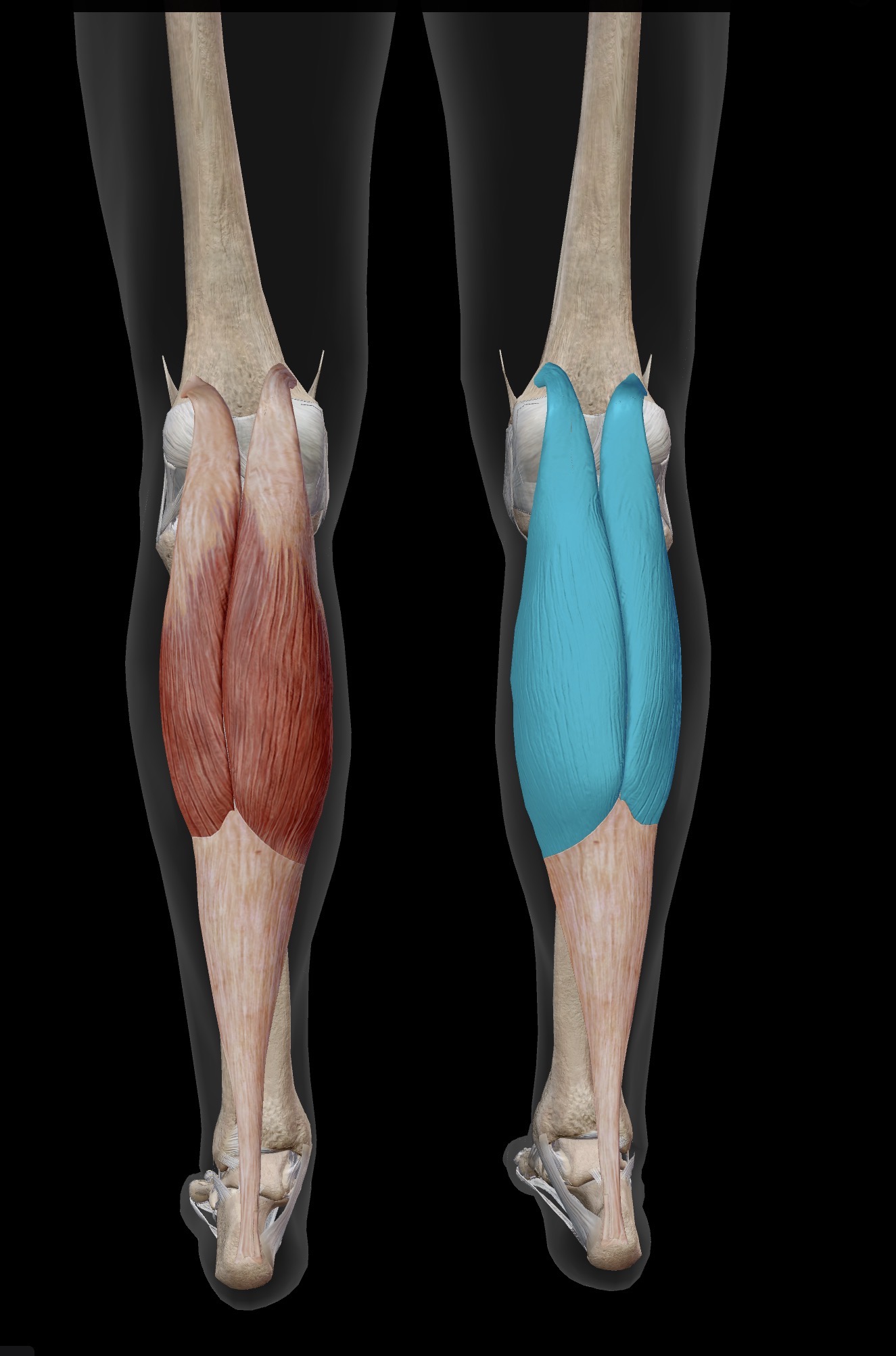
Gastrocnemius
O: Posterior aspect of medial and lateral condyles of femur
I: Calcaneal tuberosity via the Achilles tendon
A: One of 2 prime movers of plantarflexion of the talcocrural joint/ankle (Along with soleus). Synergistic of knee flexion
IN: Tibial nerve
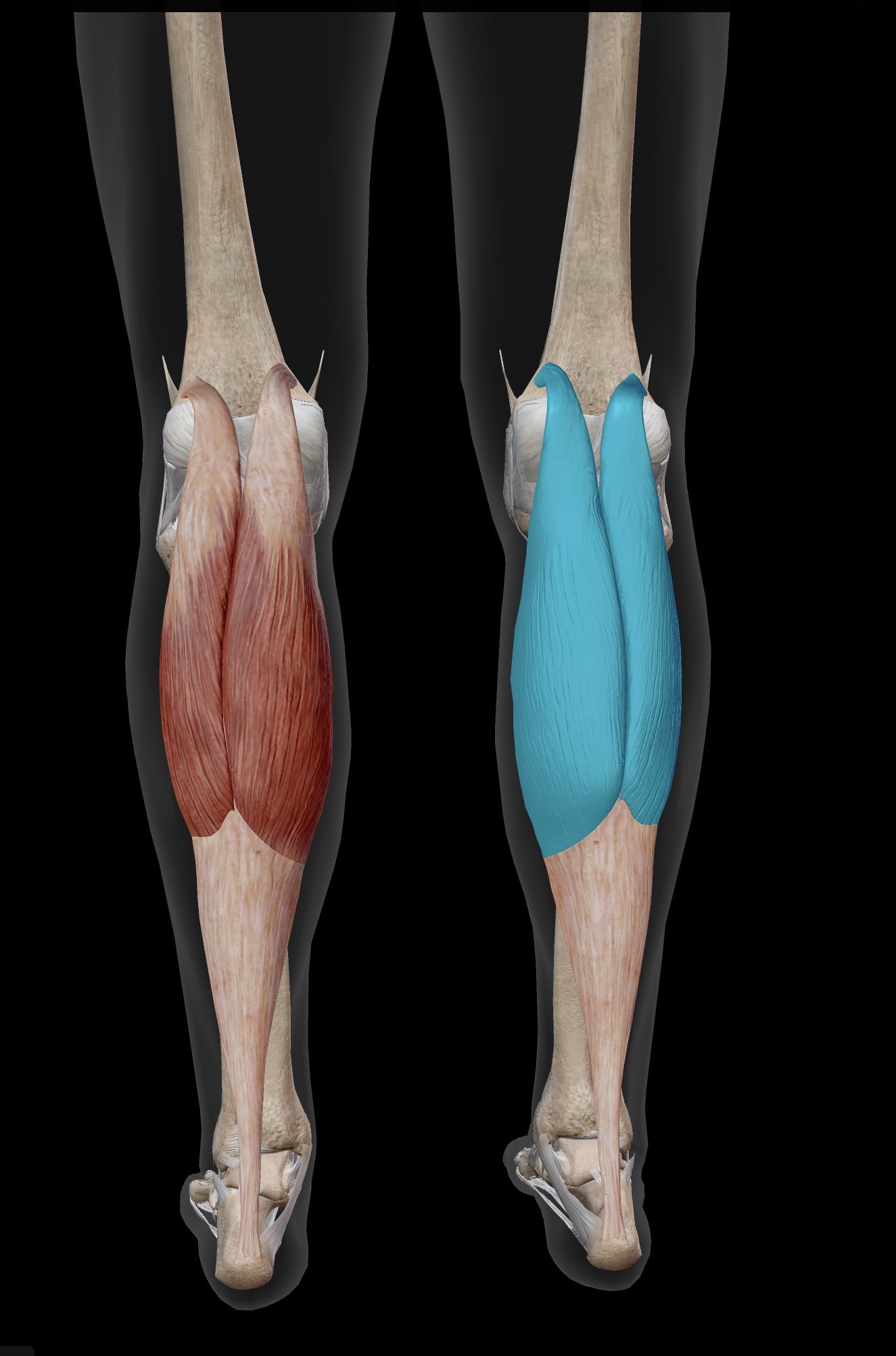
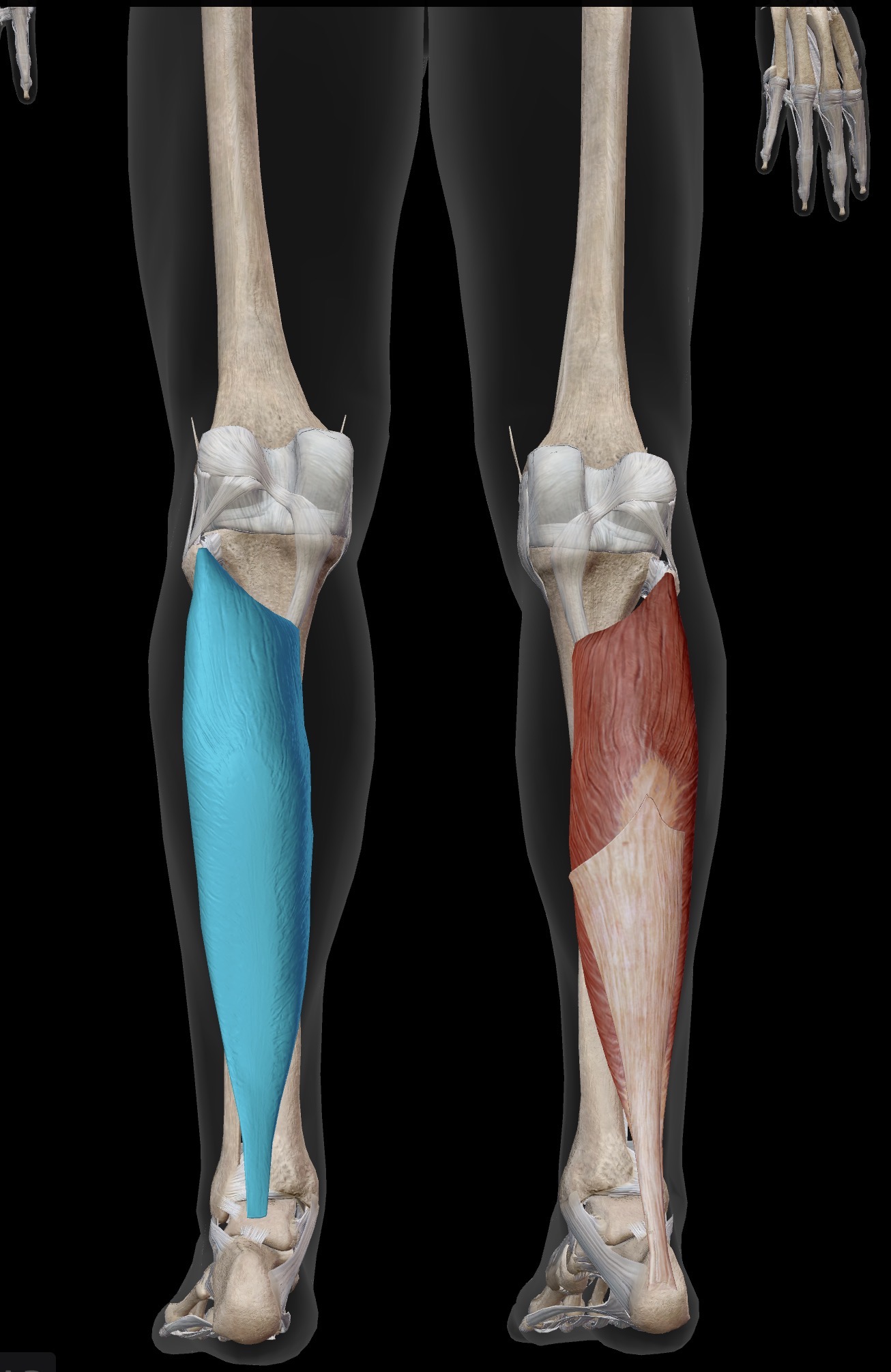
Name the muscle and corresponding letter ( O = origin, I = insertion, A = actions, IN = innervation)
O
I
A
IN
Soleus
O: Posterior aspect of soleus (soleal line) and fibula
I: Calcaneal uberosity via the Achilles tendon
A: One of 2 prime movers of plantarflexion of talocrural joint/ankle (along with gastocnemius)
IN: Tibial nerve
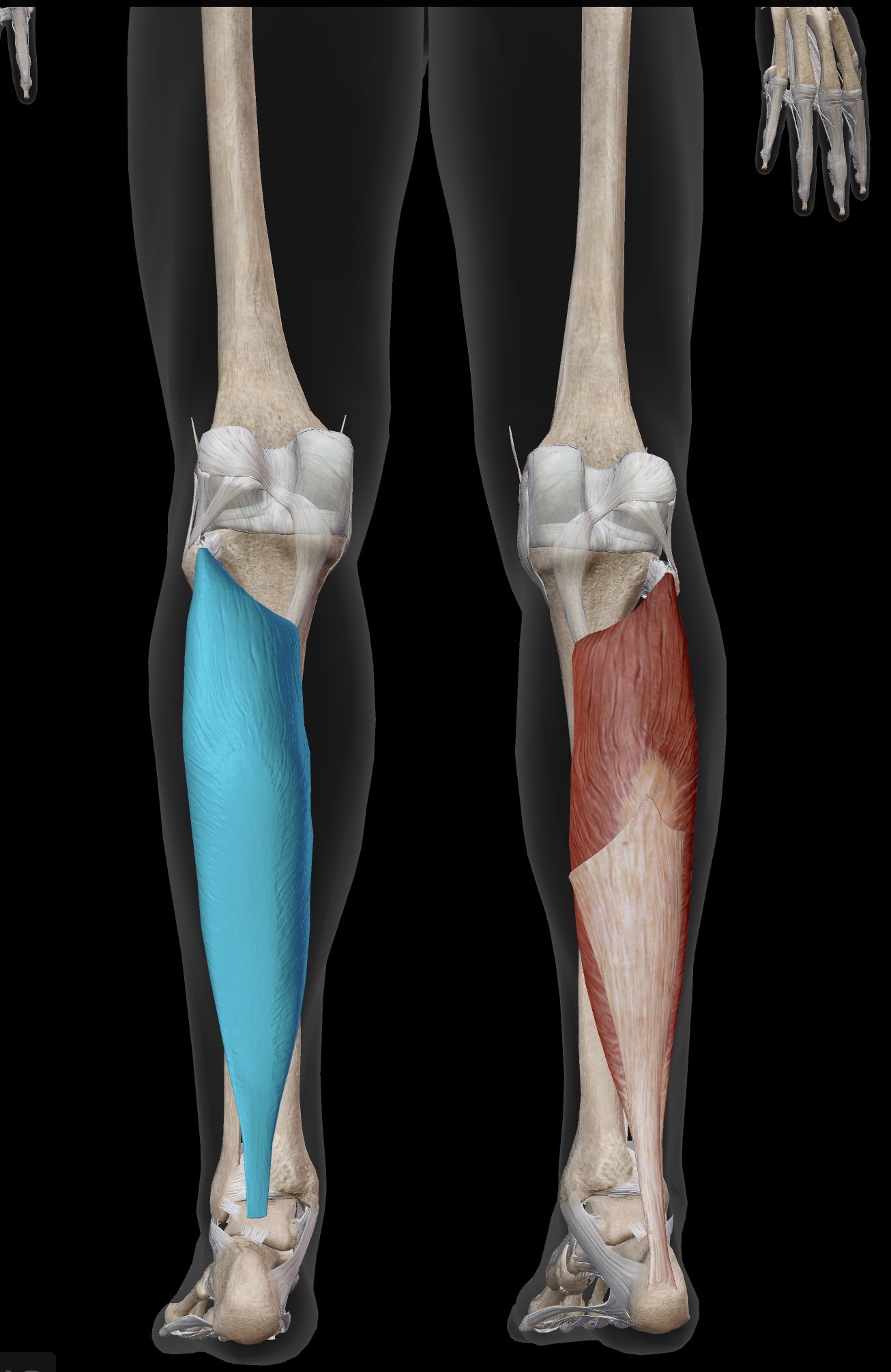
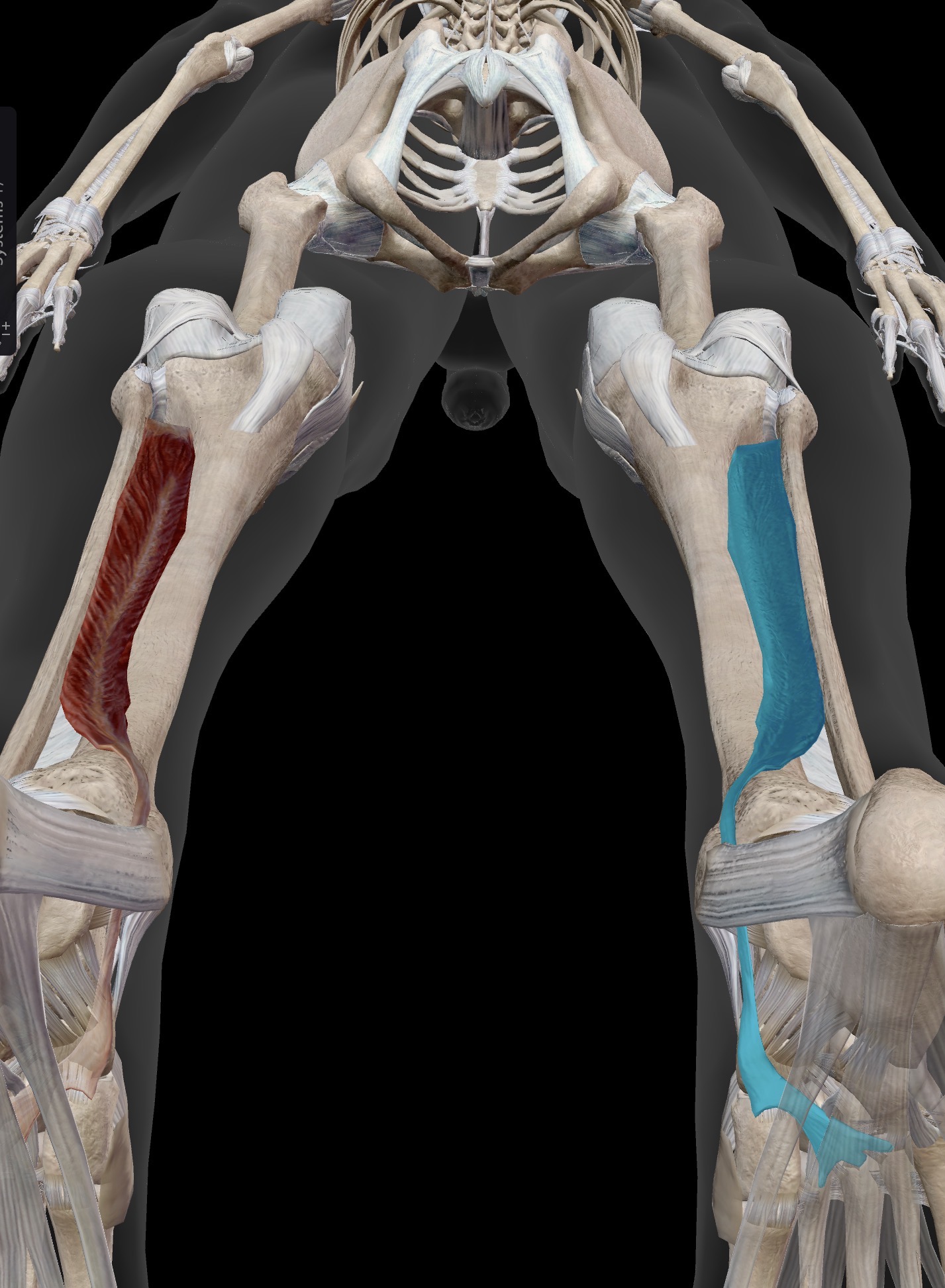
Name the muscle and corresponding letter ( O = origin, I = insertion, A = actions, IN = innervation)
O
I
A
IN
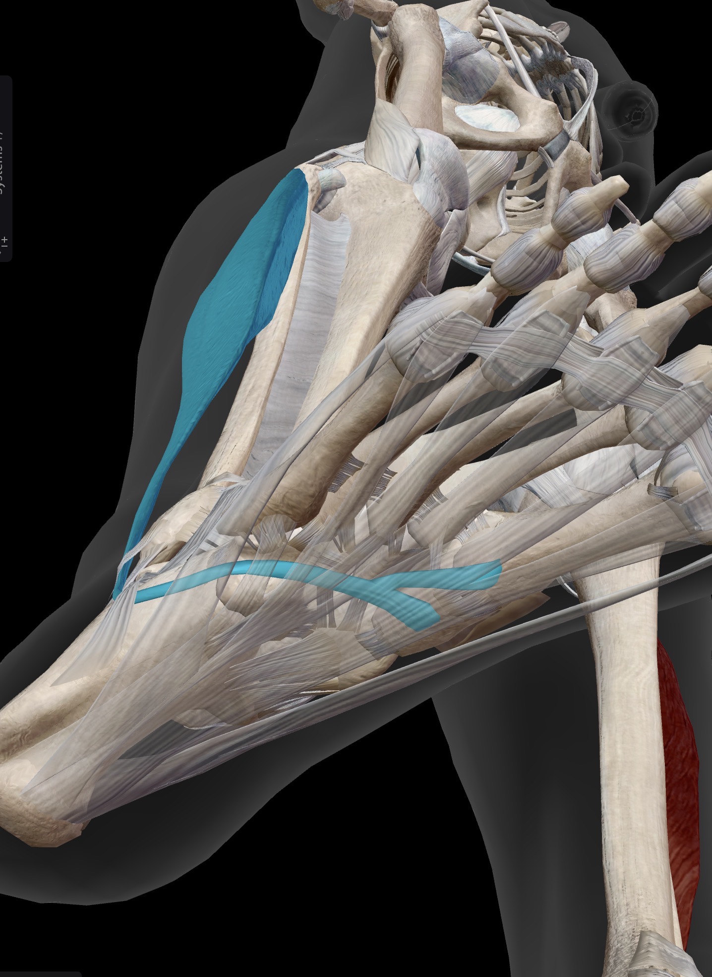
Tibialis posterior (along with tibialis anterior, this is one of the other prime movers of inversion of the foot)
O: Posterior leg, below the knee (posterior aspect of tibia and fibula)
I: Plantar aspect of several tarsal and metatarsal bones. Most important insertion of tibialis posterior is the navicular bone, on the navicular tuberosity.
A: Inversion of subtalar and intertarsal joints/foot
Because the tibialis posterior tendon travels posterior to the medial malleolus at the ankle joint, if you invert your foot in plantarflexion, you are using mainly tibialis posterior
IN: tibial nerve
Along with flexor hallucis longus and intrinsic foot muscles, tibialis posterior is an important muscle for supporting arches of the foot.
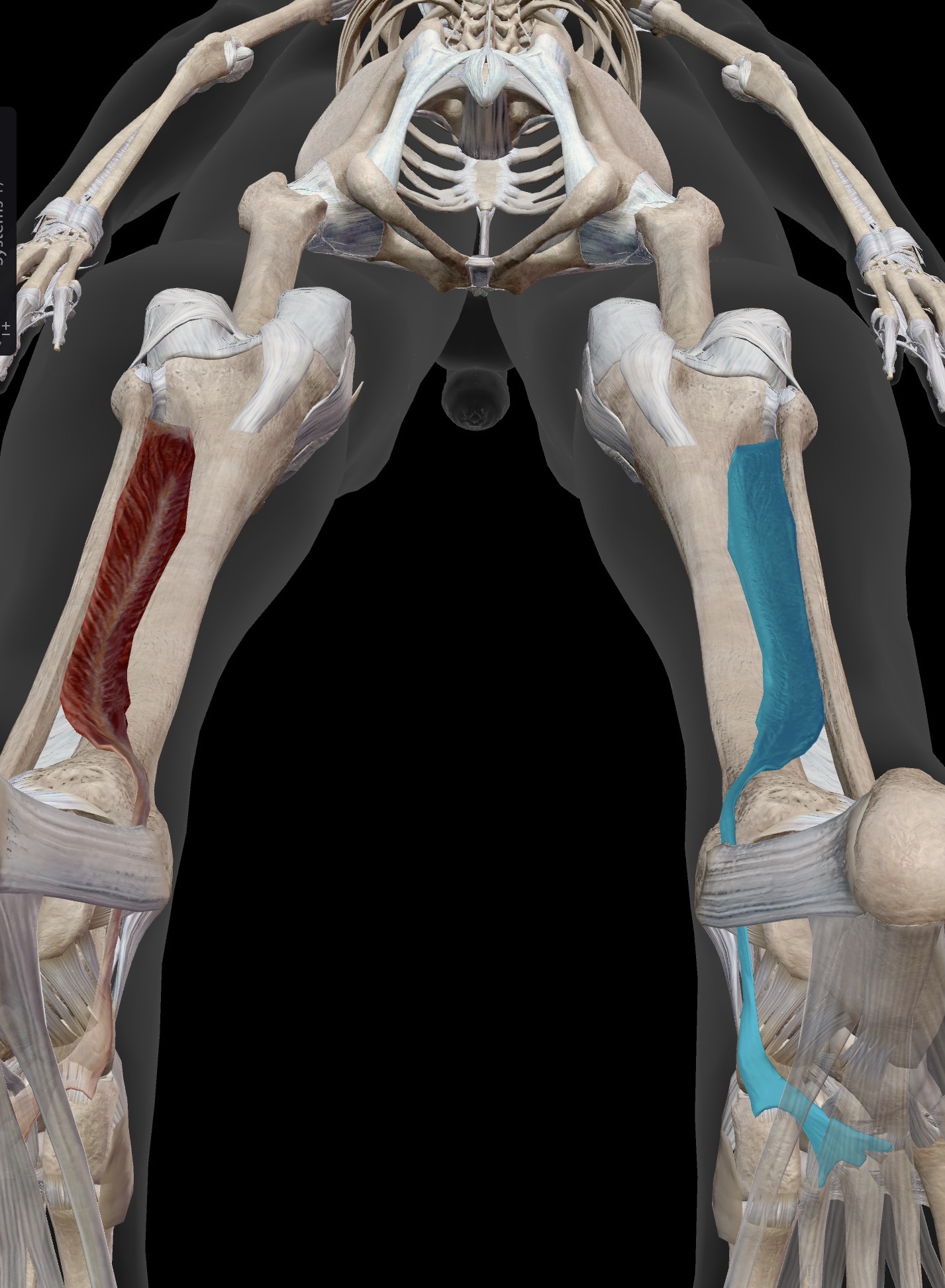
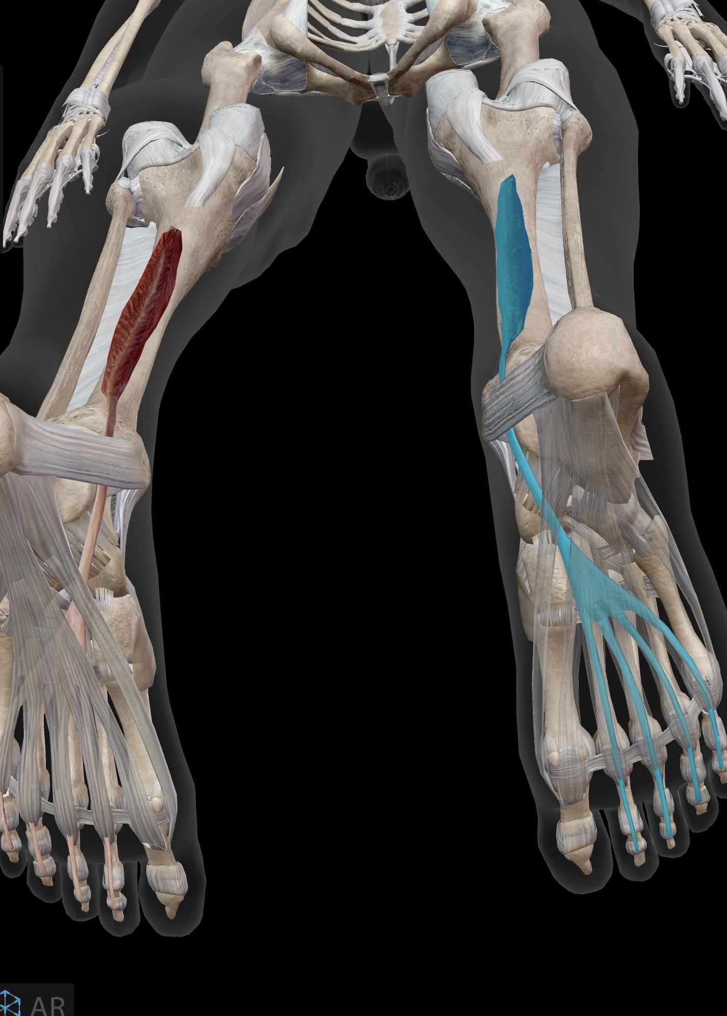
Name the muscle and corresponding letter ( O = origin, I = insertion, A = actions, IN = innervation)
O
I
A
IN
Flexor digitorum longus
O: Posterior leg, below the knee
I: Plantar aspect of distal phalanges of 2nd-5th toes via 4 tendons
A: Flexion of the 2nd-5th toes
IN: Tibial nerve
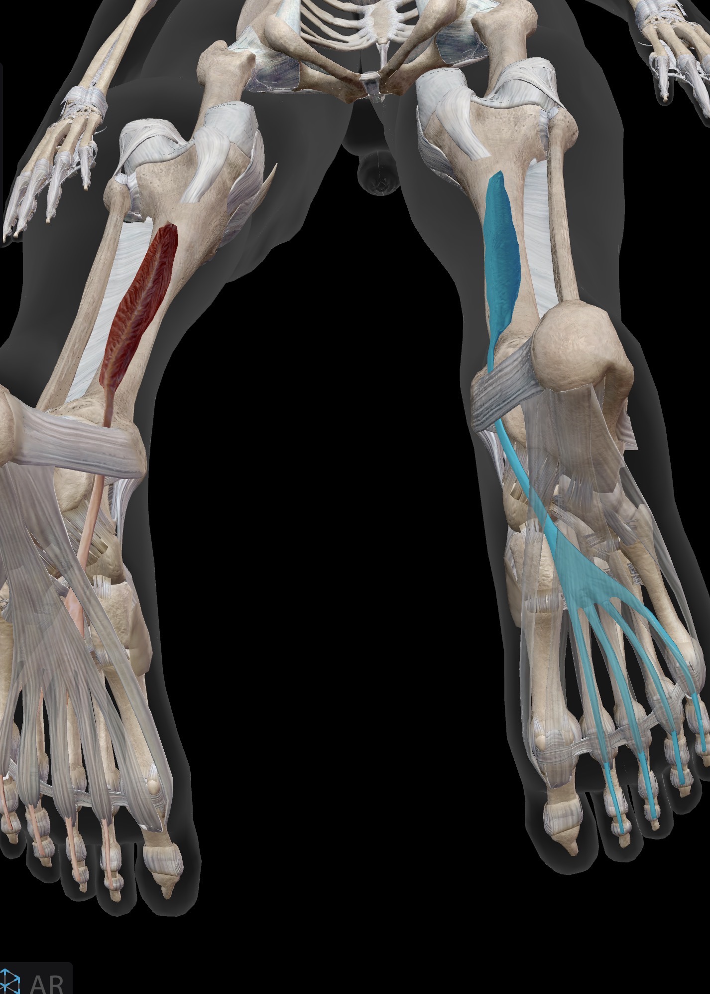
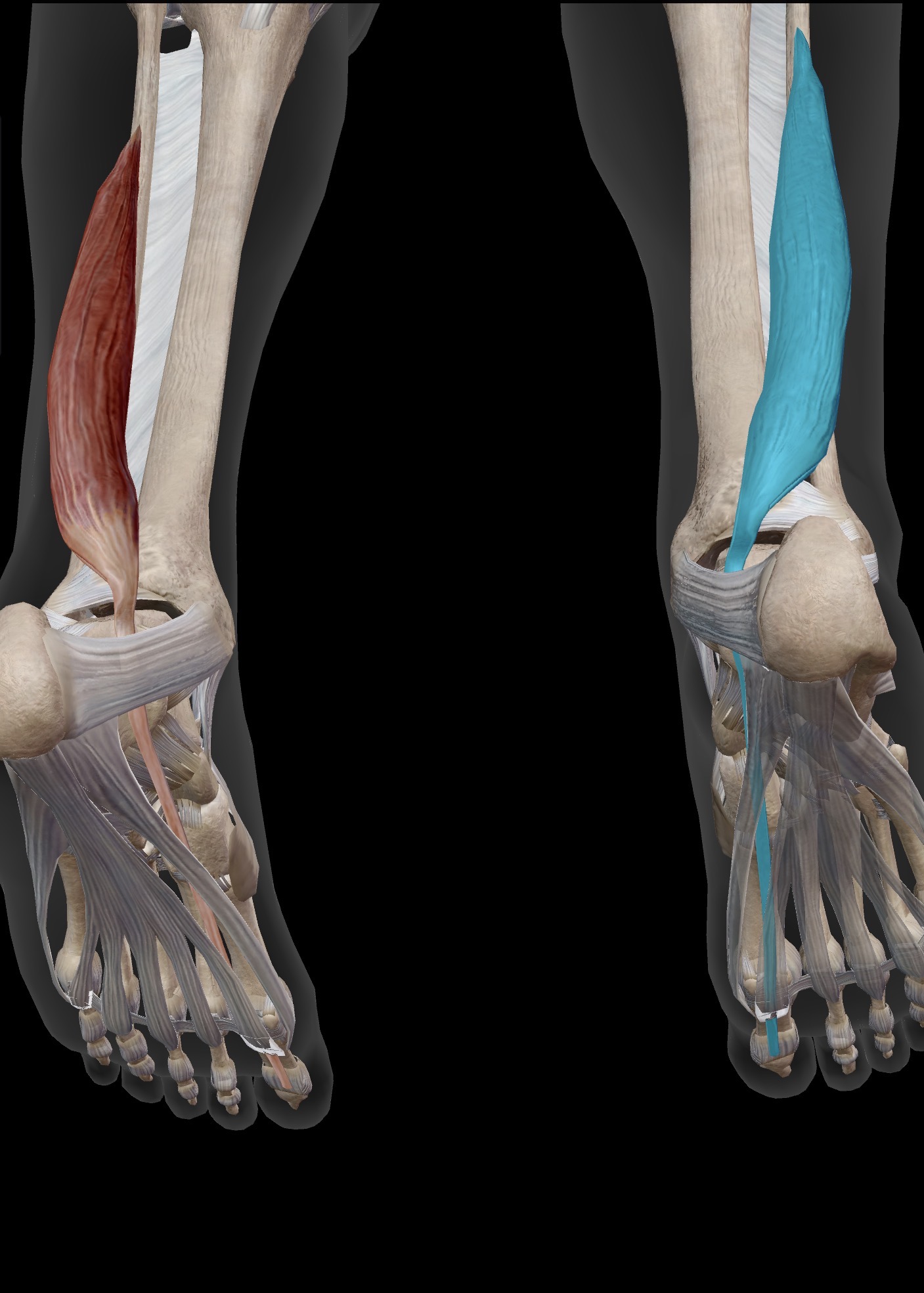
Name the muscle and corresponding letter ( O = origin, I = insertion, A = actions, IN = innervation)
O
I
A
IN
Flexor hallucis longus
O: Posterior leg, below the knee
I: Plantar aspect of distal phalanx of great toe
A: Flexion of the great toe
IN: Tibial nerve
Along with tibialis posterior and intrinsic foot muscles, flexor hallucis longus is an important muscle for supporting the arches of the foot.
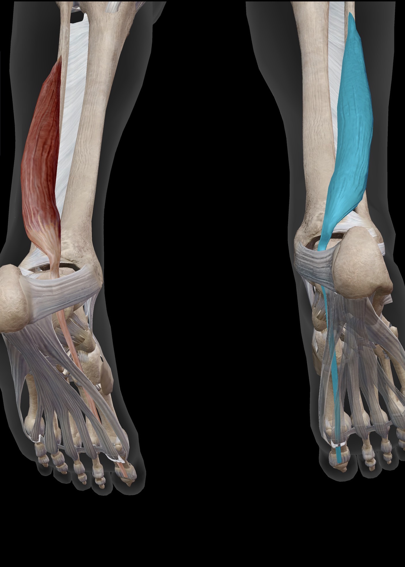


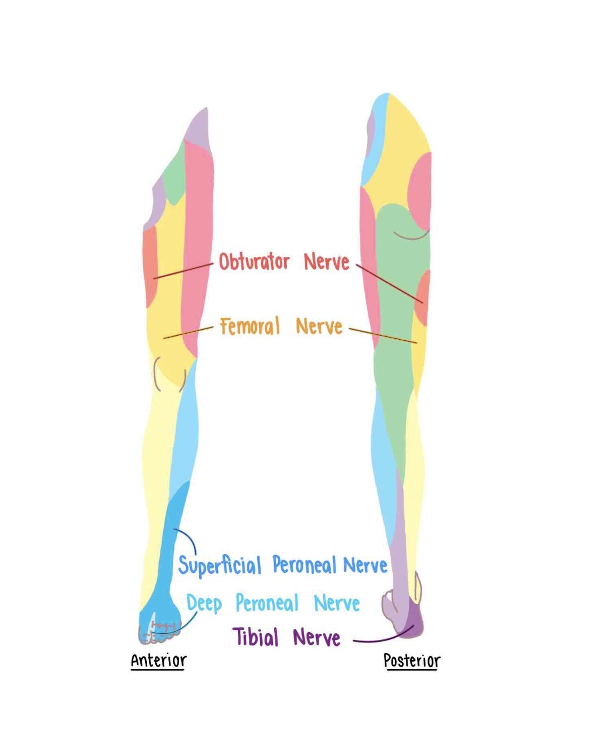
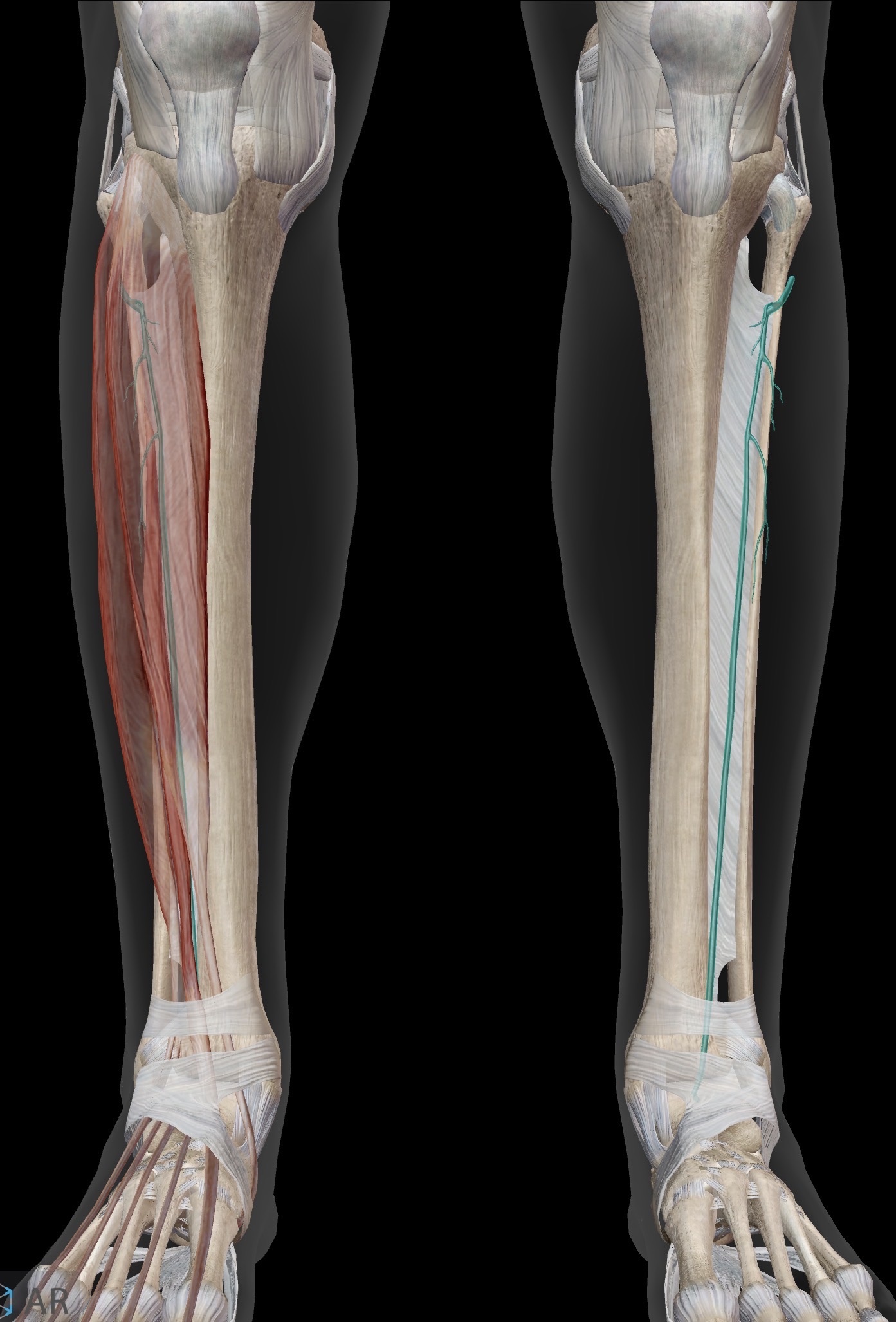
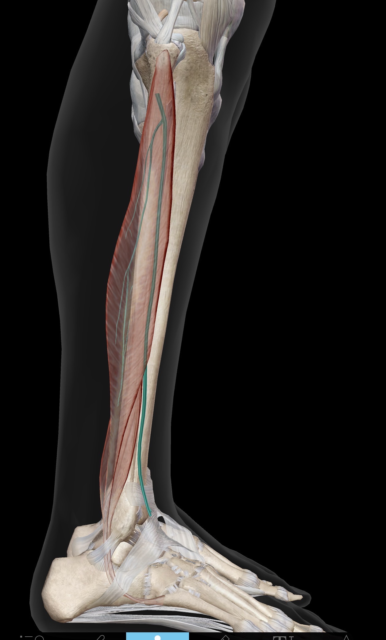
What nerve is this?
Deep peroneal (fibular) nerve
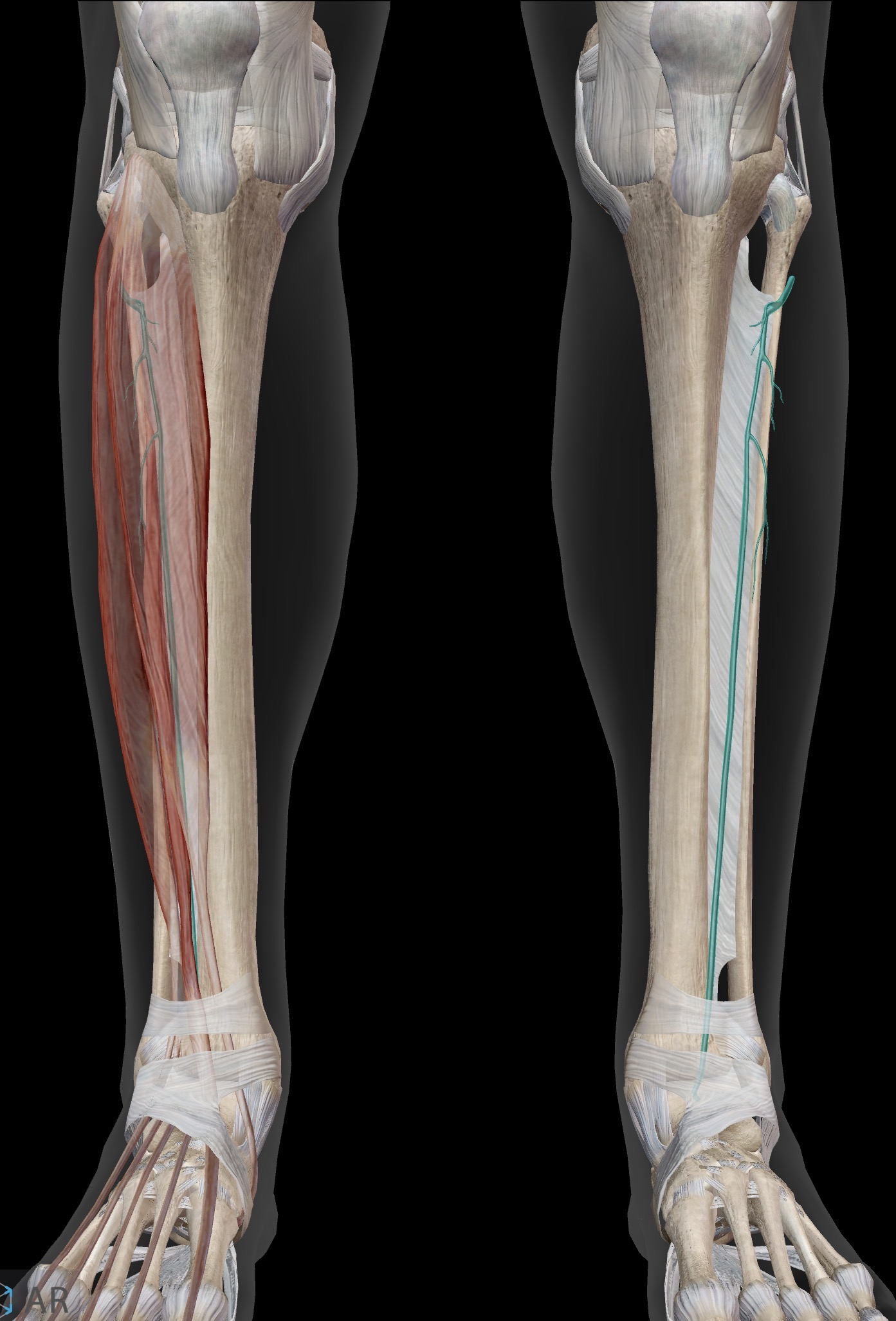
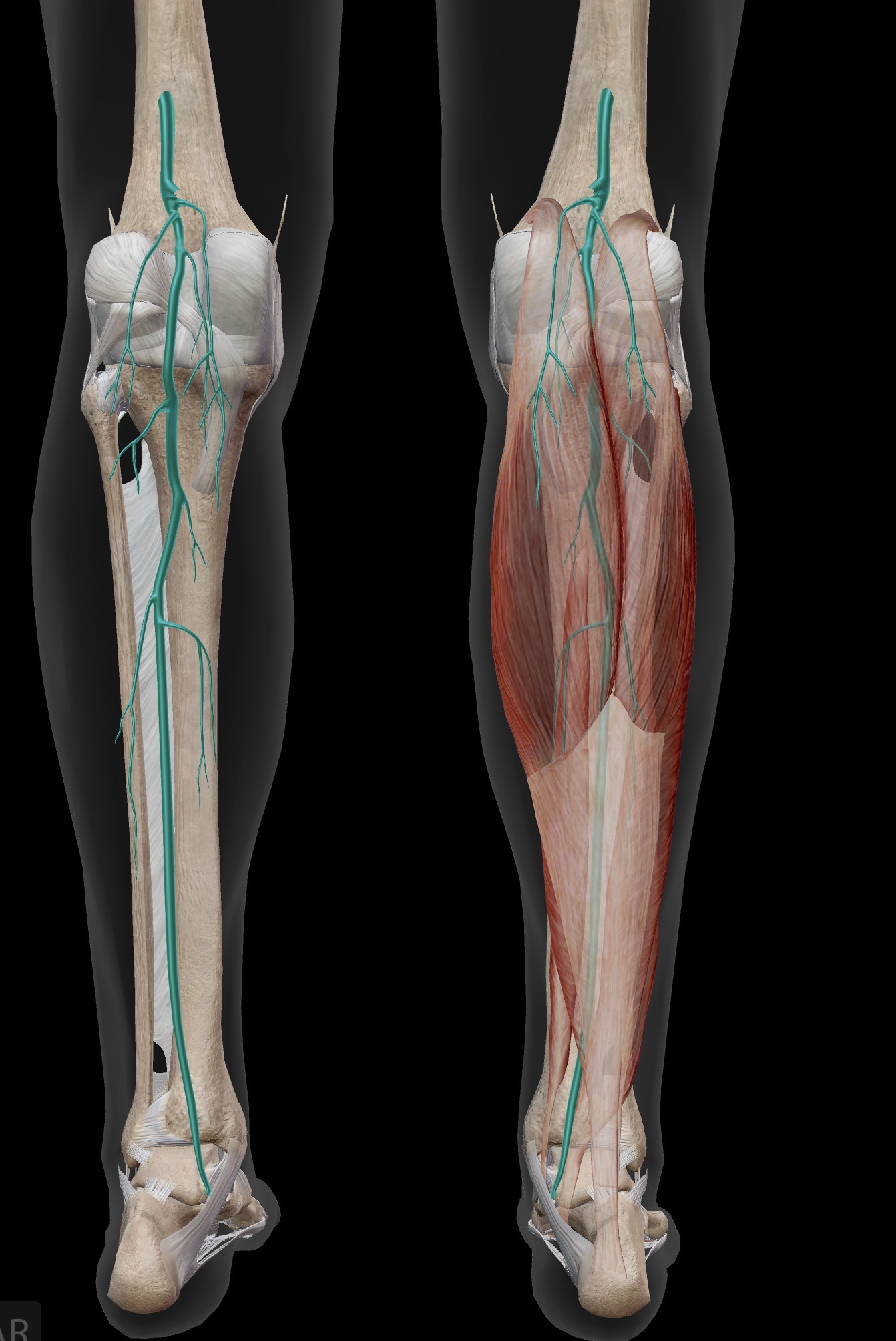
What nerve is this?
Tibial nerve
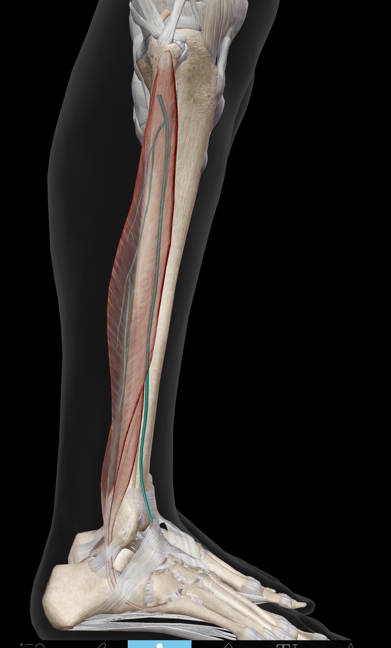
What nerve is this?
Superficial peroneal (fibular) nerve
What ligaments are these?
From left to right
Anterior talofibular fibular ligament (Fibula to anterior Talus)
Calcaneofibular ligament (fibular to calcaneus)
Posterior talofibular ligament (Fibula to posterior talus)
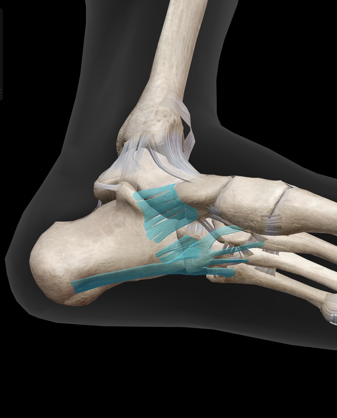
What ligaments are these?
Spring ligament (calcaneonavicular ligament)
Short plantar ligament
Long plantar ligament

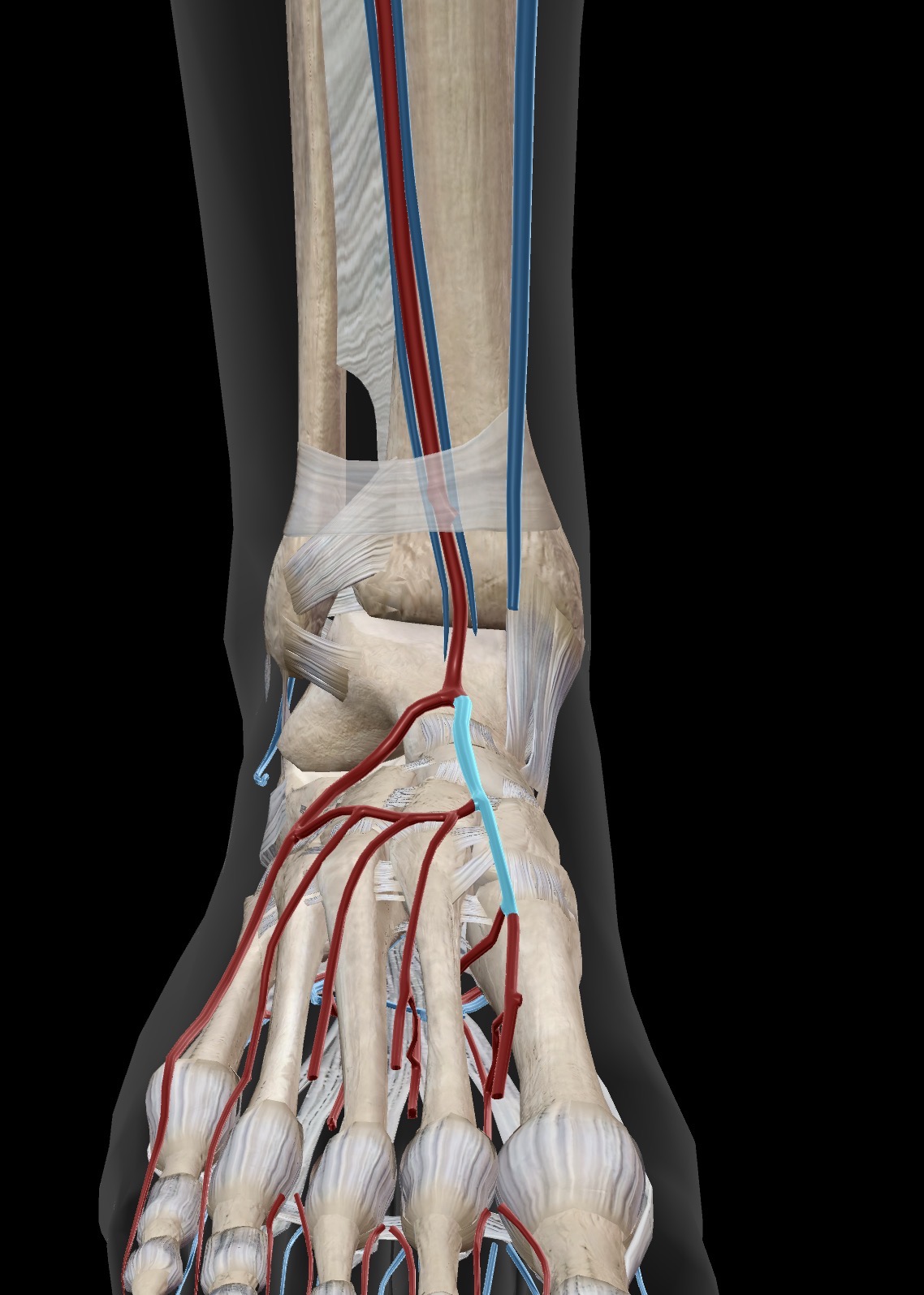
What artery is this? What is this artery useful for in physiotherapy?
Dorsalis pedis artery
Can be used to test the blood supply to the lower extremities
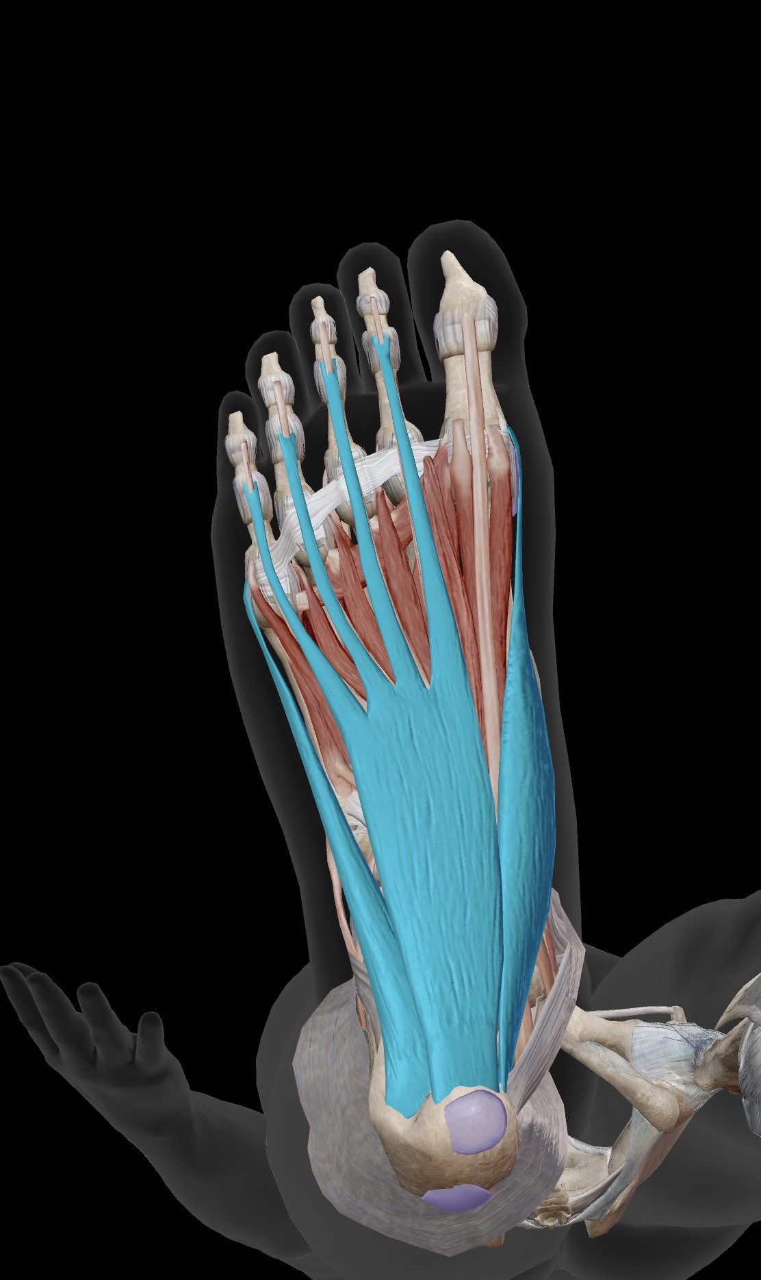
What are the muscles shown in this photo?
Origin
Insertion
Action
Abductor Hallucis
O: Medial tubercle of the calcaneus, flexor retinaculum and palmar aponeurosis
I: Medial side of the base of the proximal phalanx of the hallux
A: Abducts and flexes the great toe at the MTP joint, supports medial longitudinal arch
Flexor Digitorum brevis
O: Medial tubercle of the calcaneus and plantar aponeurosis.
I: : Middle phalanges of the lateral four toes (digits 2–5) via tendons that split around the long flexor tendons.
A: flexes the PIP and MTP joints
Abductor digiti minimi
O: Medial and lateral tubercles of the calcaneus and plantar aponeurosis.
I: Lateral side of the base of the proximal phalanx of the little toe (digit 5)
A: Abducts and flexes the little toe at the MTP joint.
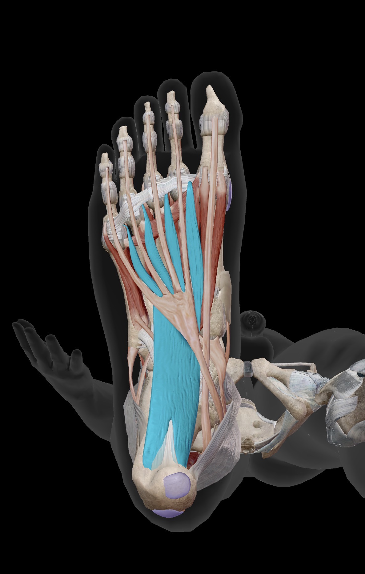
What are the muscles shown in the photo?
Origin
Insertion
Action
Quadratus plantae
O: medial and lateral surfaces of the calcaneus
I: posterolateral margin of the tendon of the flexor digitorum longus tendon (FDL)
A: Assists the FDL in flexing the terminal phalanges of the lateral 4 toes by converting its oblique pull into a straight line
Lumbricals
O: tendons of the FDL
I: Medial bases of the proximal phalanges and extensor expansions of the 4 lateral toes
Flex the MTP joint while simultaneously extending the PIP and DIP joints
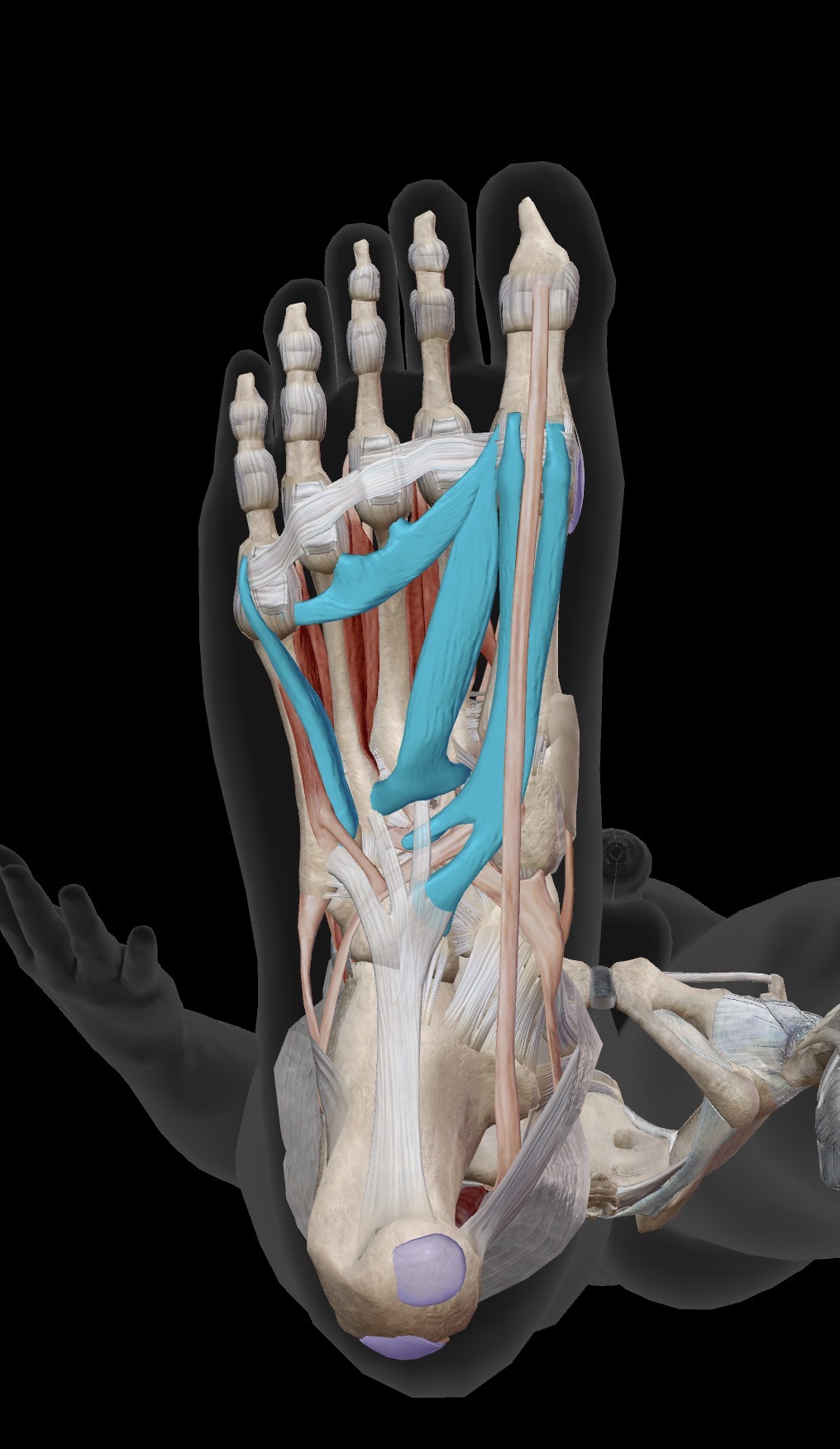
What muscles are shown in this photo
Origin
Insertion
Action
Flexor hallucis brevis (FHB)
O: Cuboid bone, lateral cuneiform bone, and the tendon of tibialis posterior
I: Medial and lateral sides of the base of the proximal phalanx of the great toe via two tendons (each containing a sesamoid bone)
A: Flexes the great toe at the MTP joint
Adductor Hallucis
O:
Oblique head: Bases of the metatarsals 2-4
Transverse head: plantar ligaments of the MTP joints of digits 3-5
I: Lateral side of the base of the proximal phalanx of the great toe
A: Adducts and assists in flexing the great toe at the MTP joint; helps maintain the transverse arch of the foot
Flexor Digiti Minimi brevis
O: Base of the 5th metatarsal bone and the long plantar ligament sheath.
I: Lateral side of the base of the proximal phalanx of the little toe (digit 5)
A: Flexes the little toe at the MTP joint
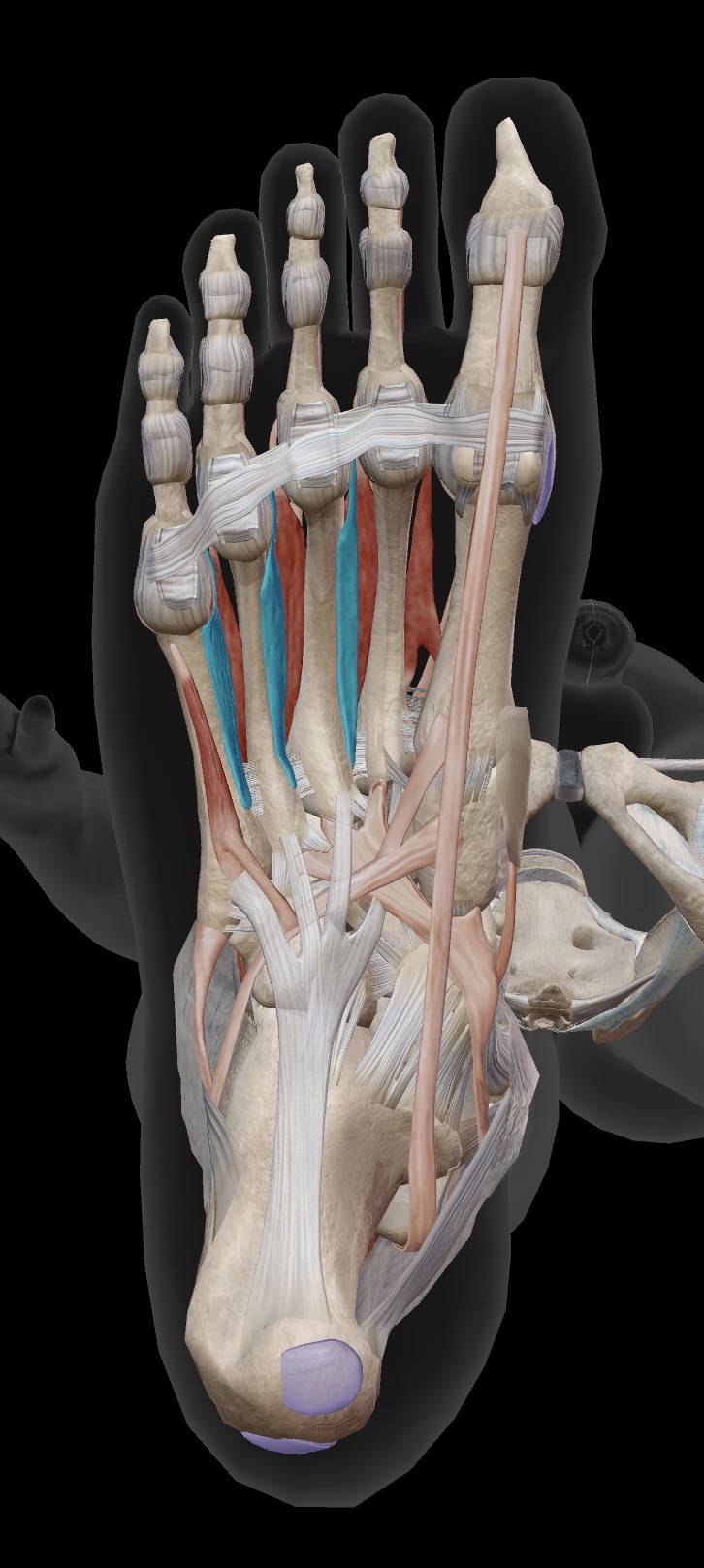
What are the muscles being shown here?
Origin
Insertion
Action
Plantar Interossei
O: Medial sides of the shafts of metatarsals 3–5.
I: Medial sides of the bases of proximal phalanges and extensor expansions of digits 3–5.
A: Adduct digits 3, 4, and 5 toward the axis (2nd toe); assist in flexing the MTP joints and extending the IP joints.
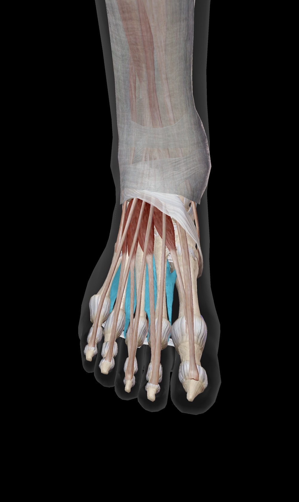
What muscles are being shown here?
Origin
Insertion
Action
Dorsal interossei
O: Bipennate muscles arising from the adjacent sides of the shafts of metatarsals 1–5.
I:
1st: Medial side of the base of the proximal phalanx of the 2nd toe.
2nd to 4th: Lateral sides of the bases of the proximal phalanges and extensor expansions of digits 2–4
A: Abduct digits 2, 3, and 4 away from the axial line (the 2nd toe is the axis of the foot, which is why it has two dorsal interossei inserting onto it to pull it in either direction). They also assist the lumbricals in flexing the metatarsophalangeal (MTP) joints and extending the interphalangeal (IP) joints.
DAB (Dorsal Abduct)
PAD (Plantar Adduct)