MUSCULOSKELETAL SYSTEM INTRO
1/25
There's no tags or description
Looks like no tags are added yet.
Name | Mastery | Learn | Test | Matching | Spaced |
|---|
No study sessions yet.
26 Terms
ILOS
Discuss the structure of the skeletal system and classification of bones • Discuss the simple classification of joints, giving relevant examples of each type • Describe the structure of bursae and explain their importance • Describe the characteristics of skeletal muscle and the process of contraction • Explain the role of Ca2+ in skeletal muscle function and how muscle force is controlled • Describe the length-tenson relationship in skeletal muscle
Bone
forms most of skeleton
Cartilage
Forms some parts of skeleton (e.g. costal cartilage)
lines articular surfaces
Tendons and ligaments
tendons attach muscles, mostly to bone
ligaments connect 2 or more bones or cartilage
Bones are classified according to their shape
long
short
flat
irregular
sesamoid = small bones resembling sesame seeds
splanchnic = collection of bones in cow heart (ossa cordis), for dogs and cats os penis & clitoral bone in females, rostral bone in pigs
pneumatic = hollow & containing air
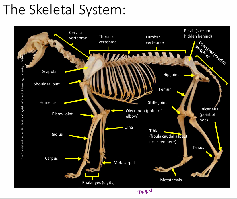
Skeletal system diagram
Bone marrow
found in medulla and in spaces between trabeculae (thin columns and plates of bone that create spongy network) of cancellous bone
(compact) cortical bone has a much denser structure compared to cancellous (spongy) bone
Where is growth plate found
Metaphysis, between epiphysis and diaphysis
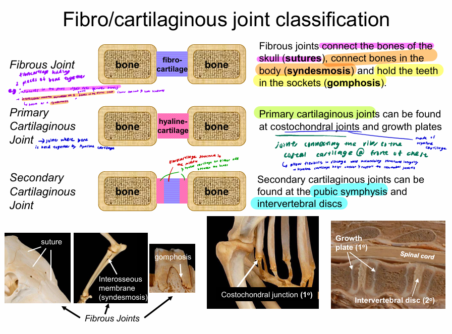
Fibro-cartilaginous joint classification
Fibrous joint = JOINT HELD TOGETHER BY FIBROCARTILAGE (bone-bone)
→ Connect bones of the skull (sutures), connect bones in the body (syndesmosis), and hold teeth in sockets (gomphosis)
Primary cartilaginous joints = JOINT HELD TOGETHER BY HYALINE CARTILAGE (bone - bone)
→ can be found at costochondral joints (connecting ribs to costal cartilage at sternum),
allows flexibility while maintaining structural integrity as hyaline cartilage helps cushion and support costochondral junction
primary cartilaginous joints also found in the growth plates of long bones.
Secondary cartilaginous joints = JOINTS HELD TOGETHER BY FIBROCARTILAGE AND HYALINE CARTILAGE (fibrocartilage in the middle and hyaline cartilage on each side between the bones) (bone-bone)
found at the pubic symphysis and intervertebral discs
Synovial joints
consist of
shaped articular cartilage surfaces
joint capsule containing synovial fluid → capsule keeps synovial fluid across surface of these cartilage structures and keeps them lubricated to reduce friction so joints can move smoothly
supporting ligaments around capsule / as part of capsule → help to hold bones together and provide stability during movement.
supporting muscles and tendon → must have muscle/tendon attached to a muscle crossing joint to actively move the joint
What does range of motion of joint depend on?
Shape of articular surfaces
Amount of muscles and tendons / ligaments across joint
What the muscles are doing
Bursae
Structures with a capsule and synovial fluid but no joint
helps to smooth passage of bones, tendons, ligaments during movement
can become inflamed “bursitis” when there is excessive friction/rubbing / infection
example:
navicular bursae in equine foot
→ around the navicular bone in equine distal foot
acts as a pouch of space for things to move smoothly across each other in that space instead of causing friction on the structures
Skeleton provides..
Support
Protection
Structure
but requires active control from muscles for movement functions of the limbs, head and neck
Muscles to maintain body posture
‘Antigravity’ extensor muscles of the limbs and spine
Muscles to move limb joints
Flexor & Extensor muscles
Muscles to move the jaw
Muscles of mastication (trigeminal muscles)
Muscles to shape the lips
Muscles of facial expression
Muscles to move food into the digestive tract
Pharyngeal and palatine muscles
Muscles to support abdominal organs
Abdominal muscles
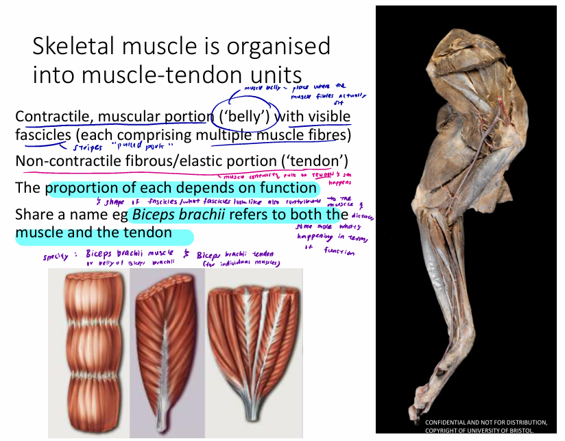
Skeletal muscle is organised into muscle tendon units
has contractile and non-contractile portion
contractile muscular ‘belly’ with visible fascicles (each comprising of multiple muscle fibres)
non-contractile fibrous elastic portion (tendon)
proportion of each depends on function
will have diff shape of fascicles which also contribute to & dictate muscle function
share a name → Biceps brachii refers to both the muscle and the tendon, so must specify Biceps brachii muscle (belly of Biceps brachii) or Biceps brachii tendon
Muscle Attachments
origin; ‘start’ of muscle, generally proximal and move less
Insertion; ‘end’ of muscle, typically distal and move more
mostly attached to bone but can also be attached to fascia (midline), or tendons
can have multiple insertion and origins per muscle
determine leverage of the muscle
intrinsic = both attachment points are within limb
extrinsic = from limb to body (e.g. attaching scapula to torso or attaching femur to pelvis)
e.g. Biceps brachii
origin = supraglenoid tubercle
insetion = proximally on the radial tuberosity and ulna
→ crosses 2 separate joints where it inserts and attaches determines how much leveragenit has around each of the joints
Skeletal muscles have
striated, cylindrical cells, multinucleate, voluntary control
contraction comes from sarcomere
fibres are arranged into visible fascicles; parallel (allow muscles to shorten quickly and changed length of muscle in more dramatic way) and pinnate
(fanned out to maximise force production within the muscle cos contraction is pulling tightly in the same direction)
motor unit = motor neuron & the skeletal muscle fibers it innervates at the neuromuscular junction
→ indiv neurons innervate a portion of the muscle belly, allowing for control cos some muscle fibers are being activated to some level, not entire muscle at once
tendons and ligaments
tendons connect muscle to bone, ligaments connect bone to bone
both connect regular connective tissue, ligaments generally have a greater elastic component
tendons act as strings or springs but for them to act as springs, attached muscle fibres need to develop force so the tendon can be stretched
reduced muscle fibre content reduces the need for energy e.g. equine superficial digital flexor tendons have an additional elastic tissue component (so more yellow) → used as an elastic storage mechanism that gives energy back in recoil, but need to contract muscle belly to make sure theres tension and muscle belly is not changing length
Skeletal muscle structure
Tendon holds muscle to bone
Skeletal muscle surrounded by epimysium / deep fascia
Individual visible fascicles surrounded by perimysium
Individual muscle fibers within fascicles surrounded by endomysium
within individual muscle fibres are myofibrils with sarcomeres
Muscle activation & contraction
MUSCLE ACTIVATION (ELECTRICAL)
Brain sends signal (AP) down motor neuron.
Nerve impulse reaches neuromuscular junction, triggering release of acetylcholine,
Ach binds to receptors on the muscle membrane, triggering an AP in the muscle cell
AP reaches sarcoplasmic reticulum and calcium is released
MUSCLE CONTRACTION
`Exposure of active sites:
Calcium binds to troponins, shifting tropomyosin away and exposing actin myofilament binding sites
Cross-bridge formation:
Myosin heads bind to the exposed actin binding sites and form cross-bridges. Phosphates are the released from the myosin heads.
Power stroke:
Energy stored in myosin heads is used to move the myosin heads, causing the actin filaments to slide past the myosin myofilaments and ADP is released from the myosin heads resulting in muscle shortening.
Cross-bridge release:
An ATP molecule binds to each of the myosin heads, causing them to detach from the actin'
Hydrolysis of ATP
The myosin ATPase portion of the myosin heads split ATP into ADP and phosphate, which remain attached to the myosin heads\
Recovery stroke
The heads of the myosin molecules return to their resting position and energy is stored in the heads of myosin molecules. If Ca2+ is still attached to troponins, cross bridge formation and movement are repeated and the cycle repeats many times during muscle contraction.
if muscle is deactivated, Ca2+ is pumped back into the sarcoplasmic reticulum. ATP then binds onto the myosin heads to allow it to let go of the actin