animal anatomy exam directional terms tissue
 Call Kai
 Learn
Learn Practice Test
 Spaced Repetition
 Match
Match Flashcards
 Knowt Play
1/107

There's no tags or description
Looks like no tags are added yet.
Name | Mastery | Learn | Test | Matching | Spaced | Call with Kai | Chat |
|---|
No analytics yet
Send a link to your students to track their progress
108 Terms
Anatomy
Focus on the form, shape and physical structure of animal organisms
Physiology
The study of the functions of living organisms and their parts
Microscopic Anatomy
Focuses on structures that can only be seen with a microscope (e.g., cells and tissues).
Macroscopic (Gross) Anatomy
Focuses on body structures seen with the unaided eye (e.g., organs, muscles, and bones).
Regional Anatomy
Examines all structures, tissue layers, vessels, and organs within a specific individual region of the body (e.g., the head, neck, or abdoment).
Systemic Anatomy
Focuses on individual body systems (e.g., cardiovascular, respiratory, or skeletal system
Anatomical planes of reference
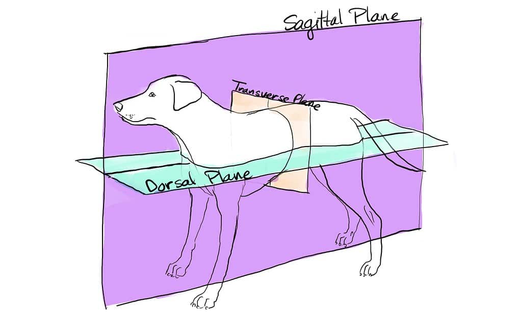
Sagittal Plane, Median Plane, Transverse Plane, Dorsal Plane
Sagittal Plane
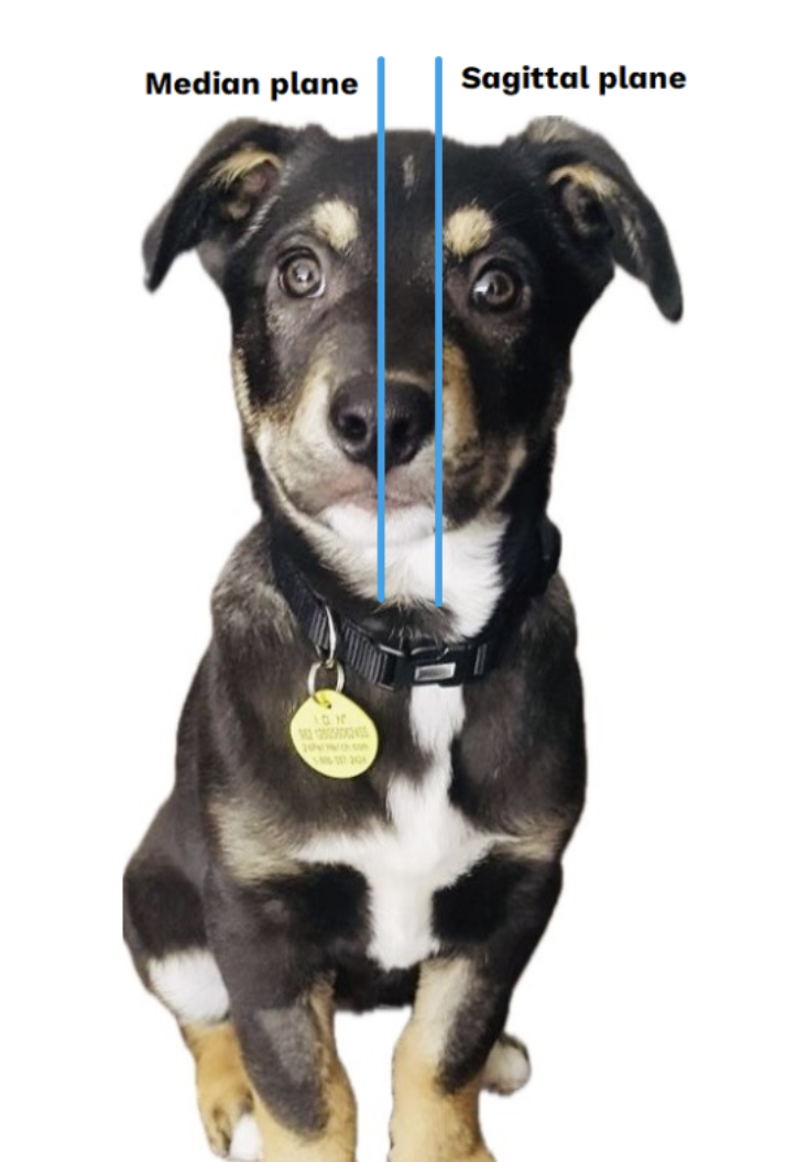
A plane running lengthwise through the body, dividing it into unequal right and left parts.

Median Plane
Runs directly along the midline, dividing the body into equal right and left halves.
Transverse Plane
A plane across the body at right angles to its long axis, dividing it into cranial (head end) and caudal (tail end) portions

Dorsal Plane
A plane dividing the body into dorsal (back) and ventral (belly) portions

Directional Terms Right and Left
Refers strictly to the animals own left and right sides, not the viewer perspective

Cranial
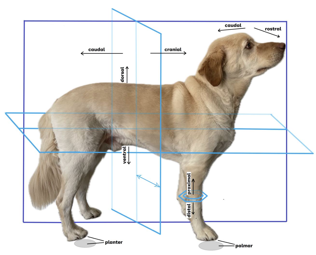
Towards the head (cranium)

Caudal
Towards the tail
Rostral
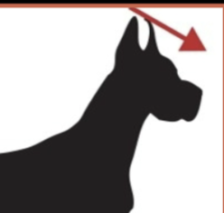
Within the head towards the nose or muzzle
Ventral
Toward the belly

Dorsal
Toward the back

Proximal
Nearer or closer to the body (refers to a limb or extremity)
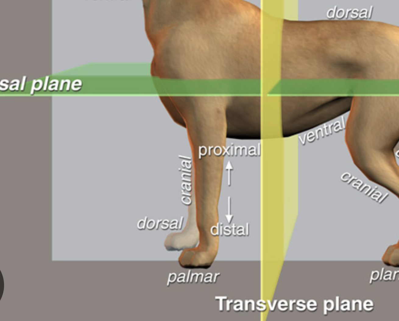
Distal
Farther or away from the body (refers to a limb or extremity)

Lateral
Farther from the midline or median plane or towards the side of the body
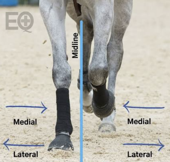
Medial
Closer to the midline or median plane or towards the middle
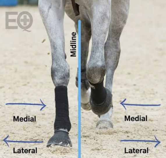
Palmar
Ground surface of front foot
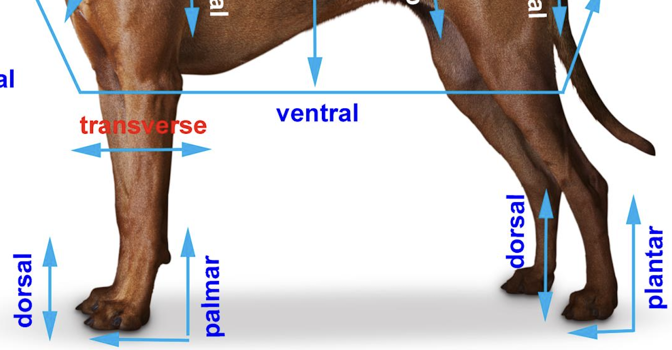
Plantar
Ground surface of hind foot

Superficial
Toward the outer surface of the body
Deep
Toward the inner core of the body

Carpus
Join and region on the front leg that corresponds to the human wrist.
Tarsus
Tarsus in a dog refers to the hock, which is the joint equivalent to the human ankle on the hind leg.
Right Lateral View
patient lies or stands with their right side closest to the image detector
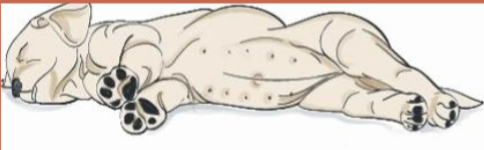
Left Lateral View
where the patient stands or lies with their left side touching the image receptor

Ventro-dorsal VD view
beam enters the front (ventral/belly) side of the body and exits through the back (dorsal/spine) side, with the patient lying on their back
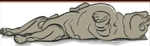
Dorso-ventral DV view
beam enters from the animal's back (dorsal side) and exits through the belly (ventral side), with the patient lying on their stomach in sternal recumbency

Common Regional Terms: Horse
Refer to Image

Dorsal Body Cavity
Central Nervous System: Cranial Cavity and Spinal Cavity

Ventral Body Cavity
CRANIAL Thoracic (chest): Enclosed by the rib cage. Contains major structures e.g. Heart, lungs, blood vessels, esophagus
CAUDAL Abdominal: Largest cavity. Contains digestive, and urinary, organs

Cranial Cavity
Formed by skull bones; houses and protects the brain.
Spinal Cavity
Formed by spinal vertebrae; houses and protects the spinal cord.
Pelvic cavity
The caudal portion of the ventral cavity enclosed by pelvic bones, housing the retum and internal reproducive organs.
Pleura
Lines thoracic cavity
Structure:
Visceral Layer: The inner layer that tightly covers the surface of the lungs.
Parietal pleura: The outer layer that attaches to your chest wall and diaphragm.
Pleural space: A tiny, fluid-filled gap separating the two layers. This fluid acts as a lubricant so the tissues glide smoothly when you breathe.

Peritoneum
Lines abdominal cavity
Visceral layer: The inner layer that wraps around and covers the abdominal organs (viscera).
Parietal layer: The outer layer that lines the inner walls of the abdominal and pelvic cavities, as well as the underside of the diaphragm.
Potential Space: The potential space between the two layers, containing a small amount of lubricating fluid that lets organs move without friction

Inflammation of the pleura
pleuritis / pleurisy
Inflammation of the peritoneum
peritonitis
Diaphragm
A dome-shaped skeletal muscle partition separating thoracic and adominal cavites, crucial for respiratiom.
Organization of the body
Cells, Tissues, Organs, Systems
Cells
Basic units of life, have specialized functions
Tissues
Groups of specialized cells, e.g. Epithelial, Connective, Muscle, Nervous
Organs
Groups of tissues working together e.g. Brain, Heart, Eyes, Lungs
Systems
Group of organs, involved in set of activities e,g, Digestive system
Major organ systems
Nervous, Cardiovascular, Respiratory, Urinary system
Homeostasis
Physiological processes maintain the bodys equilibrium (balance) in the various structures, functions, and properties.
Makes life possible.
Disease
The result when structures or functions of the body become abnormal
Ligament
strong band of dense connective tissue spanning between bones across joint, Connects bone to bone
Tendons
Connects muscle to bone
Epithelial Tissue
Cells that cover and line other tissues
Protect underlying tissue and filter biochemical substances
Absorb, secrete, or excrete biochemical substances
Play a role in reception of sensory input
Characteristics of Epithelia
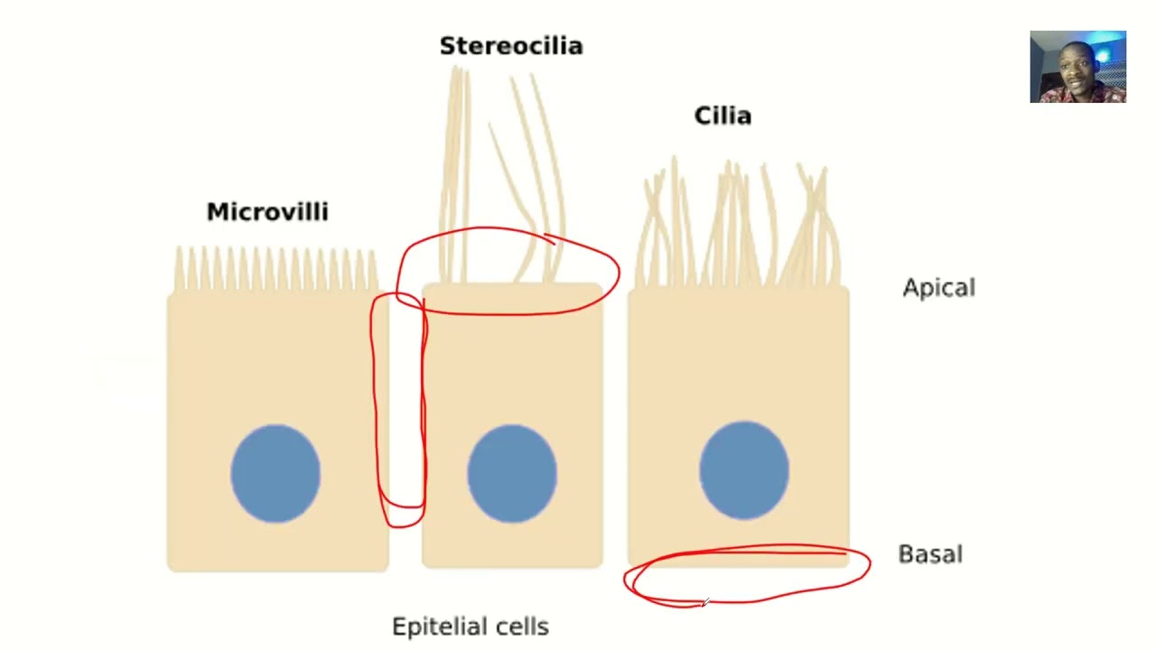
Apical surface: Faces the lumen or outside of organ
Basal surface: Faces the basal lamina and blood vessels
Lateral surface connected to cells by junctional complexes
Are avascular (without blood supply)
Are innervated (have nerve endings)

Secrete
Purpose: Produce and release substances that body needs to perform functions e.g digestion, lubrication, or regulation
Products: Hormones, enzymes, mucus, saliva, sweat, oil
e.g- Salivary glands secrete saliva to help digest food.
– Sweat glands secrete sweat to help cool the
Excretion
Purpose: To eliminate metabolic waste products and other unwanted substances from the body
Products Urine (w urea), Feces, exhaled carbon dioxide
e.g.-The lungs excrete carbon dioxide when we breathe out.
– The digestive system eliminates undigested solid waste, also called elimination or defecation
Cellular Attachments
Tight junctions
Desmosomes
Gap junctions
Tight junctions
Formed by fusion of the outermost layers of plasma membranes of cells
Found in leak proof tissues: e.g. Urinary bladder, Digestive tract

Desmosomes
Mechanical coupling made by Filaments that interlock with one another
Found in tissue that undergo repeat tension and stretching e.g skin, heart, uterus

Gap junctions
Tubular channel proteins (connexons) extend from cytoplasm of one cell to the cyto.. of another
Allow the exchange and passage of ions and nutrients
Found in intestinal epithelial cells, heart, smooth muscle tissue

Basement Membrane
also called Basal lamina a meshwork of fibers that cement epithelial cells to the underlying connective tissue and partial barrier between them
Vary in thickness
Helps prevent the cell from being torn off by the intraluminal pressures

Surface Specialization
Surface of epithelial cells vary depending on where they are located and their role: Smooth, Microvilli (brush border), Cilia, Keratin
Classification of Epithelial Tissue
Number of layers of cells: Simple, Stratified
Shape of cells: Squamous, Cuboidal, Columnar
Surface specializations: Cilia, Keratin, etc

Simple Squamous Epithelium
Found lining surfaces involved in the passage of gas or liquid, e.g Lungs
Fraglie, thin, flat, and smooth

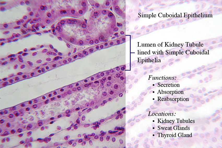
Simple cuboidal epithelium
In areas of body where secretion and absorption take place e,g Kidney
Single layer, cube shaped, round, dark nuclei aligned in a row
Simple Columnar Epithelium
Found in excretory ducts as well as digestive tract
Elongated and closely packed, Nucleui aligned in a row at base near basement membrane

Stratified Squamous Epithelium
Multilayered
Occur in areas where body is subjected to mechanical and chemical stresses e.g oral cavity, skin
Protect underlying tissue

Stratified Cuboidal Epithelium
Two layers
Found along large excretory ducts e.g salivary glands, sweat glands
Protects underlying tissue

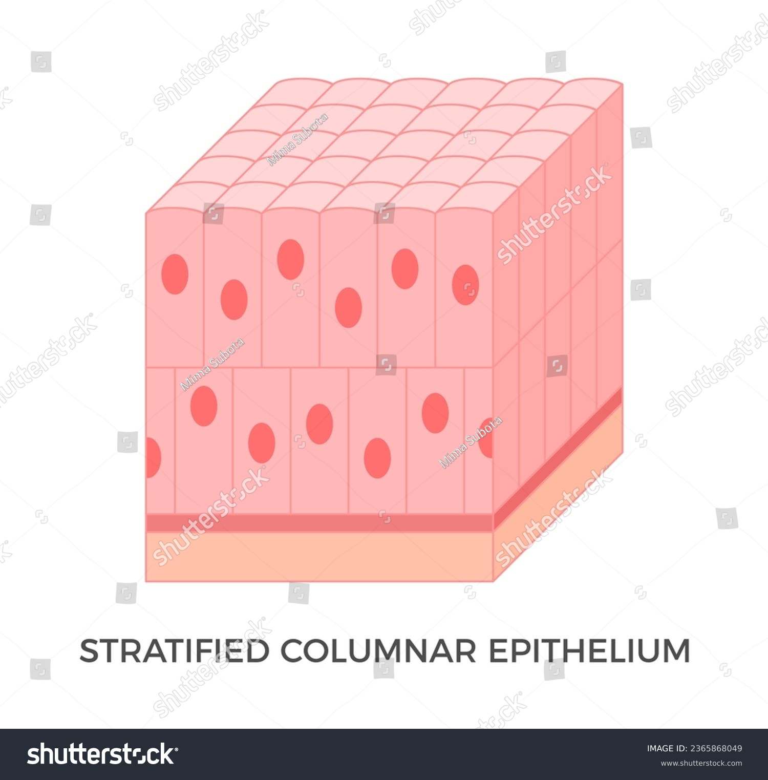
Stratified Columnar Epihelium
Found in select parts of respiratory, digestive, reproductive
Function in secretion and protection
Pseudostratified Columnar Epithelium
Cell nuclei found at different levels
Some cells do not reach the luminal surface
Found in respiratory tract and portions of the male reproductive tract
Pseudostratified Columnar Epithelium
Cell nuclei found at different levels
Some cells do not reach the luminal surface
Found in respiratory tract and portions of the male reproductive tract

Transitional Epithelium
Basal layer of cuboidal or columnar cells and superficial layer of cuboidal or squamous cells
Found in areas of body required to expand and contract as normal function e,g urinary bladder

Endocrine Glands
Do not have ducts or tubules, secretions are distributed throughout the body
Produce and secrete hormones into the bloodstream or lymphatic system
Complex biochemical network known as endocrine system

Exocrine Glands
Discharge secretions via ducts directly into local areas except for goblet cells
Unicellular or multicellular

Connective Tissue
Tissue where cells are suspended outside of the cell
Functions:
Form metabolic and structural connections between other tissues
Form protective sheath around organs and insulates the body
Reserve for energy
Frame that supports body
Composes medium that transports substances
Plays a role in the healing process and controls invading microorganisms
Connective tissue components
Extracellular Matrix
Extracellular fibers
Ground substances
Cells

Ground substance
Where cells exchange nutrients and waste with the bloodstream
Ranges in texture from liquid or gel to a calcified solid
Extracellular fibers
Strong thick strands of collagen, found in tendons and ligaments
Reticular fibers
Thin delicate networks of collagen
Provide support for high cellular organs, e,g endocrine glands, lymph modes, spleen, liver, blood vessels, nerves
Elastic fibers
Composed primarily of the protein elastin, coiled microfibrils, occurs in tissues commonly subjected to stretching e.g vocal cords, lungs, skin
Cell Types
Fixed cells: involved in production and maintenance of matrix e.g. fibroblasts, chondroblasts, osteoblasts
Transient cells: involved in the repair and protection of tissues e.g leukocytes, mast cells
Connective tissue proper
Loose connective tissue: Areolar, Adipose, Reticular
Dense connective tissue: Dense regular, Dense irregular, Elastic
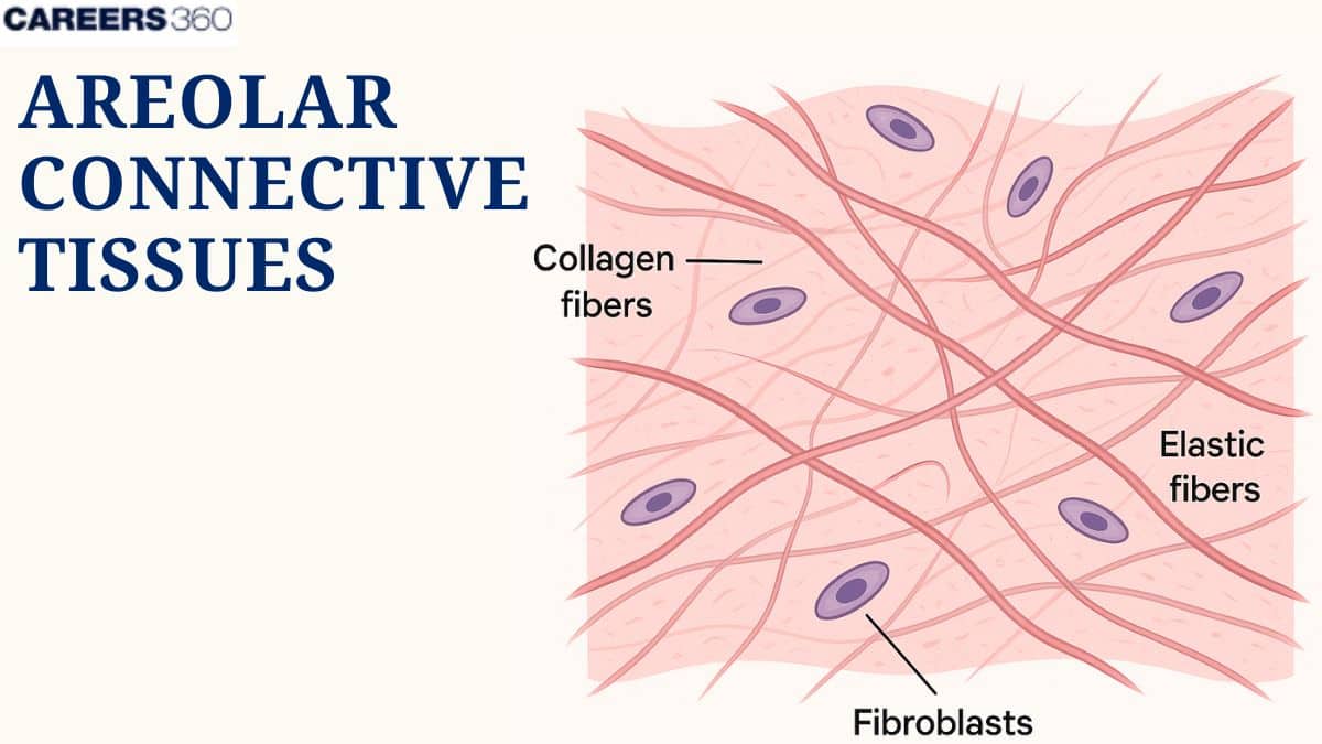
Areolar Connective Tissue
Loose, Predominant cell is the fibroblast
Manufactures the elastic, reticular, and collagenous fibers
Surrounds every organ; present in all mucous membranes
Adipose Tissue
Loose, Areolar tissue in which adipocytes predominate cell, highly vascular, Energy storehouse and thermal insulator FAT CELLS

Reticular Connective Tissue
Loose, network of thin reticular fibers, loosely arranged fibers and fibroblasts suspended, forms the stroma (framework of several organs) e.g spleen
Dense Regular Connective Tissue
Tightly packed parallel collagen fibers
Avascular (no blood supply)
Makes up the tendons ( muscles to bones) Ligaments (bone to bone)
Dense Irregular Connective Tissue
Found in the dermis of the skin, Composed primarily of collagen fibers, interwoven to form single sheet,
Elastic Connective Tissue
Elastic fibers, parallel or interwoven and collagenous fibers interspersed, Found in spaces between vertebrae and in areas of body that require stretching, e.g wall of arteries, stomach , bladder
Specialized Connective Tissue
Cartilage
Hyaline cartilage
Elastic cartilage
Fibrocartilage
Bone
Blood
Cartilage
Found in joints in ear, nose, vocal cords
Forms a framework on which bone is formed
No innervation: avascular
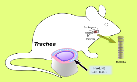
Hyaline Cartilage
Most common type of cartilage found in body, made of closely packed collagen
Found in joints at the end of long bones, tracheal rings, and connections of the ribs to the sternum, coastal cartilages
Elastic cartilage
Contains elastic fibers in dense branching bundles
Flexible: can withstand bending
Found in epiglottis of larynx and pinnae of ears
Nervous Tissue
Transmits electrical impulses throughout the body. (e.g., neurons, spinal cord).
Fibrocartilage
Merged with hyaline cartilage and dense connective tissue, found in space between vertebrae of the spine, between bones in pelvic girdle, knee joint
Bone
Combo of organic collagen fibers and inorganic calcium salts, Well vascularized (blood vessels), Haversian canal contain both vascular and nerve supply
Osteoblasts
Manufacture the fibers that are part of the matrix. specialized cells responsible for creating and forming new bone tissue.
Osteocytes
Reside in lacunae (unfilled spaces/gaps)
Blood
Matrix:
- Ground Substance: plasma
- Fibrous component: protein
Cells:
Erythrocytes
Leukocytes
Thrombocytes
Mucous Membranes (mucosae)
Line organs w connections to the outside environment (mouth, nasal passage, intestine)
Composed of stratifies squamours or simple columnar epithelium covering layer of connective tissue
Submucosa: connective tissue layer that connects muscosa to underlying structures
May contain goblet cells or multicellular glands
can produce large quantities of mucus
mucus consists primarily of water, electrolytes, and the protein mucin
Some can absorb e.g. epithelial in the intestine

Serous Membranes (serosae)
Lines the walls and cover organs e.g thorax and abdominopelvic cavities)
Consists of continuous sheet doubled over to form two layers
The portion of the membrane that lines the cavity wall is called the parietal layer
The portion of the membrane that covers the outer surface of organs is called the visceral layer
In abdominopelvic cavity, visceral layers of serosa merge to form mesenteries ( attaches the intestine to wall around stomach and holds it in place)