Lecture 1: Musculoskeletal Anatomy & Conformation
 Call Kai
 Learn
Learn Practice Test
 Spaced Repetition
 Match
Match Flashcards
 Knowt Play
1/35

There's no tags or description
Looks like no tags are added yet.
Name | Mastery | Learn | Test | Matching | Spaced | Call with Kai | Chat |
|---|
No analytics yet
Send a link to your students to track their progress
36 Terms
Why is ideal conformation and posture important?
• Supports optimal limb loading and movement patterns
• Reduces the risk for musculoskeletal injuries
Are conformational faults synonymous with lameness?
no, but they often contribute to altered limb loading and movement and increase the risk for MSK injuries
What is often a sign of MSK pain, or neurologic disease (ataxia, weakness)?
altered posture
What does ideal conformation provide?
a broad base of support
equal weight bearing across all limbs
What is conformation vs posture?
conformation: the symmetry, size, and shape of various body regions relative to each other or in general appearance (structure - not easily changed)
posture: the position in which someone holds their body when standing or sitting (function - can be modified or trained)
What are some examples of altered posture in horses?
unweighted limb: normal resting posture or lameness
laminitic stance
ataxia
severe weakness
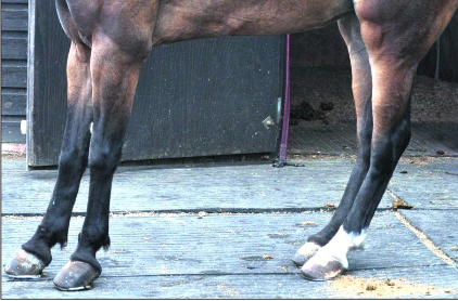
What type of altered posture is this?
laminitic stance
Conformational faults in the forelimb induce altered _____?
limb loading
What diseases are associated with conformational faults in the forelimb of horses?
splint bone disease; fetlock osteoarthritis
How would you describe this horse’s conformation?
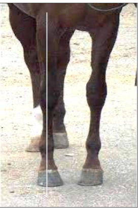
toed in
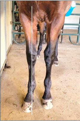
How would you describe this horse’s conformation?
toed out
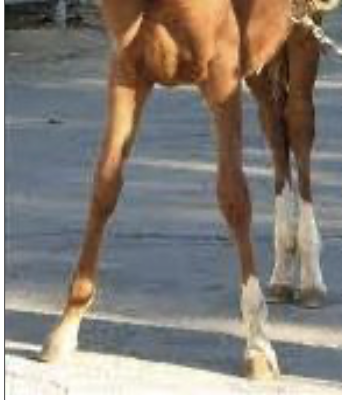
How would you describe this horse’s conformation or posture?
base wide
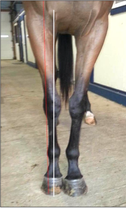
How would you describe this horse’s conformation or posture?
base narrow
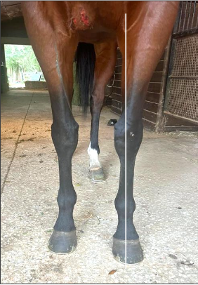
How would you describe this horse’s conformation or posture?
external rotation
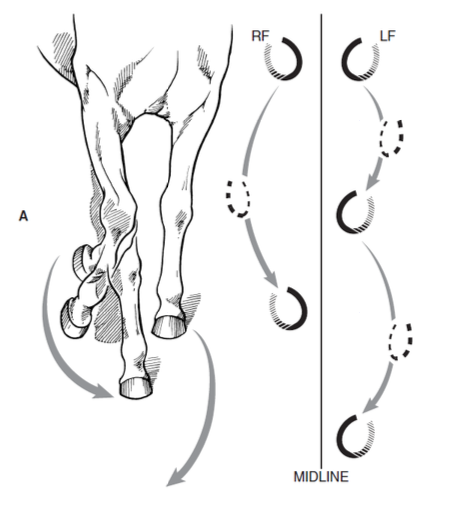
What type of limb conformation is causing this type of limb movement?
toed in → paddling
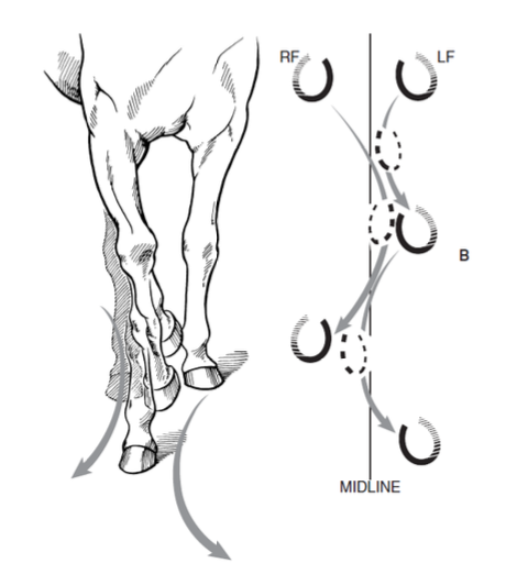
What type of limb conformation is causing this type of limb movement?
toed out → interference with contralateral limb
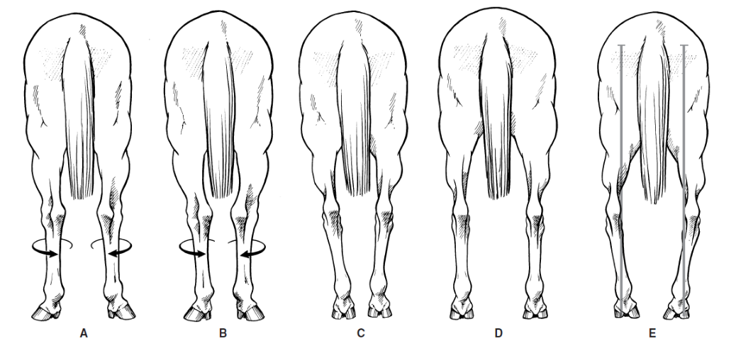
What are these hind limb conformational flaws?
A. cow hocked (external rotation) - common, not assoc. with lameness
B. cow hocked, base narrow
C. base narrow (postural fault)
D. base wide (postural fault)
E. bow legged (internal rotation)
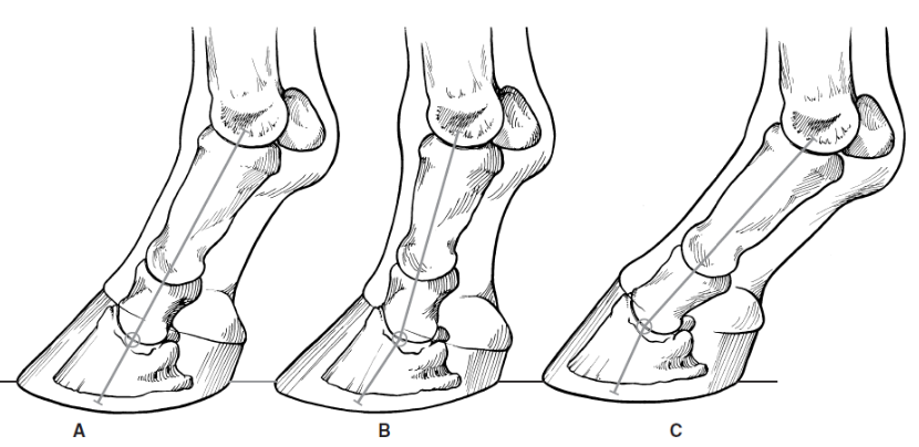
What are these 3 hoof-pastern angles?
A. Ideal
B. Upright pastern angle
C. Sloping pastern angle
What effect does an upright pastern angle have?
short stride length
prone to navicular disease and pastern OA
What effect does a sloping pastern angle have?
longer stride length
prone to soft tissue injuries → suspensory ligament injury
What allows prolonged standing with minimal muscular effort?
stay apparatus
What is the fetlock supported by?
the proximal sesamoid bones and the suspensory apparatus
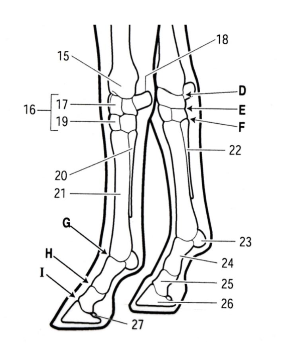
What are the joints corresponding to each letter?
D. radiocarpal joint
E. midcarpal joint
F. carpometacarpal joint
G. metacarpophalangeal joint aka fetlock
H. proximal interphalangeal joint aka pastern
I. distal interphalangeal joint aka coffin
What is the distal sesamoid bone of the coffin joint?
navicular bone
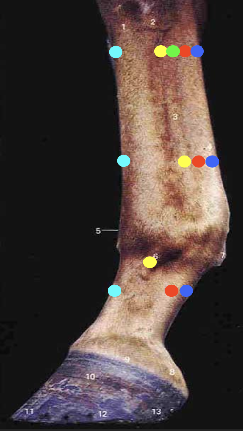
What tendons are indicated by each color?
light blue: common digital extensor tendon
yellow: suspensory ligament (body, medial & lateral branches, extensor branches)
red: deep digital flexor tendon
green: distal accessory (check ligament)
dark blue: superficial digital flexor tendon
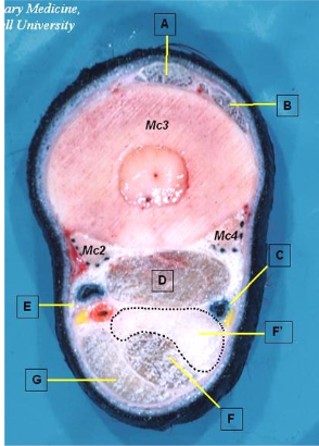
What tendons/ligaments are indicated by the letters?
A. common digital extensor tendon
B. lateral digital extensor tendon
D. interosseus (suspensory) ligament
G. superficial digital flexor tendon
F. deep digital flexor tendon
F’. distal accessory (check) ligament
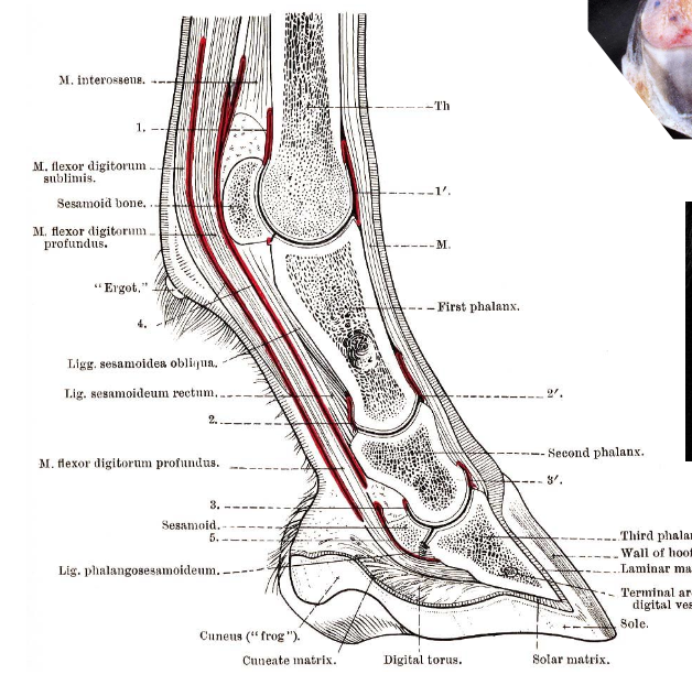
What synovial structures are indicated by the numbers?
Digital tendon sheath = 4
Fetlock joint: dorsal pouch = 1’, palmar pouch = 1
Pastern joint: dorsal pouch = 2’, palmar pouch = 2
Coffin joint: dorsal pouch = 3’, palmar pouch = 3
Navicular bursa = 5
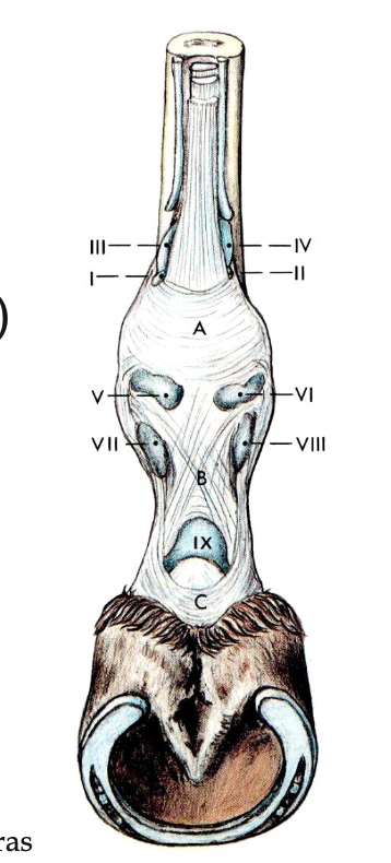
What ligaments correspond to each letter?
A. palmar annular ligament
B. proximal digital annular ligament
C. distal digital annular ligament
What structure surrounds the SDFT and DDFT, extending from mid-cannon to mid-P2?
digital tendon sheath
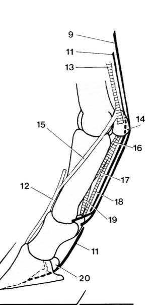
What are these numbered structures that are part of the digit stay apparatus?
superficial digital flexor tendon
deep digital flexor tendon
interosseous ligament → 15. extensor branches, 12. common digital extensor tendon
Distal sesamoidean ligaments:
straight
oblique
cruciate
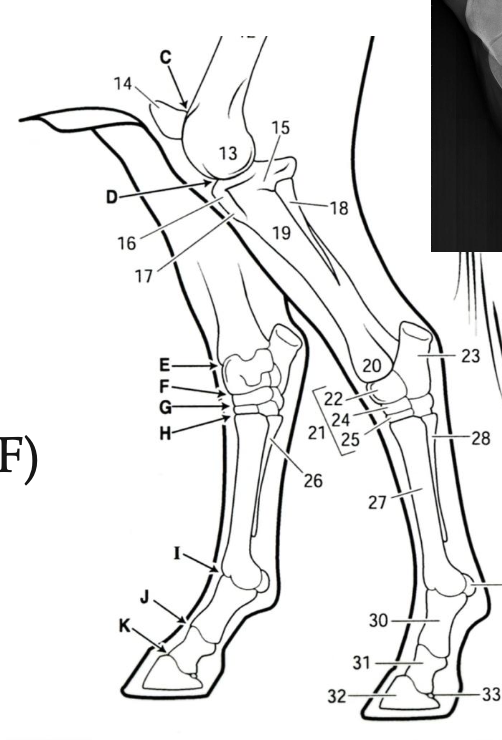
What are the pelvic limb articulations indicated by the letters?
C. patellofemoral joint
D. femorotibial joint
E. tibiotarsal joint
F. proximal intertarsal joint
G. distal intertarsal joint
H. tarsometatarsal joint
How many patellar ligaments can be found in an equine stifle joint?
3: medial, intermediate (middle), lateral
Explain the reciprocal apparatus.
important component of the stay apparatus that uses the SDF muscle and tendon and the peroneus tertius muscle to couple the movement of the stifle and the hock → when the stifle flexes the hock flexes and when the stifle extends the hock extends
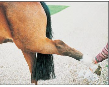
What has happened to this horse?
peroneus tertius rupture (reciprocal apparatus not maintained)
What mechanisms make up the equine pelvic limb stay apparatus?
reciprocal apparatus
patellar locking mechanism
suspensory apparatus
Explain the patellar locking mechanism.
the quadriceps extend the pelvic limb → patella rotates medially → The medial patellar ligament, along with the medial patellar fibrocartilage, hooks over the enlarged medial trochlear ridge of the femur → patella can no longer slide normally in the trochlear groove, locking the stifle and hock in an extended position without muscle contraction (allows horses to sleep while standing)