Anatomy of the Pelvic Girdle, Femur, Tibia, Fibula, and Foot Bones
1/35
There's no tags or description
Looks like no tags are added yet.
Name | Mastery | Learn | Test | Matching | Spaced |
|---|
No study sessions yet.
36 Terms
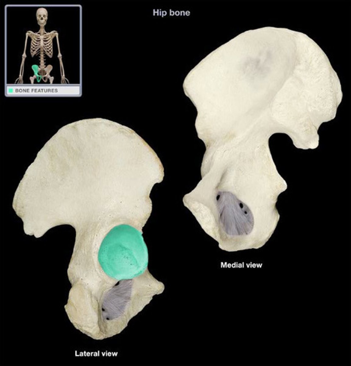
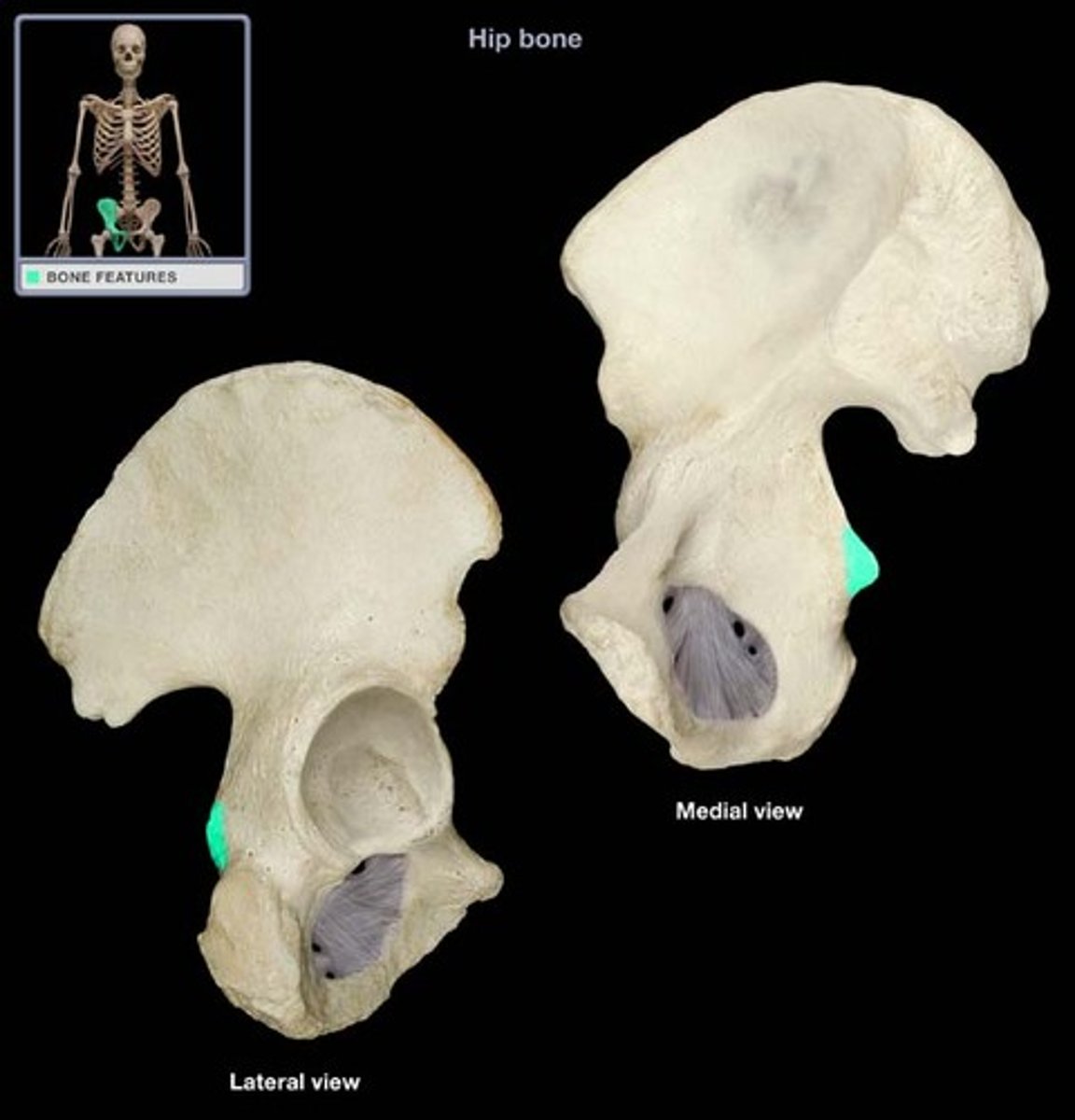
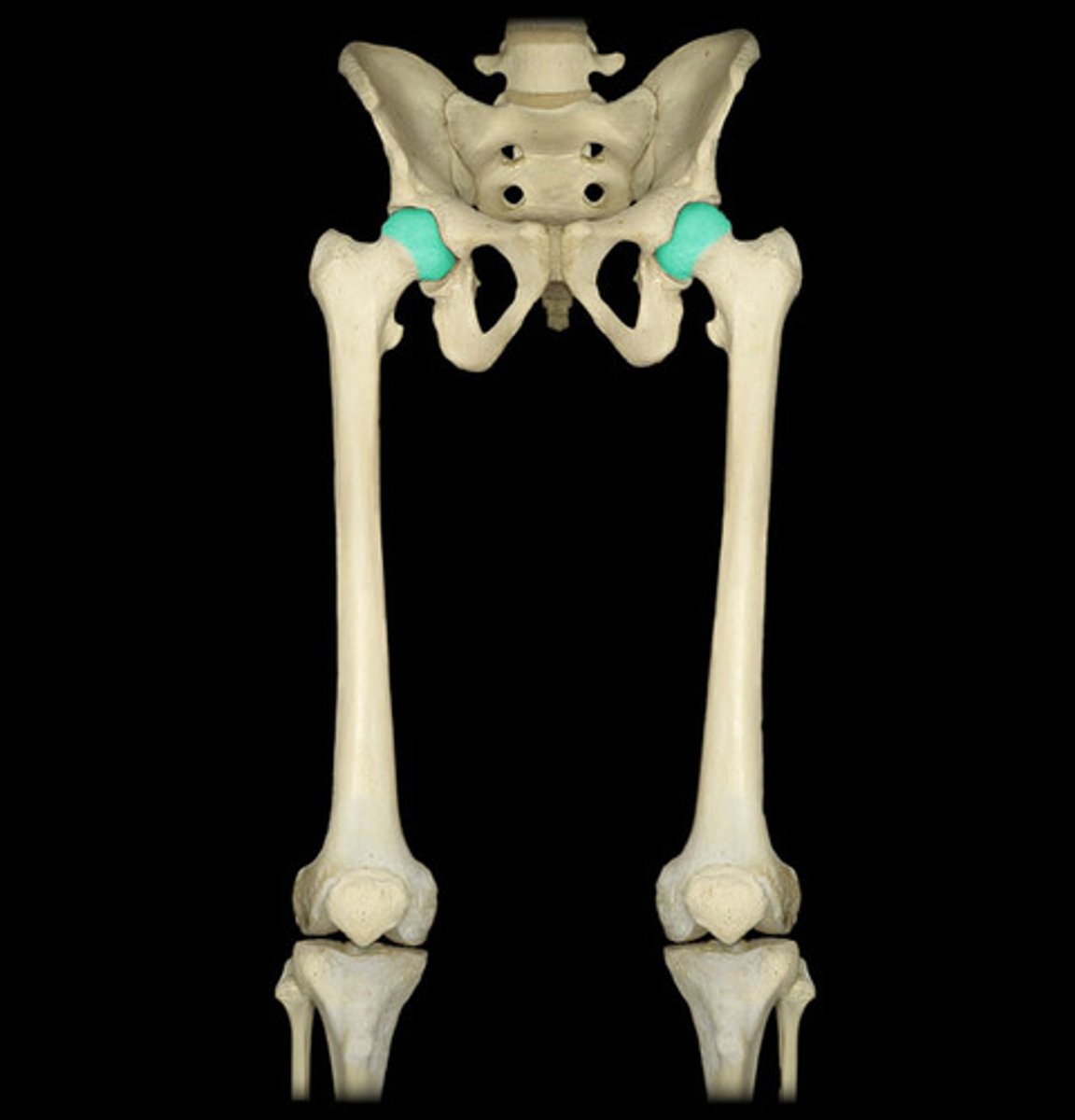
Acetabulum
The deep socket of the os coxa that articulates with the head of the femur.
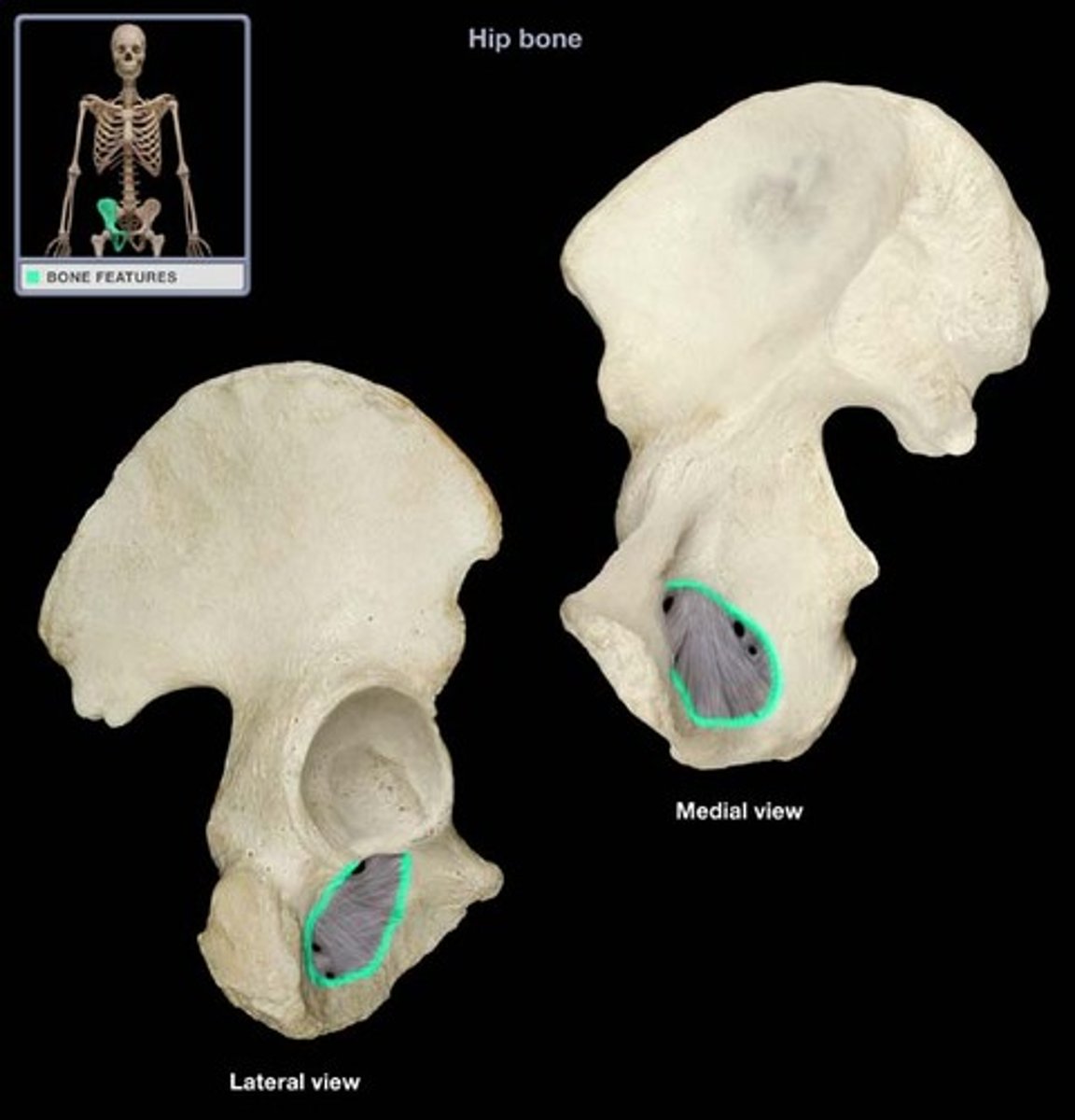
Obturator foramen
The large opening in the os coxa formed by the pubis and ischium.
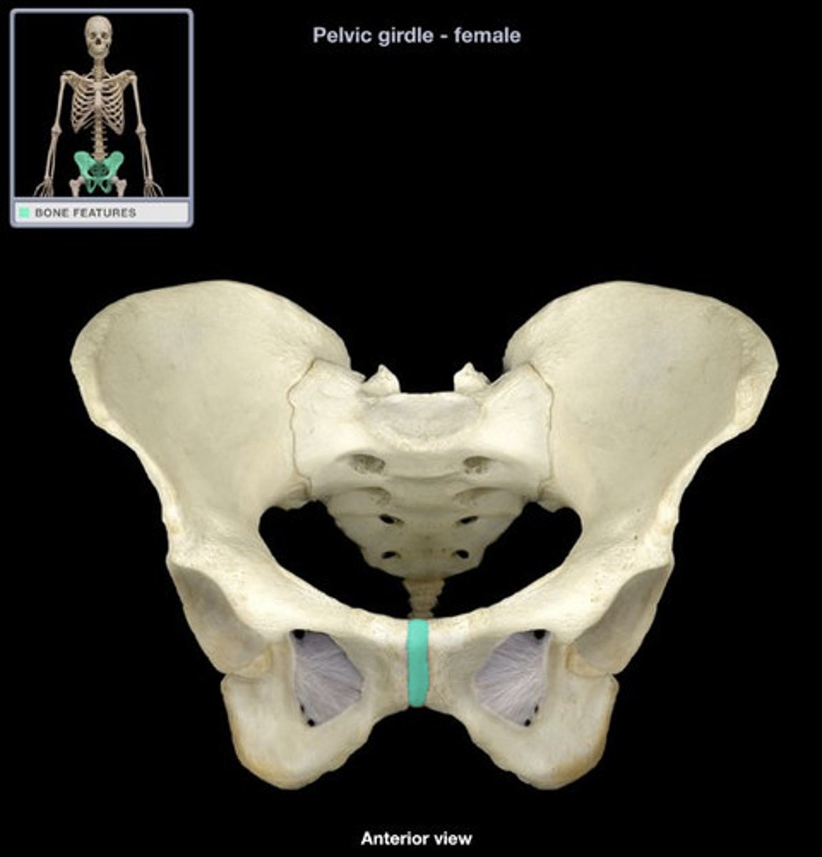
Pubic symphysis
The cartilaginous joint where the two pubic bones meet anteriorly.
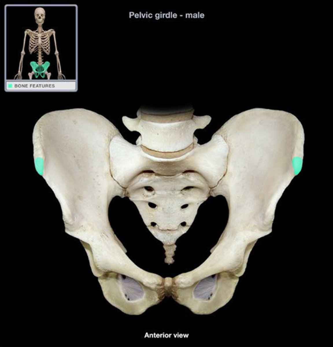
Anterior superior iliac spine (ASIS)
The prominent projection on the anterior, superior part of the ilium that serves as a landmark and muscle attachment site.
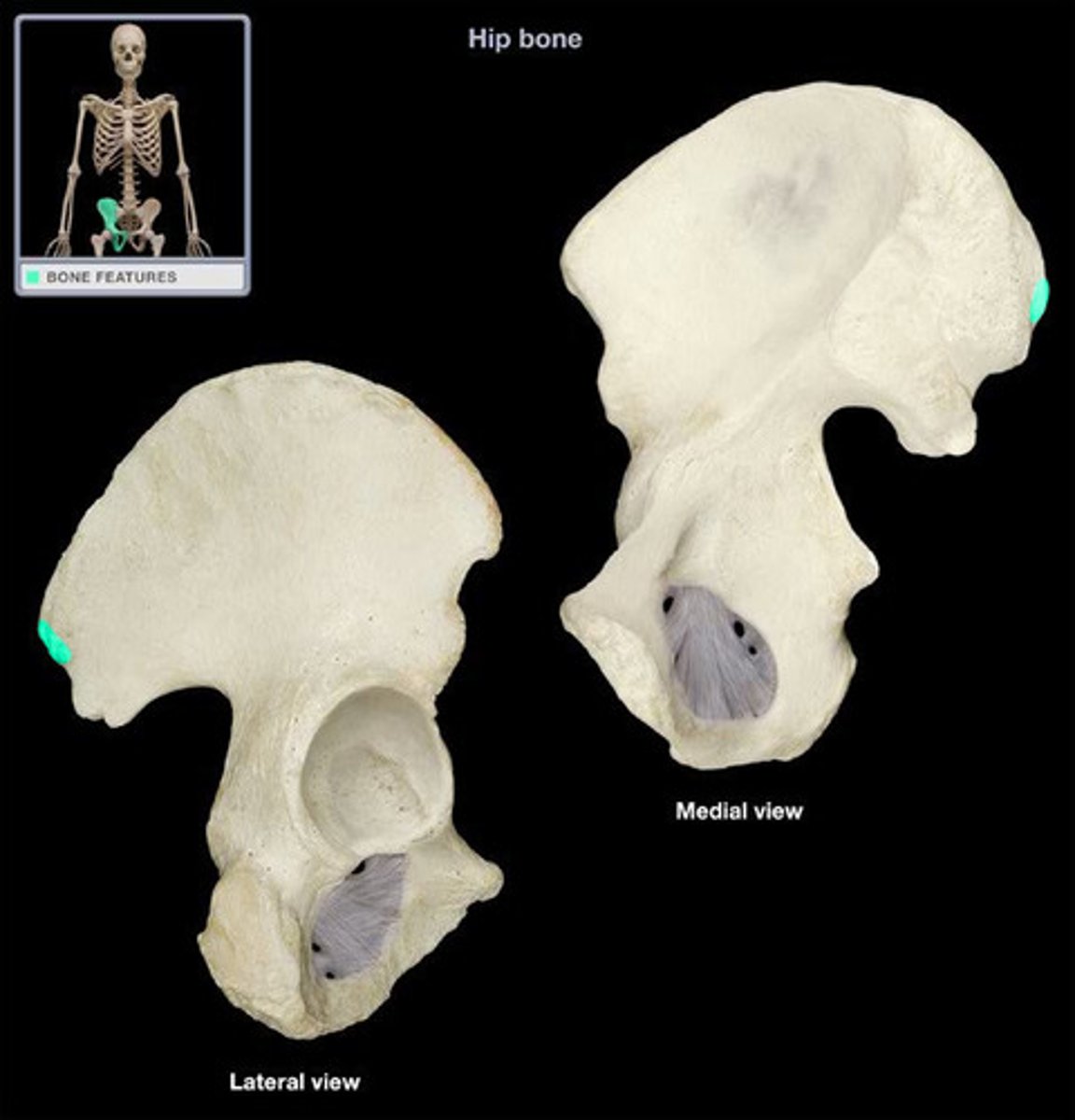
Posterior superior iliac spine (PSIS)
The projection on the posterior, superior part of the ilium.
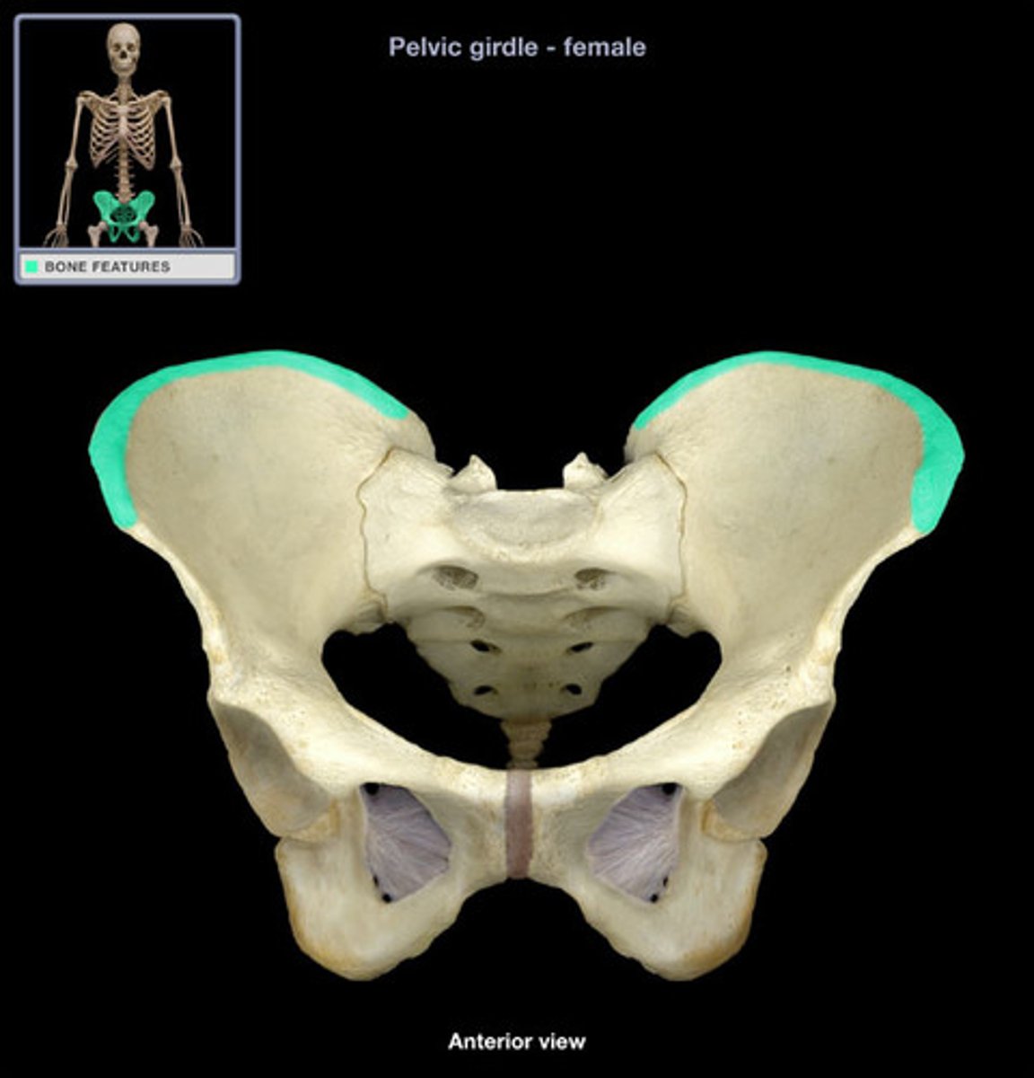
Iliac crest
The superior curved border of the ilium.
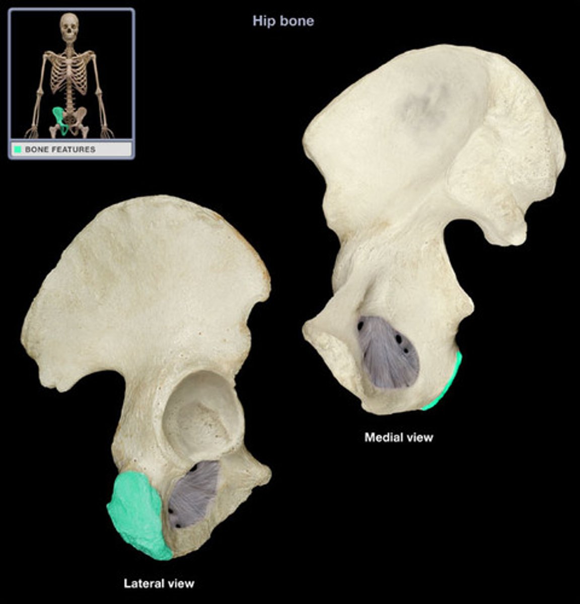
Ischial tuberosity
The roughened area on the inferior part of the ischium that is the site for hamstring attachment and the bone you sit on.
Ischial spine
The small, pointed projection located above the ischial tuberosity.
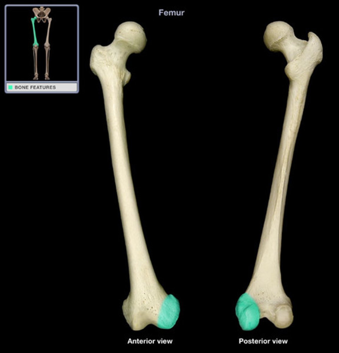
Head of the femur
The rounded proximal structure of the femur that articulates with the acetabulum of the os coxa.
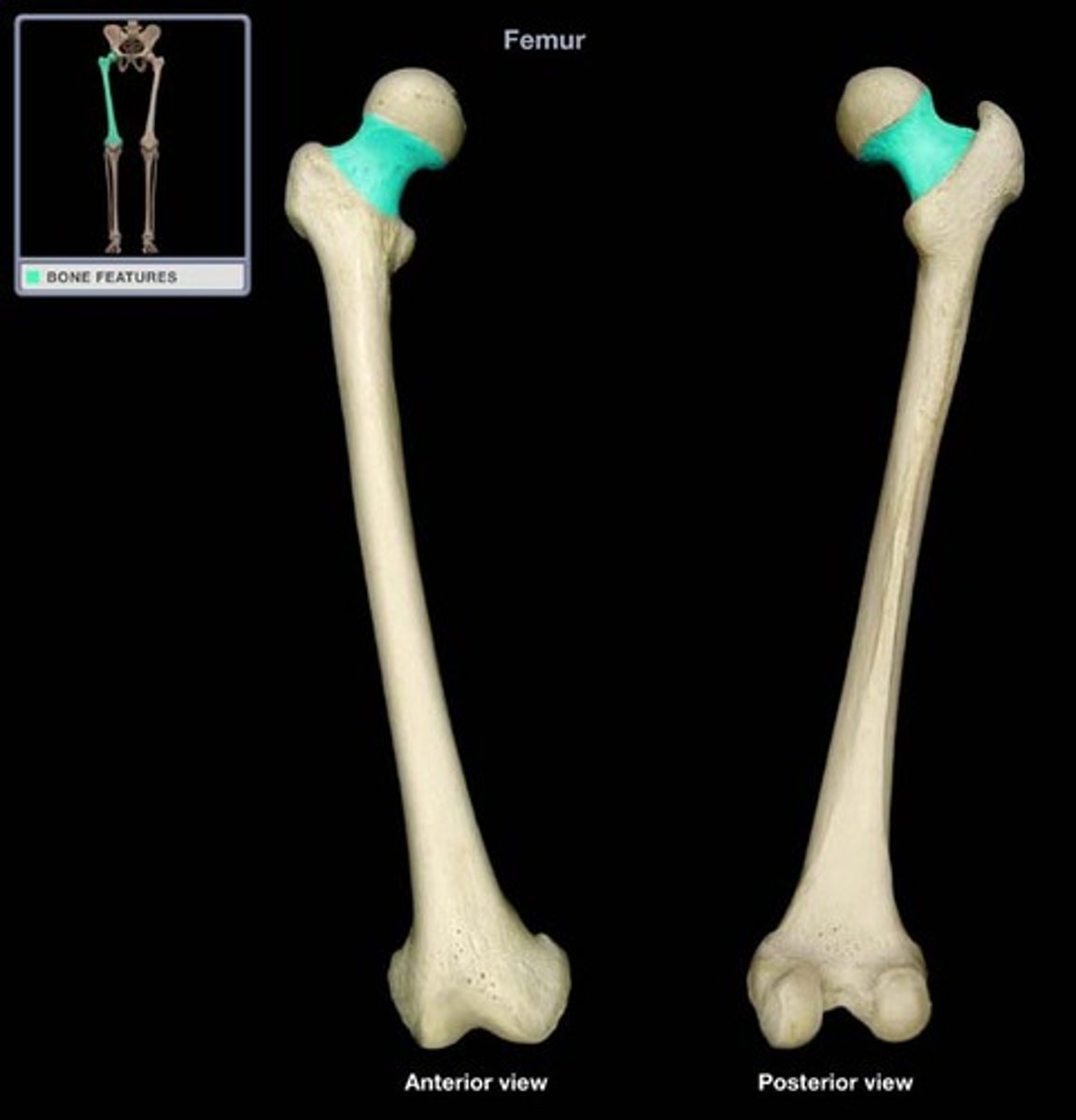
Neck of the femur
The narrowed region that connects the head of the femur to the shaft.
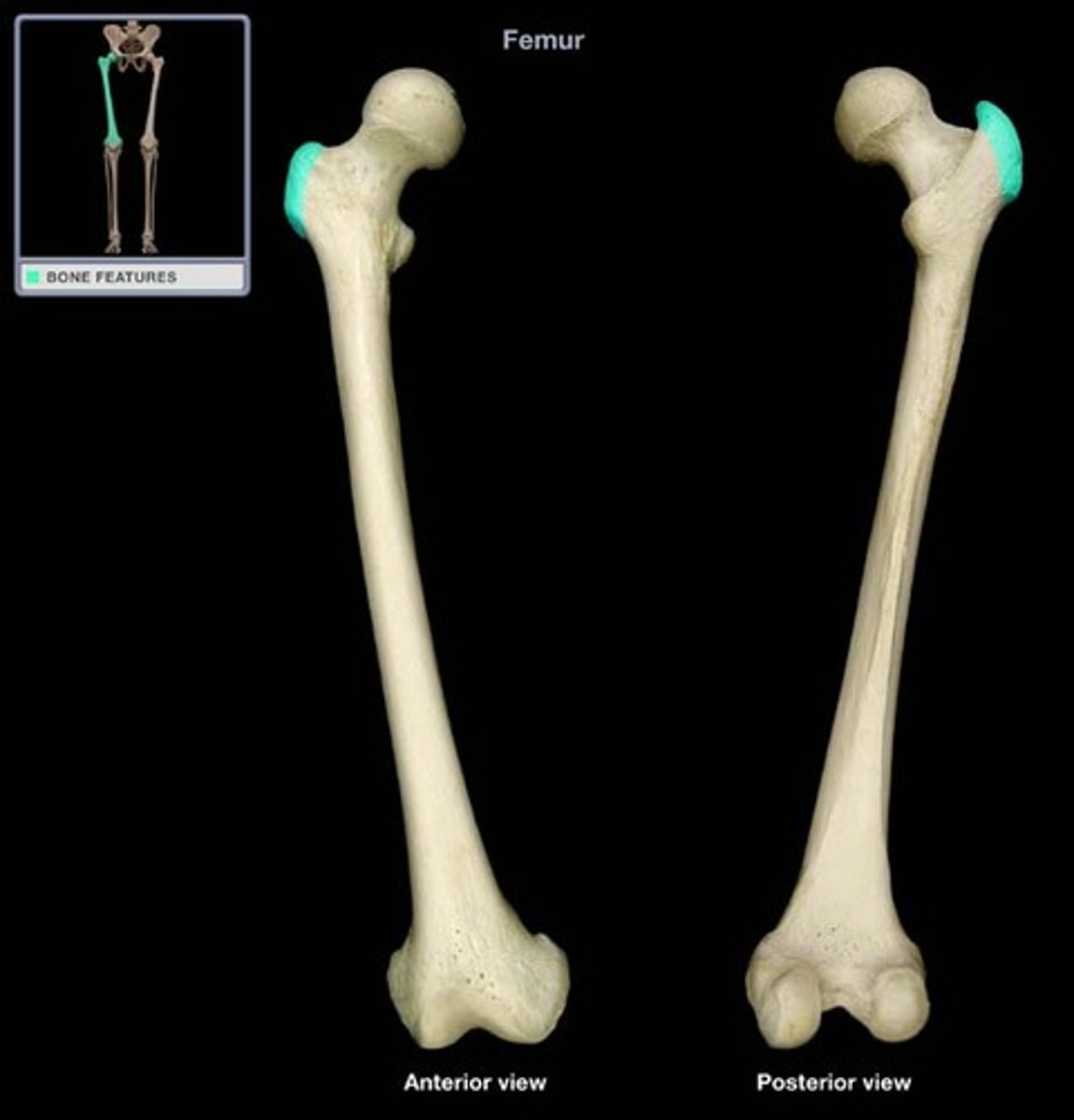
Greater trochanter
The large, lateral projection near the proximal end of the femur that serves as a major muscle attachment site.
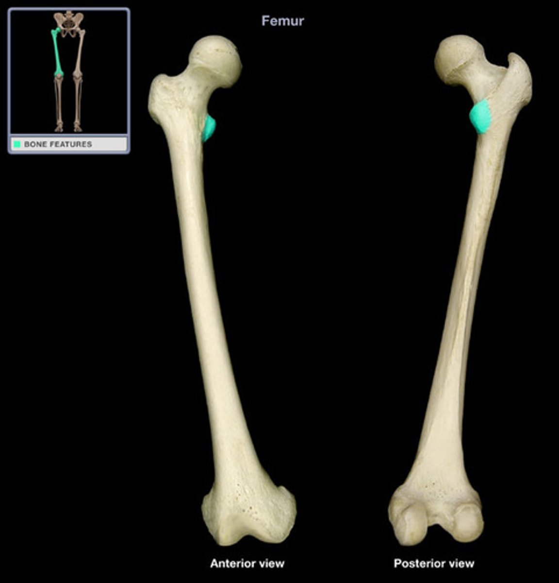
Lesser trochanter
The smaller, medial projection located inferior to the head of the femur.
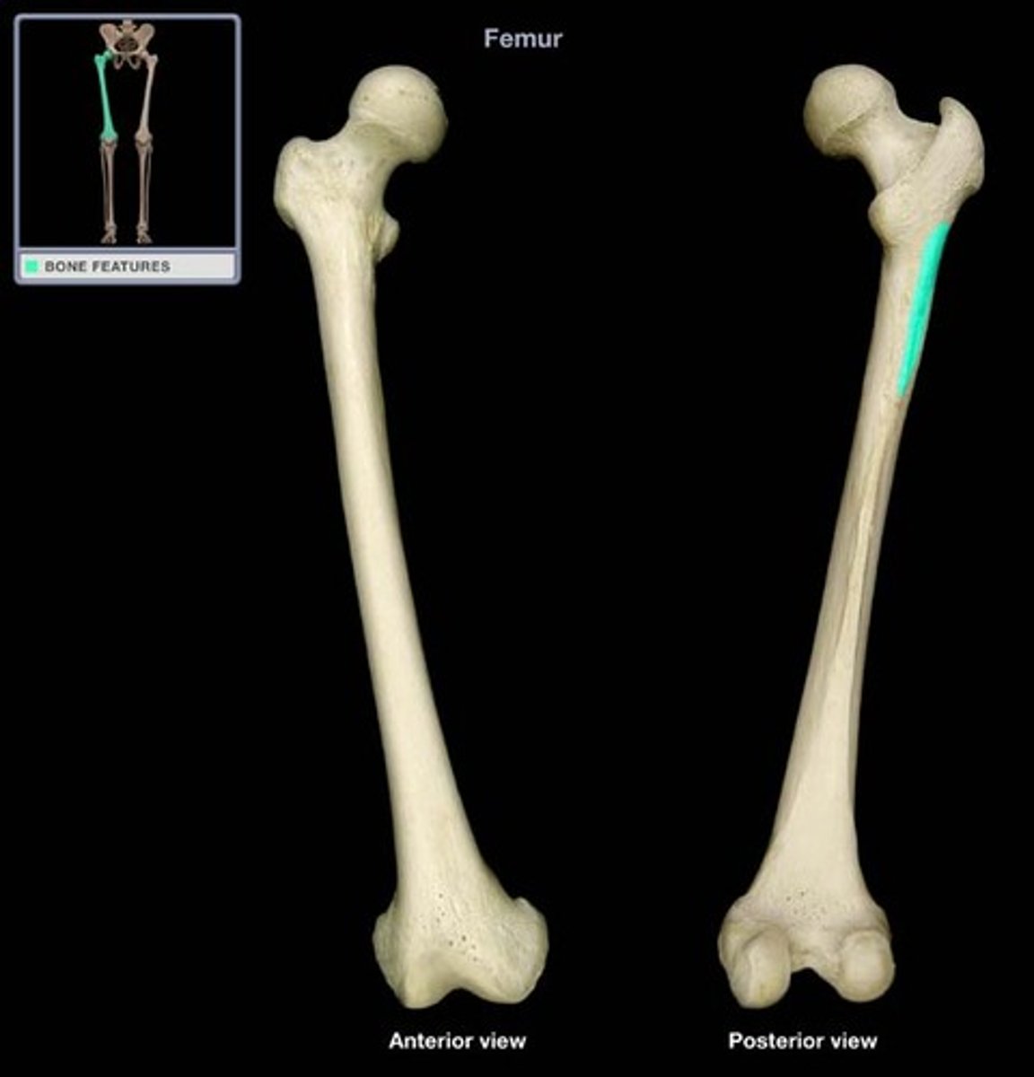
Gluteal tuberosity
The roughened vertical ridge on the posterior femur that serves as a gluteal muscle attachment.
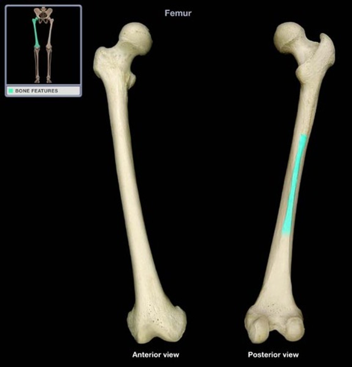
Linea aspera
The prominent ridge that runs down the posterior shaft of the femur.
Lateral condyle
The rounded distal, lateral projection of the femur that articulates with the tibia.
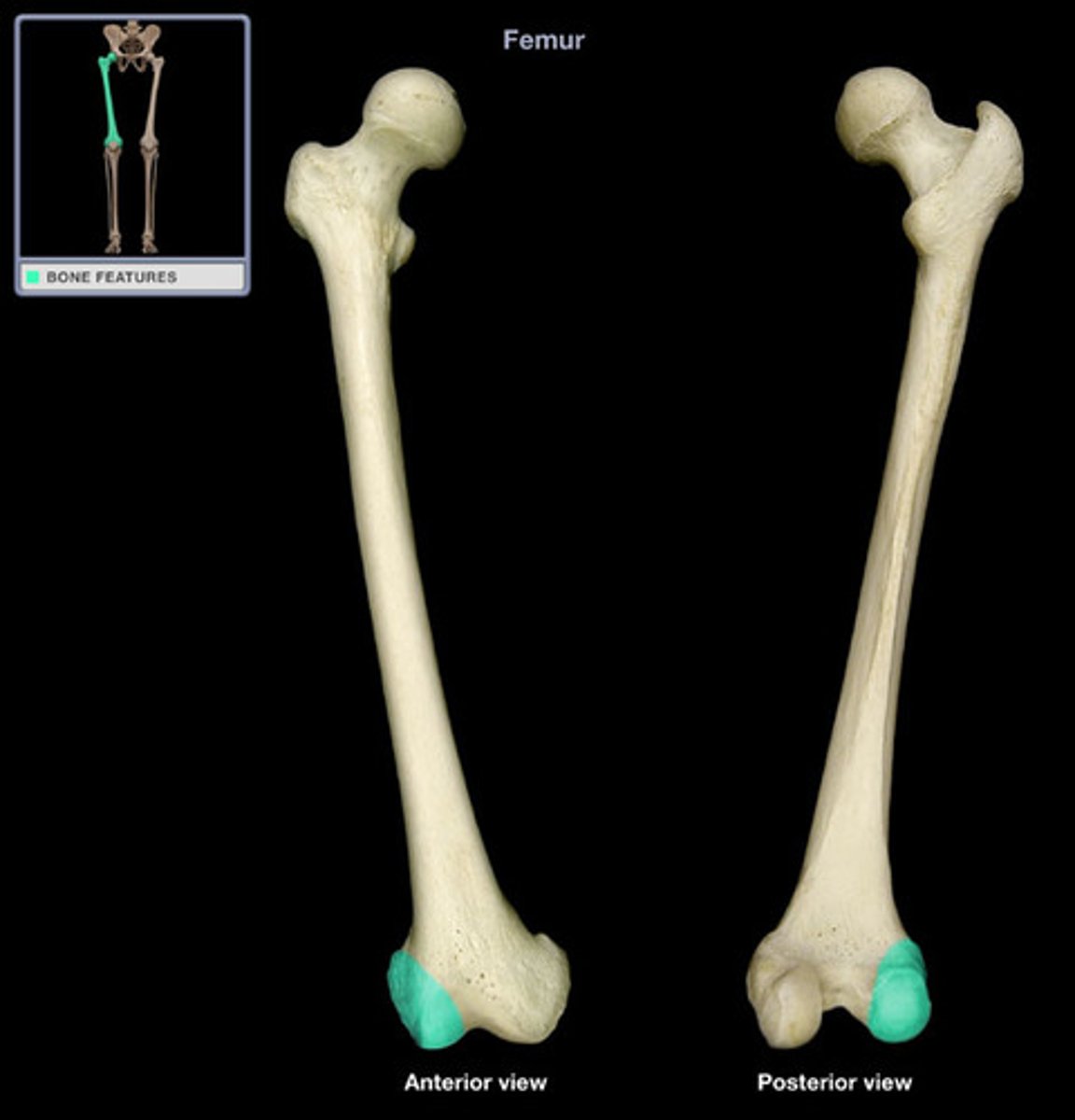
Medial condyle
The rounded distal, medial projection of the femur that articulates with the tibia.
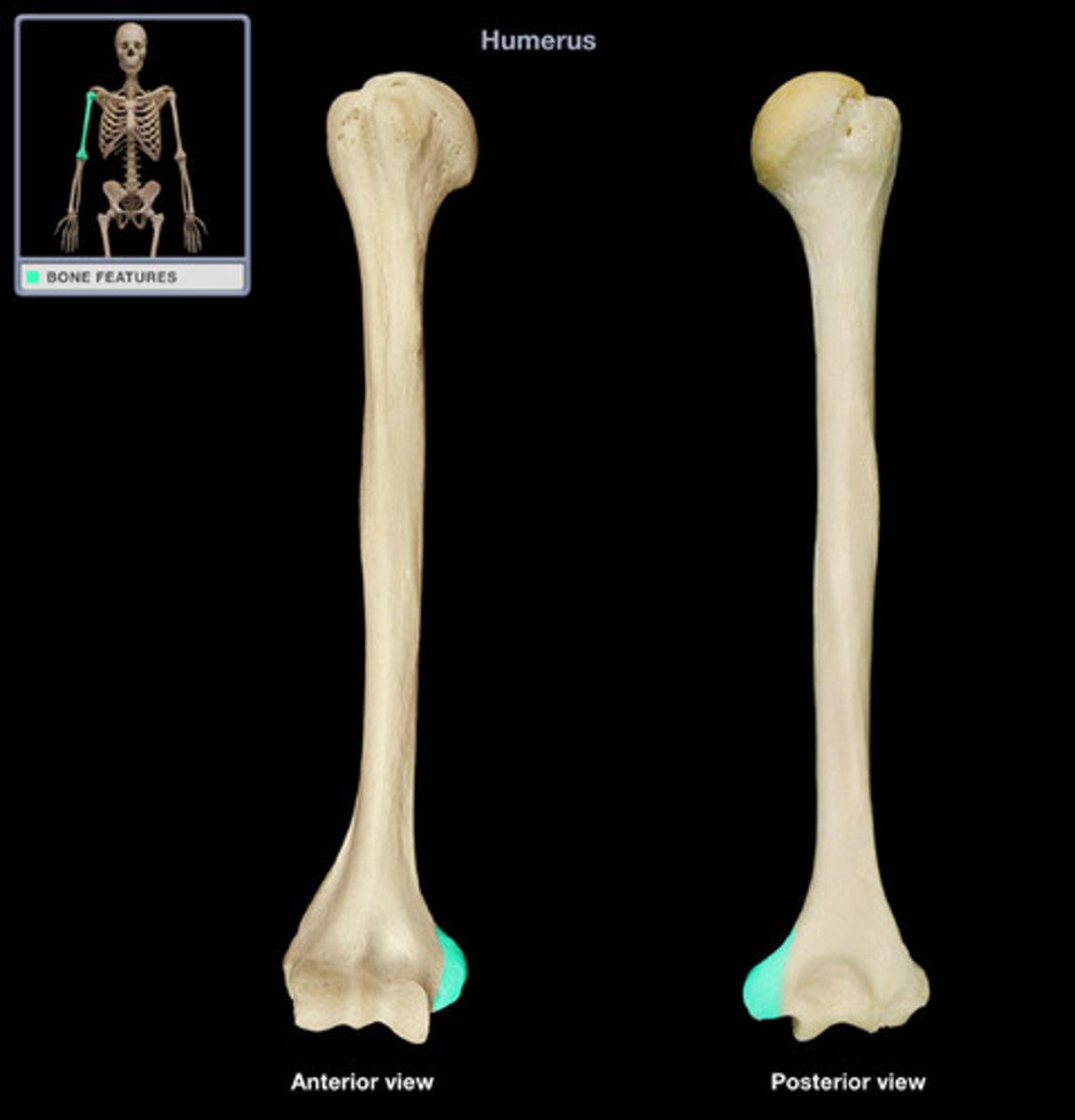
Lateral epicondyle
The smaller projection located above the lateral condyle that serves as a ligament attachment site.
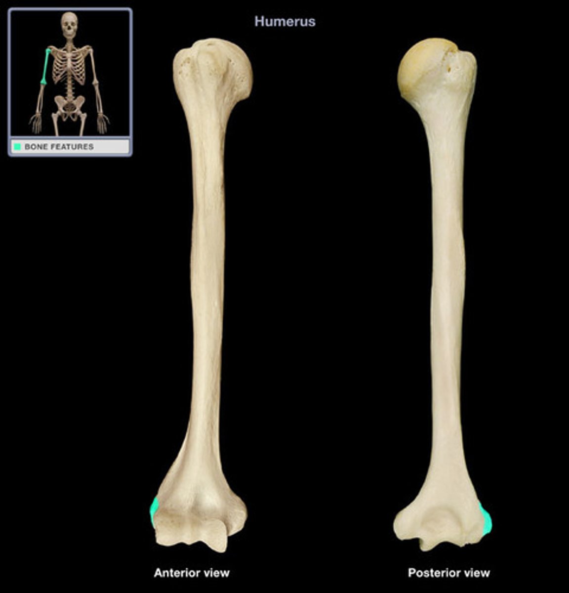
Medial epicondyle
The smaller projection located above the medial condyle that serves as a ligament attachment site.
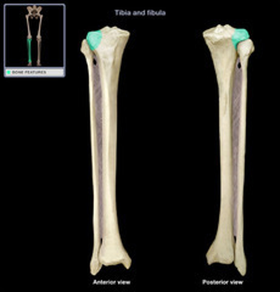
Medial condyle (tibia)
The large medial projection on the proximal tibia that articulates with the femur.
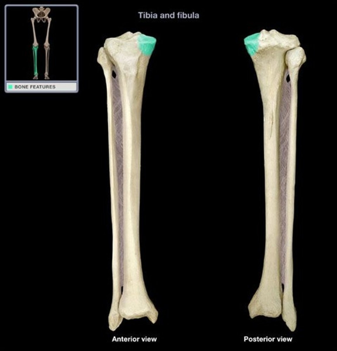
Lateral condyle (tibia)
The large lateral projection on the proximal tibia that articulates with the femur.
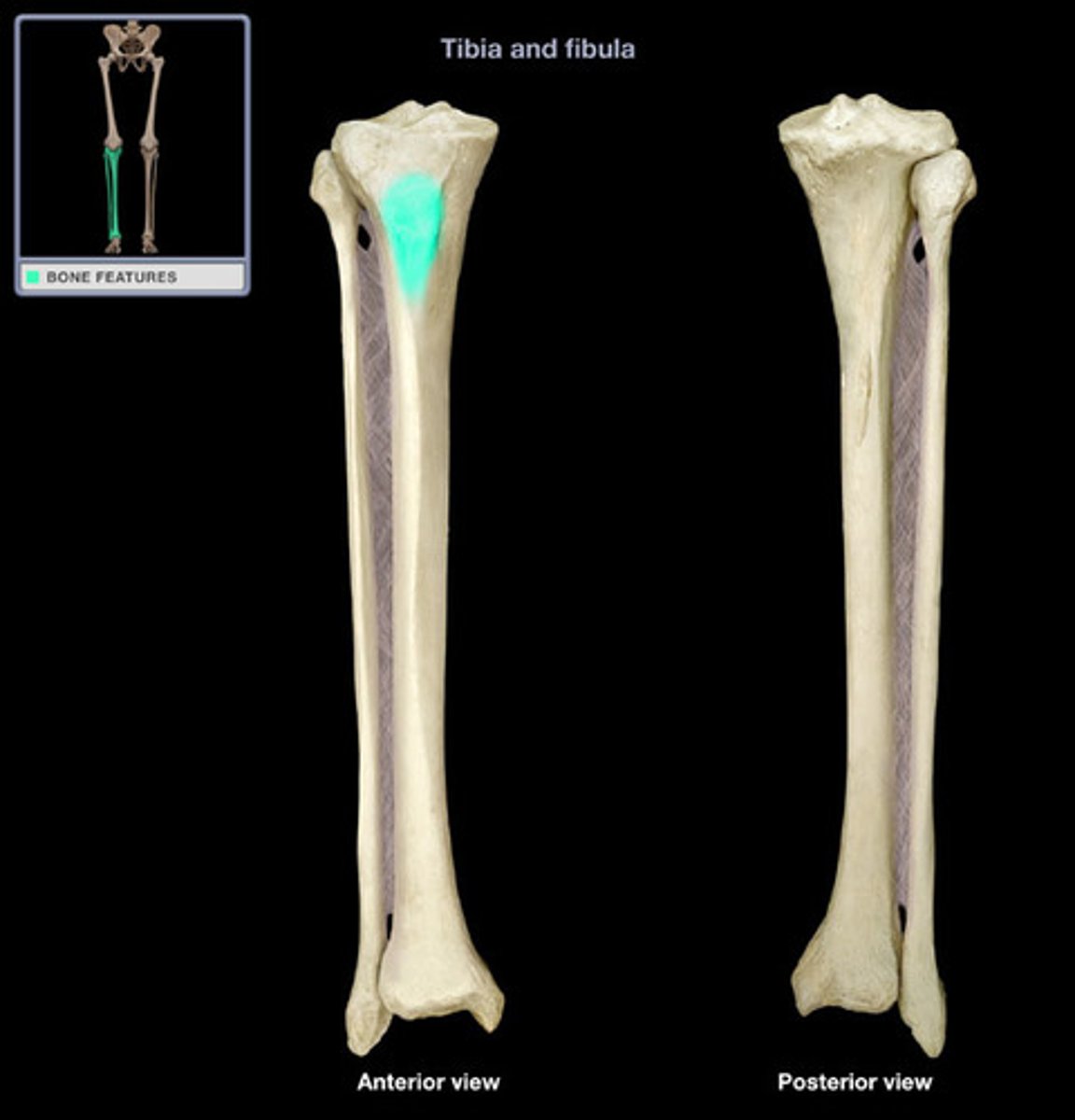
Tibial tuberosity
The roughened bump on the anterior proximal tibia that serves as the attachment for the patellar ligament.
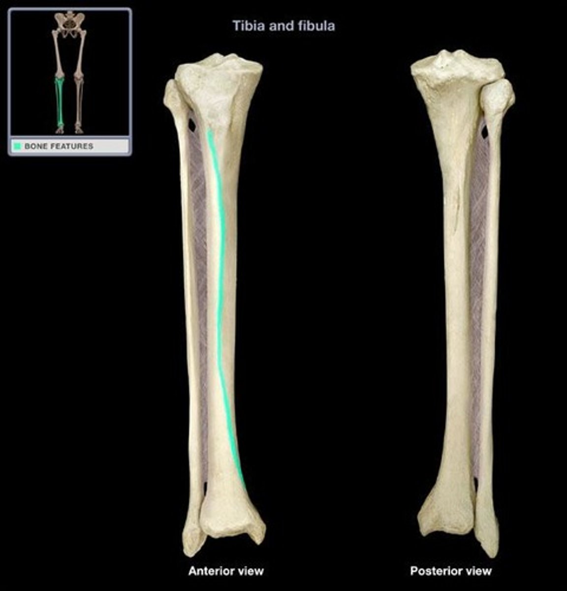
Anterior crest
The sharp ridge that runs down the anterior shaft of the tibia and is commonly called the 'shin'.
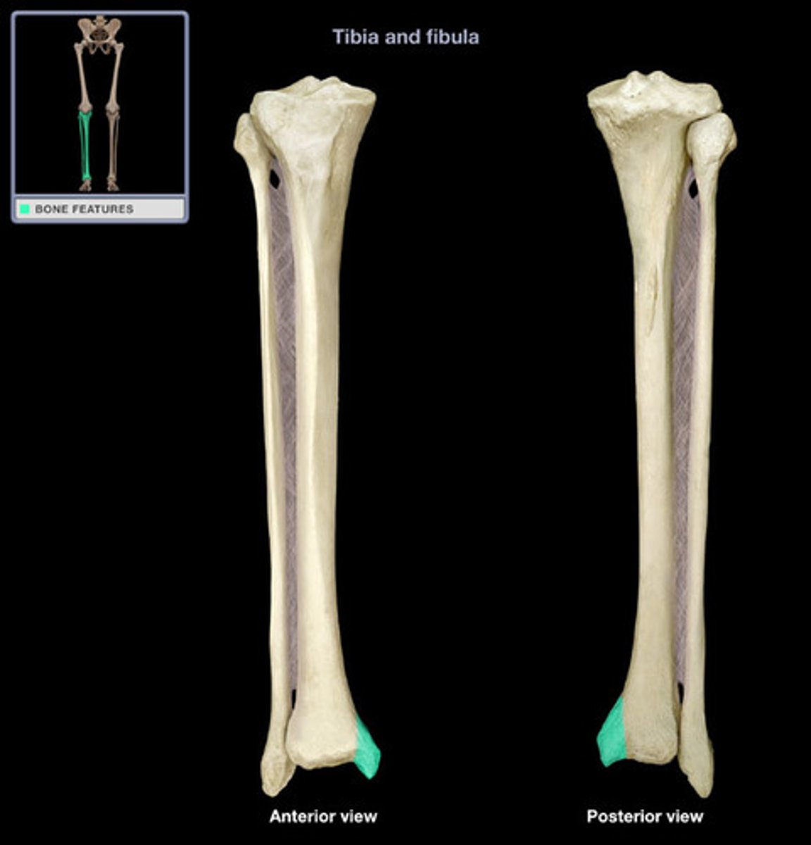
Medial malleolus
The distal, medial projection of the tibia that forms the inner ankle bump.
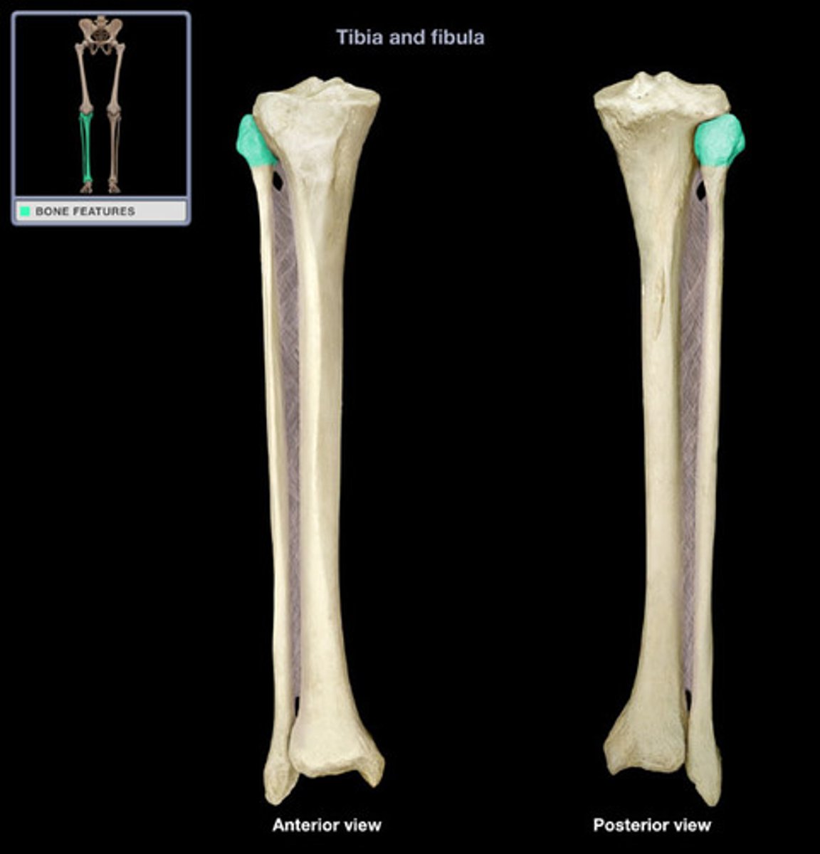
Head of the fibula
The expanded proximal end of the fibula that articulates with the tibia (not the femur).
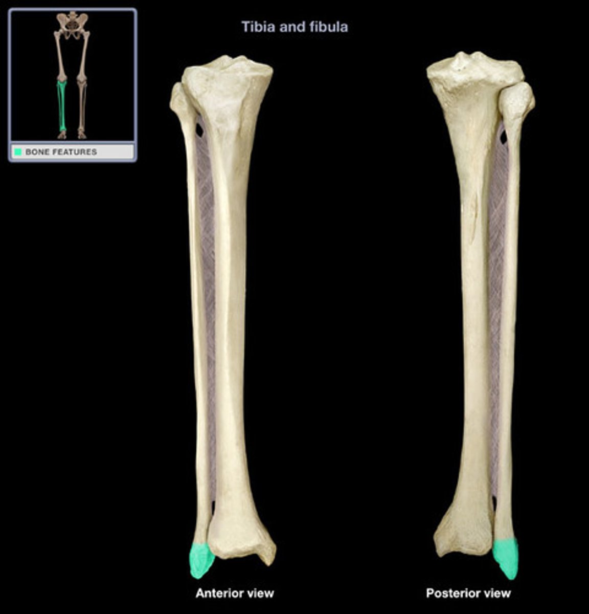
Lateral malleolus
The distal, lateral projection of the fibula that forms the outer ankle bump.
Talus
The bone that sits on top of the calcaneus and connects with the tibia and fibula.
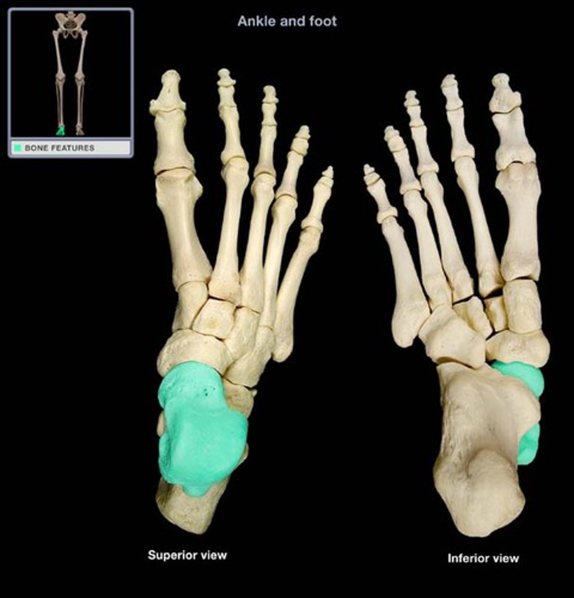
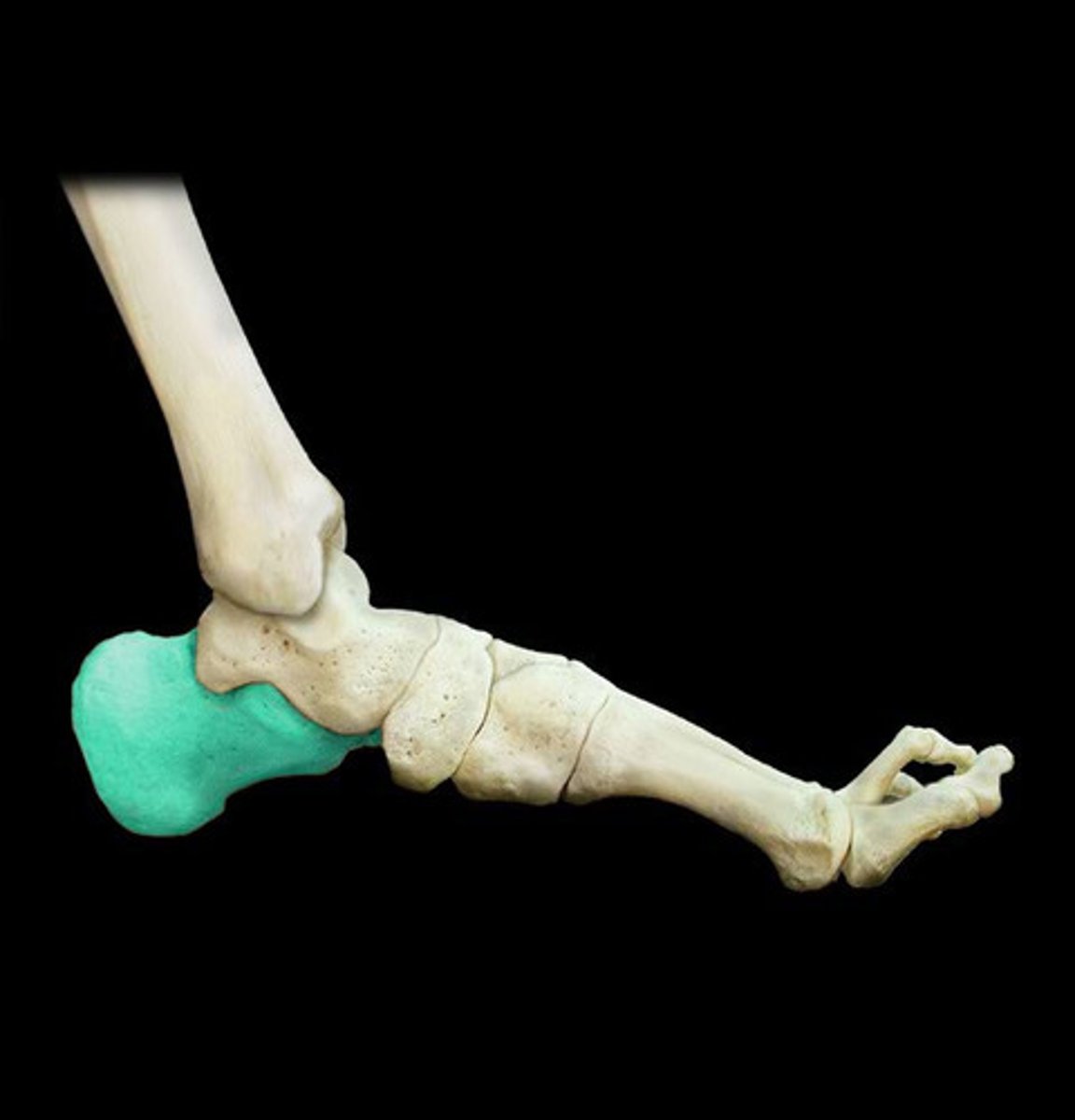
Calcaneus
The largest tarsal bone, forming the heel.
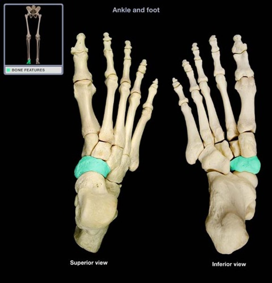
Navicular
The tarsal located in front of the talus on the medial side.
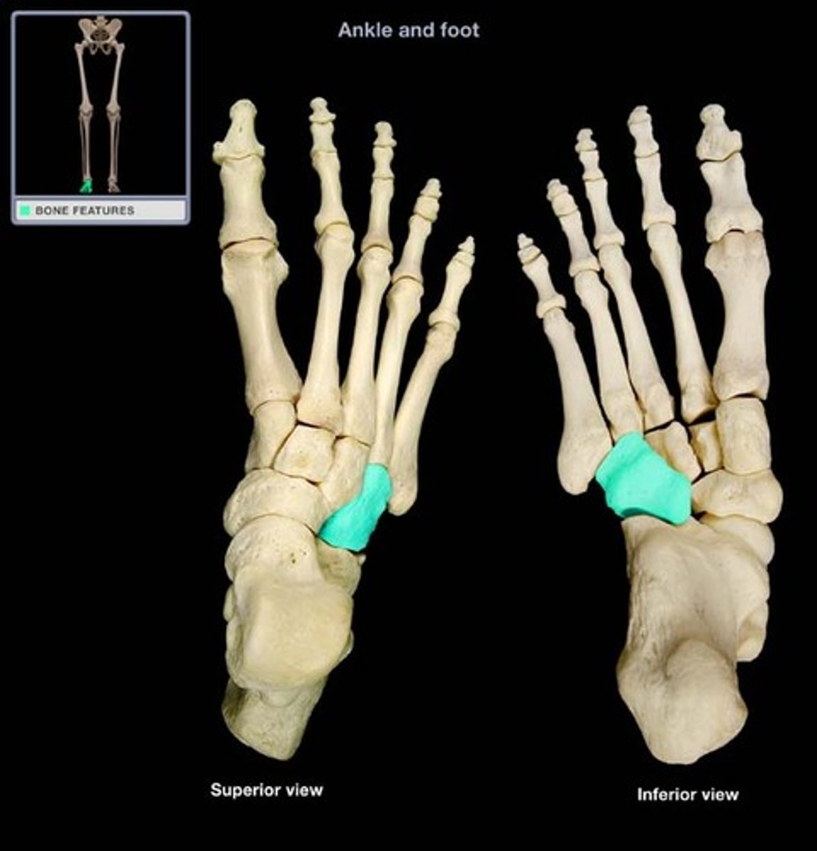
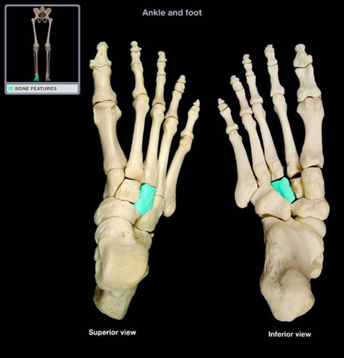
Cuboid
The tarsal on the lateral side in front of the calcaneus.
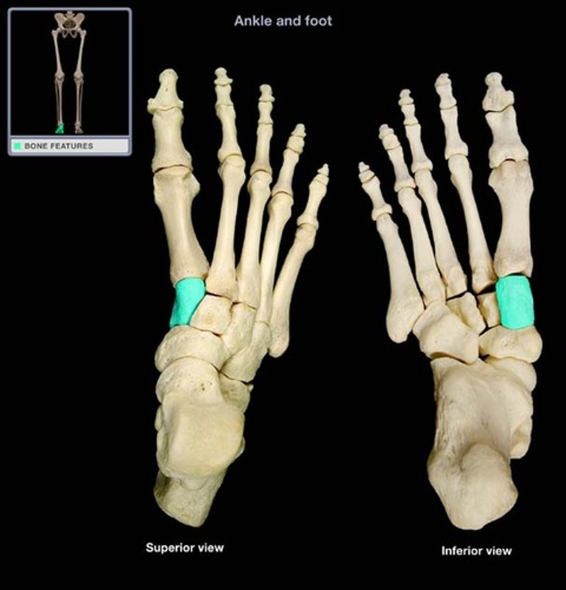
Medial cuneiform
The tarsal in front of the navicular on the medial side of the foot.
Intermediate cuneiform
The tarsal located between the medial and lateral cuneiforms.
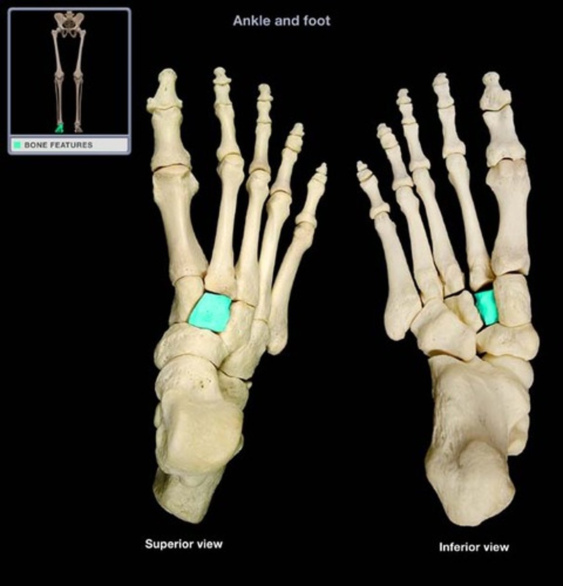
Lateral cuneiform
The cuneiform next to the intermediate cuneiform in line with the 3rd metatarsal.
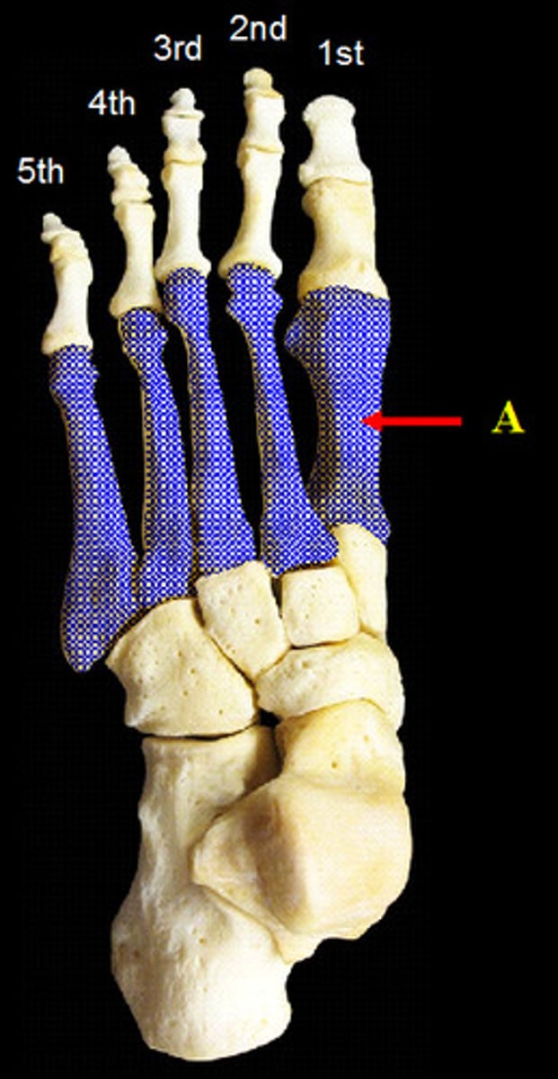
Metatarsals 1-5
The five long bones in the midfoot, numbered medial to lateral.
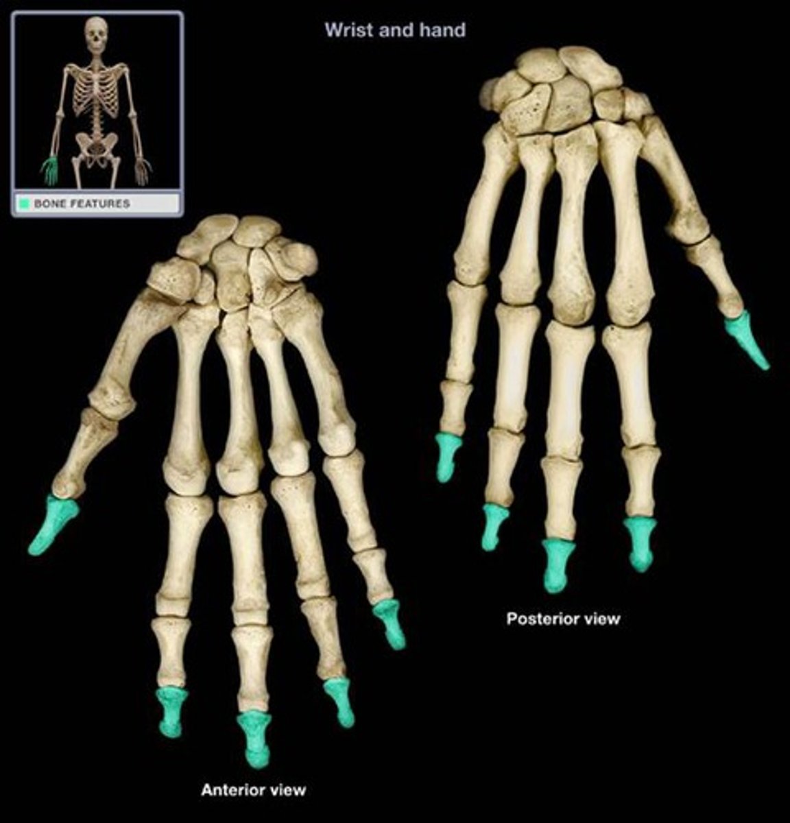
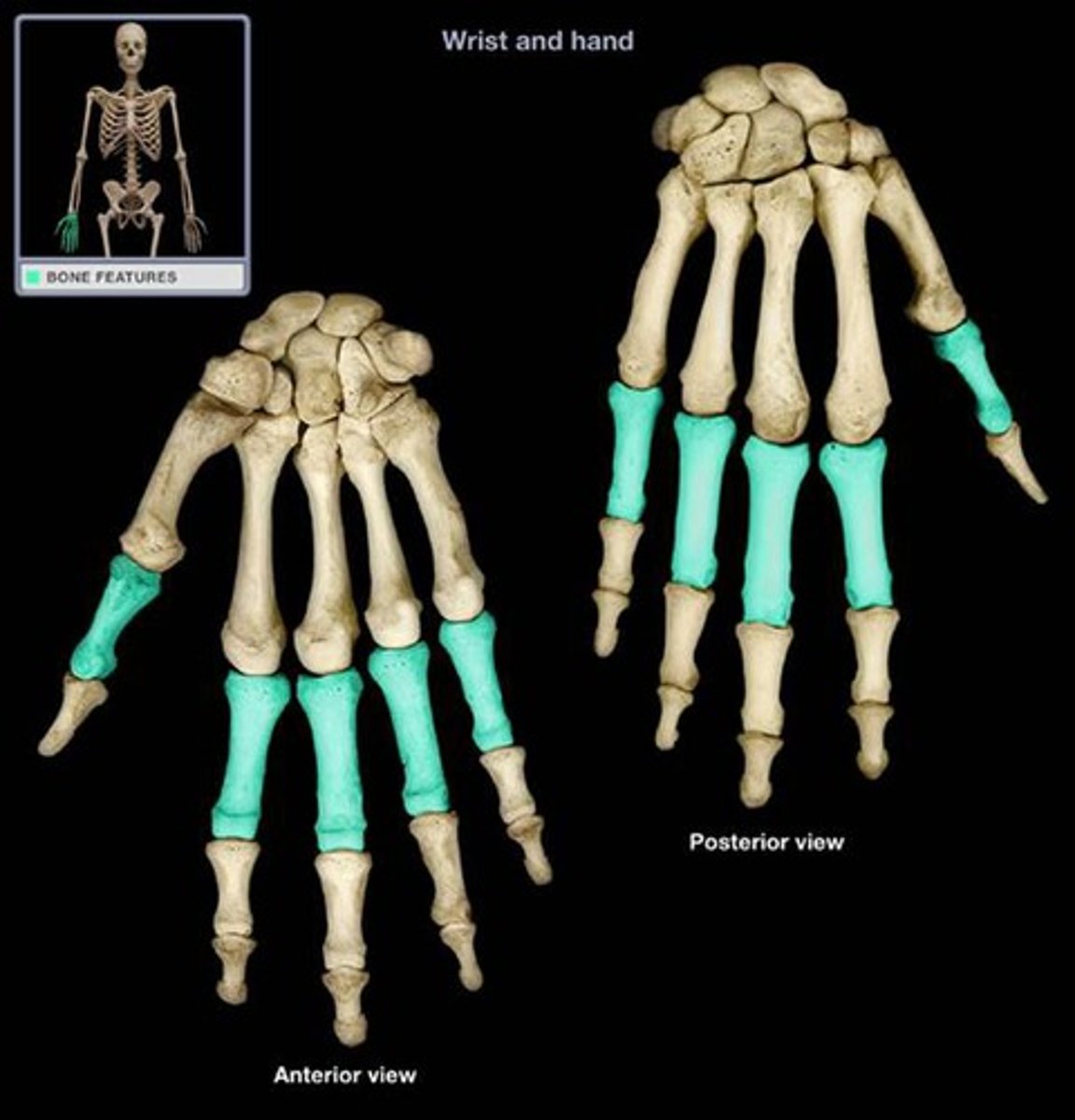
Proximal phalanges
The bones in the toes closest to the metatarsals.
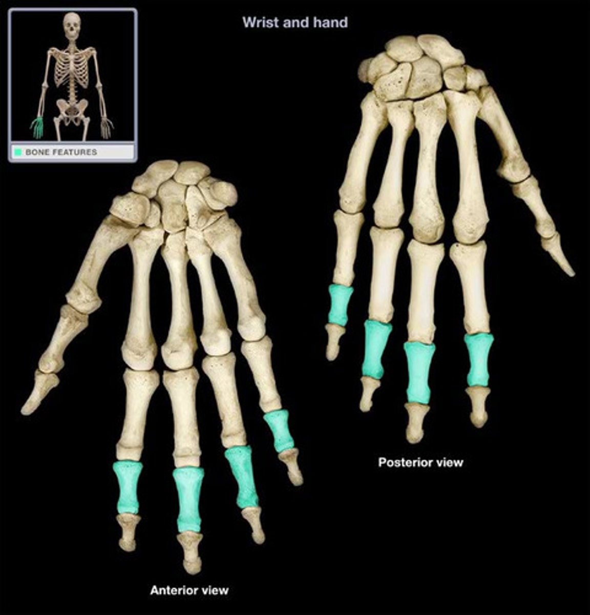
Intermediate phalanges
The middle bones of toes 2-5.
Distal phalanges
The bones at the tip of all the toes.