Cells Terminology Practice
 Call Kai
 Learn
Learn Practice Test
 Spaced Repetition
 Match
Match Flashcards
 Knowt Play
1/43

Earn XP
Description and Tags
Comprehensive practice flashcards for Anatomy and Physiology I Lab, focusing on cell structure, the cell cycle, membrane transport, and histology (epithelial, connective, muscle, and nervous tissues).
Name | Mastery | Learn | Test | Matching | Spaced | Call with Kai | Chat |
|---|
No analytics yet
Send a link to your students to track their progress
44 Terms
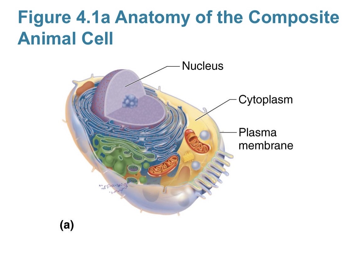
Nucleus
The 'control center' of the cell. Appearance: Large, spherical organelle containing nucleoli and chromatin, enclosed by a double-membrane nuclear envelope. Identification: Usually the most prominent, dark-staining structure in the cell on a diagram or slide.
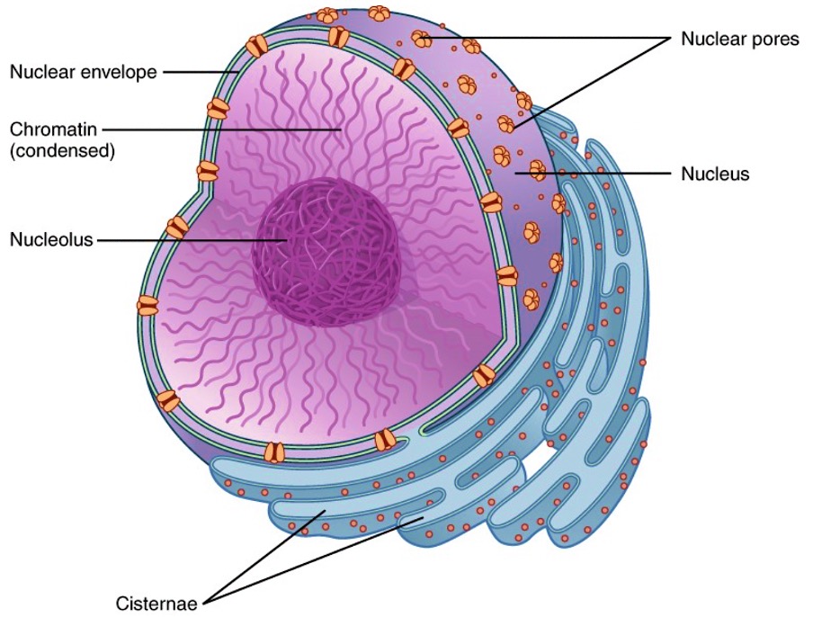
Nuclear Envelope and Nuclear Pores
Structure: A double-layer membrane surrounding the nucleus. The pores are openings that regulate the passage of proteins and RNA. Function: To separate the nucleoplasm from the cytoplasm and facilitate transport between the two.
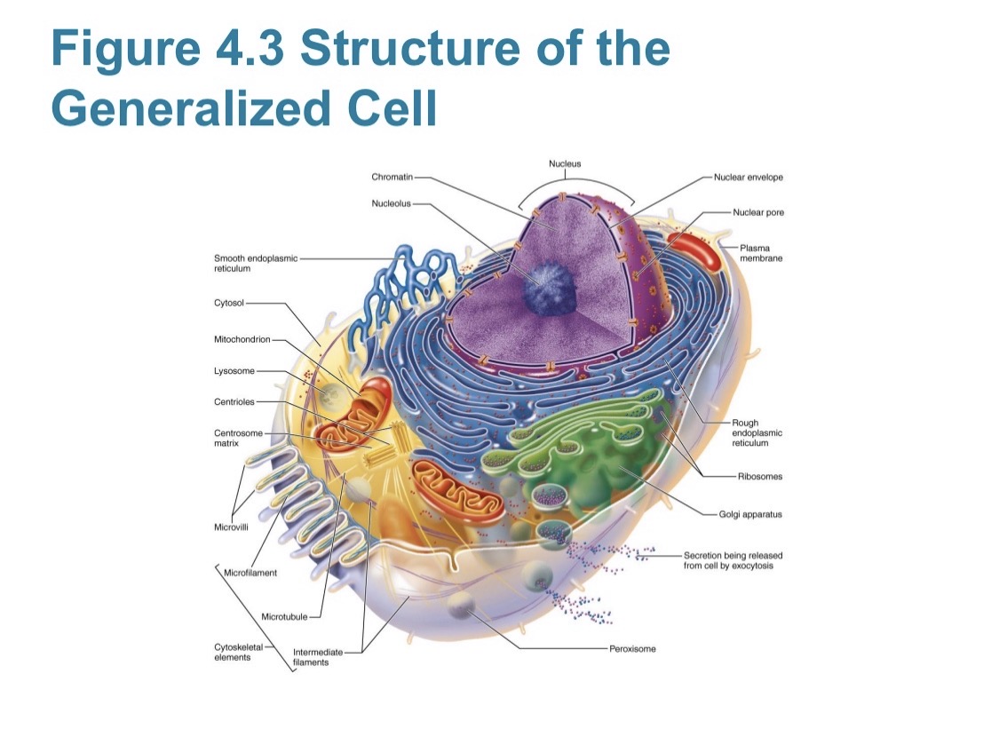
Nucleoli
Appearance: Small, dense bodies found within the nucleus. Function: Site of ribosome subunit synthesis. Identification: Darker-staining spots inside the nucleus itself.
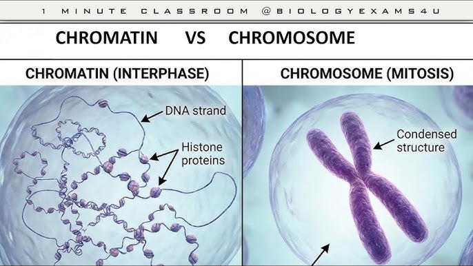
Chromatin vs. Chromosomes
Chromatin is the loose, thread-like form of DNA and proteins found during interphase. Chromosomes are highly condensed, bar-like bodies of DNA and protein formed during mitosis. Chromatin is for transcription/replication; chromosomes are for transport/movement during division.
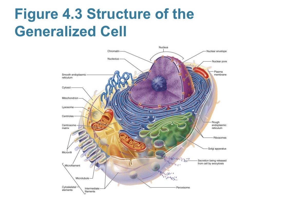
Rough Endoplasmic Reticulum (RER)
Structure: Membranes studded with ribosomes, often continuous with the nuclear envelope. Appearance: Flattened sac-like appearance. Function: Protein synthesis and modification. Identification clue: Looks like 'studded' or 'bumpy' sheets near the nucleus.
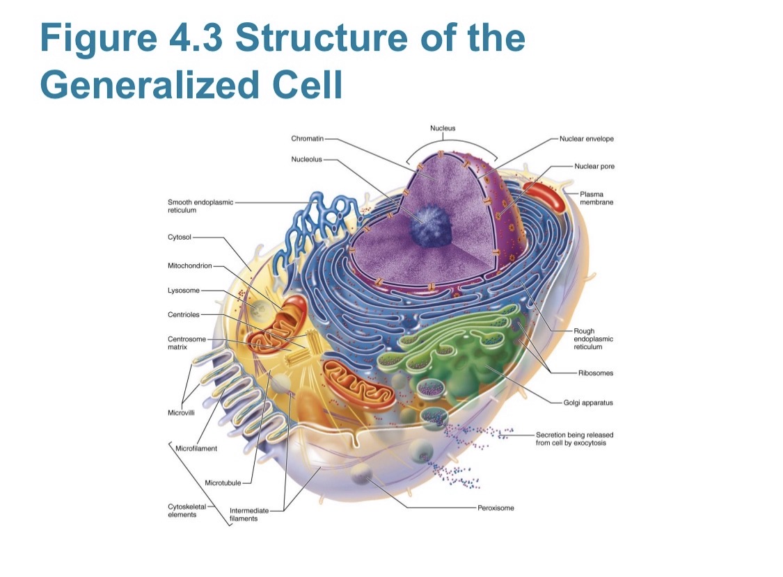
Smooth Endoplasmic Reticulum (SER)
Structure: Membranous tubules without ribosomes. Function: Lipid and steroid synthesis, detoxification of drugs, and calcium storage. Identification clue: More tubular and lacks the 'bumps' seen on the RER.
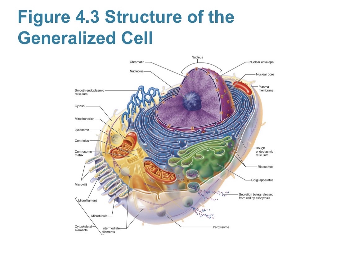
Golgi Apparatus
Appearance: A stack of flattened membranous sacs (resembles a stack of pita bread). Function: Modifies, concentrates, and packages proteins and lipids into vesicles. Location: Often found between the RER and the plasma membrane.
Mitochondria
Structure: Double-membrane organelle; inner membrane has folds called cristae. Function: The 'powerhouse' of the cell; produces ATP via aerobic cellular respiration. Identification: Elongated shape with internal 'zig-zag' lines (cristae).
Lysosomes vs. Peroxisomes
Lysosomes contain digestive enzymes to break down old organelles and foreign substances. Peroxisomes contain oxidases and catalases to neutralize toxins like free radicals and hydrogen peroxide. Both look like small spherical sacs, but lysosomes are generally involved in digestion while peroxisomes handle detox.
Centrosome and Centrioles
The centrosome is the microtubule-organizing center near the nucleus. Each centrosome contains a pair of centrioles, which are small, barrel-shaped organelles (triplets of microtubules). Function: They organize the spindle fibers during mitosis.
Microvilli vs. Cilia vs. Flagella
Microvilli: Small, finger-like extensions of the plasma membrane used to increase surface area for absorption (found in intestines). Cilia: Hair-like motile projections that move substances across the cell surface (found in the trachea). Flagella: Long, singular tail-like projection used to propel the cell itself (found in sperm cells).
Cytoskeleton: Microtubules, Intermediate Filaments, Microfilaments
Microtubules: Largest; hollow tubes composed of tubulin; determine cell shape and move organelles. Intermediate Filaments: Stable, rope-like proteins; provide mechanical strength (e.g., keratin). Microfilaments: Smallest; composed of actin; involved in cell movement and muscle contraction.
Interphase (G1, S, G2)
The period from cell formation to cell division. G1: Phase of vigorous growth and metabolism. S: Phase where DNA is replicated. G2: Preparation for division; enzymes/proteins are synthesized. Visual Clue: The nucleus contains chromatin; the nuclear envelope is intact.
Prophase (Early vs. Late)
Early: Chromatin condenses into chromosomes (paired sister chromatids); centrosomes begin moving to opposite poles. Late (Prometaphase): The nuclear envelope breaks up; spindle fibers attach to the kinetochores of chromosomes.
Metaphase
Identification: Chromosomes line up along the equator (metaphase plate) of the cell. Clue: Centromeres are perfectly aligned in the middle.
Anaphase
Identification: The centromeres split and sister chromatids are pulled apart toward opposite poles. These moving chromatids are now called daughter chromosomes. Visual clue: Chromosomes look V-shaped as they are dragged through the cytosol.
Telophase
Identification: Movement stops. Chromosomes uncoil back into chromatin, new nuclear envelopes form around each set of chromosomes, and spindle fibers disappear. Differences: It is the exact reverse of prophase.
Cytokinesis
The division of the cytoplasm. Identification clue: Appearance of a cleavage furrow (a ring of actin microfilaments) that pinches the cell into two daughter cells. This process typically begins during late anaphase and completes through telophase.
Simple Diffusion
Type: Passive transport. Mechanism: Movement of nonpolar/lipid-soluble substances (e.g., O2, CO2) directly through the phospholipid bilayer. ATP required: No. Direction: Down the concentration gradient (high to low).
Facilitated Diffusion
Type: Passive transport. Mechanism: Movement of polar or charged molecules (e.g., glucose, ions) across the membrane using channel or carrier proteins. Identification: Diffusion that requires a 'helper' protein.
Osmosis
Type: Passive transport. Definition: The diffusion of water (solvent) through a selectively permeable membrane or through aquaporins. Water moves from low solute concentration to high solute concentration (high water concentration to low water concentration).
Passive vs. Active Transport
Passive transport (diffusion, osmosis) moves substances down their concentration gradient and requires no ATP. Active transport moves substances against their concentration gradient (low to high) and requires ATP.
Hypertonic Solution Scenario
Concentration: The solution has a higher solute concentration than the cell. Water movement: Water leaves the cell. Result: The cell shrinks and shrivels, a process called crenation.
Hypotonic Solution Scenario
Concentration: The solution has a lower solute concentration than the cell. Water movement: Water enters the cell. Result: The cell swells and may eventually burst (lyse).
Isotonic Solution Scenario
Concentration: Solute concentration is the same inside and outside the cell. Water movement: No net movement of water. Result: The cell maintains its normal shape and volume.
Vesicular Transport: Endocytosis (Phagocytosis vs. Pinocytosis)
Endocytosis: Bringing matter into the cell. Phagocytosis: 'Cell eating'; engulfing large particles like bacteria. Pinocytosis: 'Cell drinking'; taking in droplets of extracellular fluid containing dissolved solutes. Both require ATP and vesicles.
Exocytosis
Type: Vesicular transport. Process: A secretory vesicle fuses with the plasma membrane to release contents (like hormones or waste) outside the cell. Visual clue: A vesicle merging with the outer membrane to expel contents.
Simple Squamous Epithelium
Description: Single layer of flat, scale-like cells. Function: Rapid diffusion and filtration. Location: Kidney glomeruli, air sacs of lungs (alveoli), lining of heart and blood vessels. Identification: Profile view looks like thin lines with bulging nuclei.
Simple Cuboidal Epithelium
Description: Single layer of cube-shaped cells with large, central nuclei. Function: Secretion and absorption. Location: Kidney tubules and ovary surface. Identification: Look for a ring of square-shaped cells surrounding a central lumen (opening).
Simple Columnar Epithelium
Description: Single layer of tall, narrow cells with oval nuclei usually near the basement membrane. Function: Absorption and secretion of mucus. Location: Non-ciliated type lines the digestive tract (stomach to rectum). Identification: Tall columns; may contain goblet cells for mucus production.
Pseudostratified Columnar Epithelium
Description: Single layer of cells of varying heights, making it appear layered. All cells touch the basement membrane. Function: Secretion and propulsion of mucus. Location: Ciliated version lines the trachea and upper respiratory tract. Identification: Nuclei at different 'levels', often ciliated on the apical surface.
Stratified Squamous Epithelium
Description: Multiple layers; apical cells are flat, basal cells are often cuboidal/columnar. Function: Protection against abrasion. Location: Non-keratinized in the esophagus/mouth; keratinized in the skin (epidermis). Identification: Thickest epithelium with many layers of flat cells at the surface.
Transitional Epithelium
Description: Resembles both stratified squamous and stratified cuboidal; cells can change shape (distend). Function: Permits stretching and distension of urinary organs. Location: Lines the ureters and urinary bladder. Identification: Plump, 'dome-shaped' cells on the apical surface when the organ is empty.
Areolar Connective Tissue
Type: Loose Connective Tissue. Fibers: Random arrangement of collagen and elastic fibers. Matrix: Gel-like ground substance. Function: Wraps and cushions organs; plays a role in inflammation. Location: Widely distributed under epithelia. Identification: Looks like a loose 'web' of criss-crossing strings.
Adipose Tissue
Type: Loose Connective Tissue. Description: Closely packed adipocytes (fat cells); nucleus is pushed to the side by a large fat droplet. Function: Energy storage, insulation, and protection. Location: Under skin (hypodermis), around kidneys, and within breasts. Identification: Looks like clear, empty bubbles or 'chicken wire'.
Dense Regular Connective Tissue
Description: Primarily parallel collagen fibers with a few fibroblasts. Function: Attaches muscles to bones (tendons) or bones to bones (ligaments). Identification: Wavy, organized, parallel fibers. Difference from Irregular: Fibers go in the same direction.
Hyaline Cartilage
Appearance: Amorphous but firm matrix; collagen fibers are not visible. Cells: Chondrocytes in lacunae. Function: Supports and reinforces; cushions. Location: Costal cartilage of ribs, nose, trachea, larynx, ends of long bones. Identification: Smooth, 'glassy' matrix with cells in 'little houses' (lacunae).
Elastic Cartilage
Appearance: Similar to hyaline but with many visible dark-staining elastic fibers in the matrix. Function: Maintains shape while allowing great flexibility. Location: External ear (pinna) and epiglottis.
Fibrocartilage
Appearance: Matrix similar to but less firm than hyaline; thick collagen fibers predominate. Function: Tensile strength with the ability to absorb compressive shock. Location: Intervertebral discs, pubic symphysis, discs of knee joint. Identification: Distinct 'wispy' blue or pink collagen fibers.
Skeletal Muscle
Description: Long, cylindrical, multinucleated cells with obvious striations. Control: Voluntary. Location: Attached to bones. Identification: Parallel 'rods' with visible stripe patterns (striations) and nuclei at the periphery.
Cardiac Muscle
Description: Branching, striated, usually uninucleate cells. Identification: Presence of intercalated discs (specialized junctions). Control: Involuntary. Location: Walls of the heart. Difference: Found only in the heart; branching fibers distinguish it from skeletal muscle.
Smooth Muscle
Description: Spindle-shaped cells with central nuclei; no striations. Control: Involuntary. Location: Walls of hollow organs (e.g., digestive tract, blood vessels). Identification: 'S' shaped nuclei and a lack of stripes.
Nervous Tissue: Neuron Parts
Cell Body: Biosynthetic center containing the nucleus. Dendrites: Branching processes that receive signals. Axon: Single long process that generates/conducts nerve impulses away from the cell body. Identification: Look for 'star-shaped' cells with long extensions.
Connective Tissue Fibers: Collagen vs. Elastic vs. Reticular
Collagen: Thickest, strongest, white fibers; high tensile strength. Elastic: Long, thin, yellow fibers; allow for stretch and recoil. Reticular: Short, fine, highly branched collagenous fibers; form internal 'skeletons' (stroma) of soft organs like the spleen.