BSC2085L E.16 Special Senses
0.0(0)
Card Sorting
1/52
Study Analytics
Name | Mastery | Learn | Test | Matching | Spaced |
|---|
No study sessions yet.
53 Terms
1
New cards
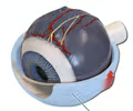
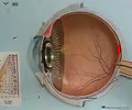
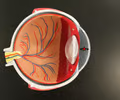
sclera
• The outer layer of the eye superficial to the choroid.
• Appears as a white, external covering.
• Appears as a white, external covering.
2
New cards
cornea
The transparent anterior portion of the eye covering the iris and pupil.
3
New cards
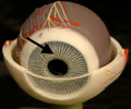
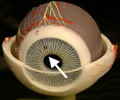
iris
• The colored circle of the eye surrounding the pupil.
• A ring of smooth muscle controlling the diameter of the pupil in response to light exposure.
• A ring of smooth muscle controlling the diameter of the pupil in response to light exposure.
4
New cards
pupil
• The black circle of the eye surrounded by the iris.
• Dilates and contracts in response to light exposure via the iris.
• Dilates and contracts in response to light exposure via the iris.
5
New cards
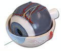
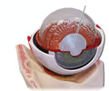
ciliary body
• A structure arising at the junction of the cornea and sclera.
• Consists of the ciliary muscle and ciliary muscles.
• Composes the ora serrata at its posterior end and the suspensory ligaments attaching to the lens.
• Consists of the ciliary muscle and ciliary muscles.
• Composes the ora serrata at its posterior end and the suspensory ligaments attaching to the lens.

6
New cards
ora serrata
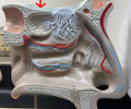
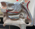
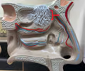
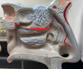
The serrated boundary between the ciliary muscle and the retina.

7
New cards
suspensory ligaments
Fibrous extensions of the ciliary body holding the lens in place.
8
New cards
lens
• The transparent structure behind the pupil that focuses light onto the retina.
• Held by the suspensory ligaments.
• Held by the suspensory ligaments.
9
New cards
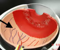
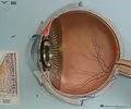
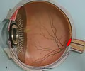
choroid
• The middle layer of the eye deep to the sclera and superficial to the retina.
• Highly vascular.
• Highly vascular.

10
New cards
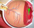
retina
• The inner layer of the eye deep to the choroid.
• The photosensitive region containing rods and cones.
• The photosensitive region containing rods and cones.
11
New cards
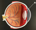
macula lutea
• A region of the retina containing only cone cells.
• Appears as a small depression on the posterior portion of the retina.
• Appears as a small depression on the posterior portion of the retina.
12
New cards
fovea centralis
• A small depression in the macula lutea.
• Contains the highest concentration of cone cells.
• Contains the highest concentration of cone cells.
13
New cards
optic disc
• The area of the retina where the optic nerve exits.
• Lacks photoreceptors.
• Also known as the blind spot.
• Lacks photoreceptors.
• Also known as the blind spot.
14
New cards
optic nerve
• The nerve carrying visual information from the retina to the brain.
• Located at the optic disc or blind spot.
• Located at the optic disc or blind spot.
15
New cards
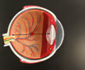
anterior chamber
• Part of the anterior cavity between the cornea and the iris.
• Contains aqueous humor.
• Contains aqueous humor.
16
New cards
posterior chamber
• Part of the anterior cavity between the iris and the lens.
• Contains aqueous humor.
• Contains aqueous humor.
17
New cards
posterior cavity
• Extends from the lens to the retina.
• Contains vitreous humor.
• Contains vitreous humor.
18
New cards
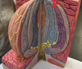
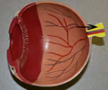
tapetum lucidum
A pigmented layer of the choroid seen in the cow eye.

19
New cards
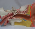
auricle
• Part of the outer ear.
• The external cartilaginous appendage of the ear that channels sound waves.
• The external cartilaginous appendage of the ear that channels sound waves.
20
New cards
helix
• Part of the outer ear.
• The outer rim of the auricle.
• The outer rim of the auricle.
21
New cards
lobule
• Part of the outer ear.
• The inferior feature of the auricle.
• The inferior feature of the auricle.
22
New cards
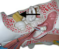
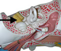
external auditory canal
• The opening and passage of the ear from the auricle to the tympanic membrane.
• Contains ceruminous glands.
• Contains ceruminous glands.
23
New cards
tympanic membrane
• Part of the outer ear.
• A thin membrane of skin separating the outer and middle ear.
• Converts sound waves to mechanical vibrations.
• Also known as the eardrum.
• A thin membrane of skin separating the outer and middle ear.
• Converts sound waves to mechanical vibrations.
• Also known as the eardrum.
24
New cards
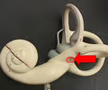
auditory ossicles
• Part of the middle ear.
• The three small bones within the tympanic cavity: the malleus, incus, and stapes.
• The three small bones within the tympanic cavity: the malleus, incus, and stapes.
25
New cards
malleus
• Part of the middle ear.
• The lateral of the three auditory ossicles.
• Also known as the hammer.
• The lateral of the three auditory ossicles.
• Also known as the hammer.
26
New cards
incus
• Part of the middle ear.
• The intermediate of the three auditory ossicles.
• Also known as the anvil.
• The intermediate of the three auditory ossicles.
• Also known as the anvil.
27
New cards
stapes
• Part of the middle ear.
• The medial of the three auditory ossicles.
• Also known as the stirrup.
• The medial of the three auditory ossicles.
• Also known as the stirrup.

28
New cards
auditory tube
• Part of the middle ear.
• Connects the tympanic cavity to the nasal cavity.
• Also known as the Eustachian tube or pharyngotympanic tube.
• Connects the tympanic cavity to the nasal cavity.
• Also known as the Eustachian tube or pharyngotympanic tube.
29
New cards
oval window
• Part of the middle ear.
• Small membrane-covered opening that separates the middle ear and inner ear.
• Often not visible because it is covered by the stapes.
• Small membrane-covered opening that separates the middle ear and inner ear.
• Often not visible because it is covered by the stapes.
30
New cards
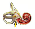
round window
• Part of the middle ear.
• Small membrane-covered opening between the middle ear and cochlea.
• Small membrane-covered opening between the middle ear and cochlea.
31
New cards
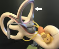
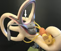
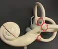
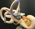
anterior semicircular canal
• Part of the inner ear.
• The semicircular canal facing the cochlea.
• The semicircular canal facing the cochlea.
32
New cards
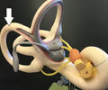
posterior semicircular canal
• Part of the inner ear.
• The semicircular canal perpendicular to the anterior semicircular canal.
• The semicircular canal perpendicular to the anterior semicircular canal.
33
New cards
lateral semicircular canal
• Part of the inner ear.
• The semicircular canal lying on the transverse plane.
• The semicircular canal lying on the transverse plane.
34
New cards
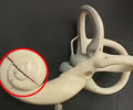
ampulla
• Part of the inner ear.
• The expanded ends of each semicircular canal.
• The expanded ends of each semicircular canal.
35
New cards
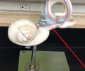
vestibule
• Part of the inner ear.
• The two sacs of membranous labyrinth.
• The two sacs of membranous labyrinth.
36
New cards
saccule
• Part of the inner ear.
• The anterior sac of the vestibule, closer to the cochlea.
• The anterior sac of the vestibule, closer to the cochlea.
37
New cards
utricle
• Part of the inner ear.
• The posterior sac of the vestibule, closer to the anterior semicircular canal.
• The posterior sac of the vestibule, closer to the anterior semicircular canal.
38
New cards
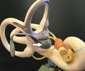
cochlea
• Part of the inner ear.
• The anterior chamber of the inner ear resembling a spiral snail shell.
• The anterior chamber of the inner ear resembling a spiral snail shell.
39
New cards
vestibular nerve
• The nerve connecting to the vestibule and semicircular canals.
• Forms the vestibulocochlear nerve (CN VIII).
• Forms the vestibulocochlear nerve (CN VIII).
40
New cards
cochlear nerve
• The nerve connecting to the cochlea.
• Forms the vestibulocochlear nerve (CN VIII).
• Forms the vestibulocochlear nerve (CN VIII).
41
New cards
vestibulocochlear nerve
• The nerve associated with hearing and equilibrium.
• Formed from the vestibular and cochlear nerves of the inner ear.
• Abbreviated CN VIII.
• Formed from the vestibular and cochlear nerves of the inner ear.
• Abbreviated CN VIII.

42
New cards
olfactory tract
The path extending from the olfactory bulb to the brain.
43
New cards
olfactory bulb
Located just above the nasal cavity.
44
New cards
olfactory nerves
The fibers of the olfactory bulb perforating the cribriform foramina and into the nasal cavity.
45
New cards
olfactory hairs
The dendrites of the olfactory nerves.
46
New cards
filiform papillae
Sharp, spiky bumps of the tongue.
47
New cards
fungiform papillae
Mushroom-like bumps of the tongue.
48
New cards
circumvallate papillae
• Large circular bumps of the tongue.
• Also referred to as vallate.
• Also referred to as vallate.
49
New cards
taste bud
• Located within the papillae of the tongue.
• A sense receptor in the tongue responding to chemical particles from food.
• A sense receptor in the tongue responding to chemical particles from food.
50
New cards
gustatory cell
• An epithelial cell or chemoreceptor for taste inside a taste bud.
• Has gustatory hairs on the apical surface to detect molecules.
• Has gustatory hairs on the apical surface to detect molecules.
51
New cards
supporting cell
An epithelial cell on the side of a taste bud.
52
New cards
basal cell
• A stem cell in a taste bud.
• Replaces gustatory cells as they die.
• Replaces gustatory cells as they die.
53
New cards
gustatory hairs
• Located on the apical surface of gustatory cells.
• Detects molecules for taste.
• Detects molecules for taste.