Clinical Box & Surface Anatomy - Shoulder
 Call Kai
 Learn
Learn Practice Test
 Spaced Repetition
 Match
Match Flashcards
 Knowt Play
1/20

Earn XP
Description and Tags
+ High yield comparison cards
Name | Mastery | Learn | Test | Matching | Spaced | Call with Kai | Chat |
|---|
No analytics yet
Send a link to your students to track their progress
21 Terms
What is the most common direction of glenohumeral dislocation?
Anterior
Why is anterior glenohumeral dislocation the most common?
The joint is least reinforced inferiorly
In what direction does the humeral head move during an anterior glenohumeral dislocation?
Antero-infero-medially
What structures can be damaged during an anterior glenohumeral dislocation?
Glenoid labrum
Axillary nerve
Joint capsule.
When does posterior glenohumeral dislocation typically occur?
It is rare and is usually associated with:
Electric shock
Epileptic seizure.
What is the "light bulb sign"?
An abnormal appearance of the humeral head on an AP radiograph associated with posterior shoulder dislocation.
Why does the humeral head appear like a "light bulb" in posterior dislocation?
The humerus becomes internally rotated, causing the humeral head to project with a light-bulb-like contour on the AP radiograph
What is a bursa?
A bubble/sac of synovial membrane containing synovial fluid.
What is the function of a bursa?
It acts as a soft cushion, helping prevent bone pressing against bone during movement.
What is the subacromial bursa?
A bursa around the shoulder that helps reduce friction during movement.

What is bursitis?
Inflammation of a bursa, usually resulting from overuse or trauma.
What type of movement can contribute to subacromial bursitis?
Activities requiring a lot of shoulder abduction, which can irritate the subacromial bursa.
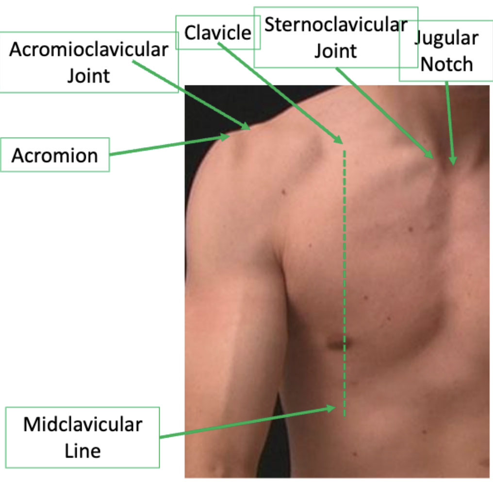
What surface landmarks of the pectoral girdle are identified in the lecture?
Jugular notch
Sternoclavicular joint
Clavicle
Acromioclavicular joint
Midclavicular line
Acromion
What is the midclavicular line?
A vertical surface-anatomy reference line running approximately through the midpoint of the clavicle.
Where can you locate the acromion?
It is the prominent bony projection of the scapula forming the top/lateral aspect of the shoulder.
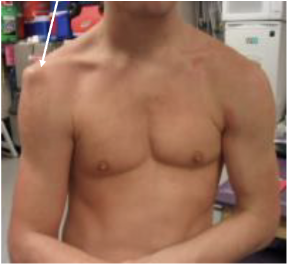
What is the sulcus sign?
An abnormal visible depression below the acromion associated with glenohumeral joint dislocation. The lecture illustrates this with an acromion appearing displaced in a dislocated shoulder
Compare the three major joints of the pectoral girdle.
Joint | Type | Articulations |
|---|
SC joint | Atypical saddle synovial | Clavicle + manubrium + 1st costal cartilage |
AC joint | Atypical plane synovial | Clavicle + acromion |
GH joint | Ball-and-socket synovial | Humeral head + glenoid fossa |
What are the key stabilising structures of the glenohumeral joint?
Passive stabilisers:
Glenoid labrum
Joint ligaments
Active stabilisers:
Rotator cuff muscles.
Why is the glenohumeral joint both highly mobile and relatively unstable?
Its large humeral head, shallow glenoid fossa and lax joint capsule allow a huge range of motion but provide relatively little bony stability.
What are the major clinical nerve associations from the humerus/shoulder lecture?
Surgical neck fracture → axillary nerve
Radial groove/shaft injury → radial nerve
Anterior shoulder dislocation → axillary nerve can be damaged.
Label