MG U5/AP U4: The Mitotic Cell Cycle (Mitosis)
1/51
There's no tags or description
Looks like no tags are added yet.
Name | Mastery | Learn | Test | Matching | Spaced |
|---|
No study sessions yet.
52 Terms
mitosis
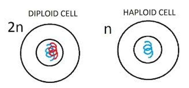
a form of nuclear division (not the only form of cell division - meiosis, binary fission) that maintains the diploid number of chromosomes
- even when the chromosomes split during metaphase, the diploid number stays the same
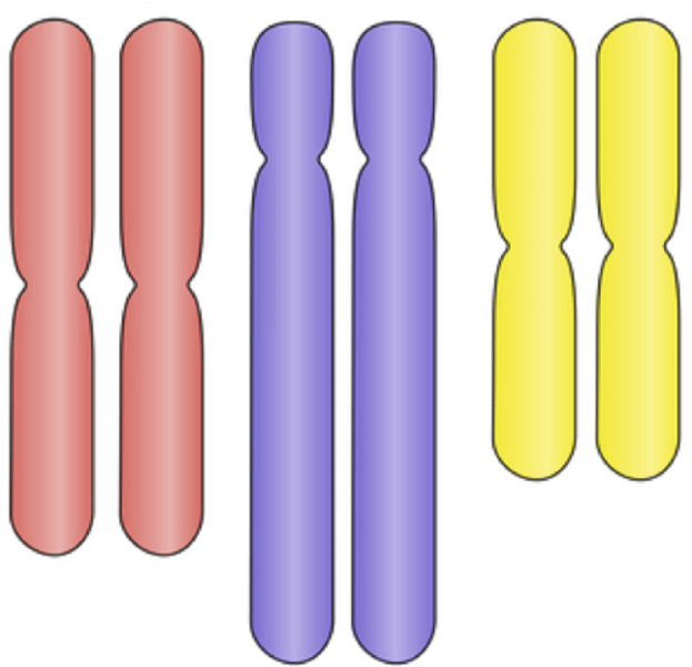
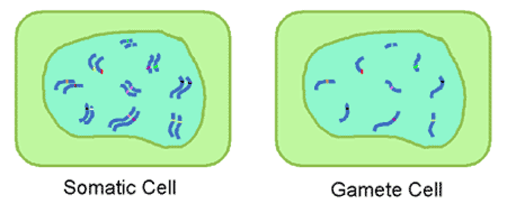
diploid (2n)
there are two copies of each type of chromosome
diploid organisms
have two copies of each chromosome, so two copies of each gene, of which a copy comes from both a paternal and maternal source
somatic cells
Any cells in the body other than reproductive cells; found systemically throughout body in blood, liver, skin cells, etc.
these are the cells that divide through mitosis to create more somatic cells
Why does a cell need to divide?
as cells increase in volume, the surface area decreases and demand for material resources increases which limits cell size
better words: cells need to maintain an adequate SA:V and Genome:V ratio, and they divide to avoid becoming too large
What is the purpose of mitosis?
- for unicellular Eukaryotes: reproduction
- for multicellular Eukaryotes - growth (which can mean an increase in the number of cells OR an increase in the size of cells), repairs/replacement, and development (from fertilized cell)
What are the products of mitosis?
two genetically identical, 2n daughter cells
- separates the original and the copy of chromosomes into their respective daughter cells
TRUE/FALSE: There is only one round of division in ONE cycle of Mitosis.
TRUE; there is only one round of PPMAT, but it is not for any genetic variation
Cell Cycle
the specific series/sequence of events a cell will go through from conception to its division
the two major parts of the Cell Cycle are Interphase, the longer of the two), and the Mitotic Stage (M-stage), where mitosis and cytokinesis occur
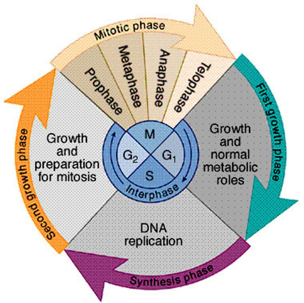
Interphase
- the longer period of the Cell Cycle in which the cell generally grows, undergoes protein synthesis, and doubles its organelles to prepare for its next division
- consists of three subphases: G1, S, and G2 Stages (growth occurs during all 3 but DNA replication is only during S Stage)
- "G" stands for Gap, "S" stands for Synthesis
- DNA is relaxed and appears as chromatin
- prepares for division
Structure:
- nuclear envelope intact, nucleolus present
- centrosomes/centrioles* doubled (evidence of organelles doubling)
- DNA present as chromatin
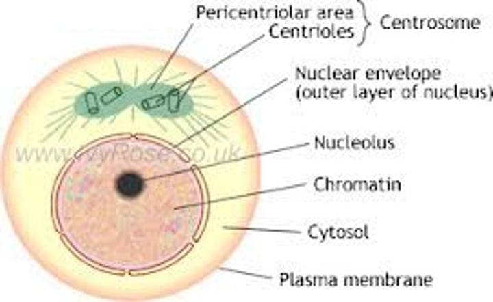
centrosomes
MTOC = MicroTubule Organizing Center
an organelle near the nucleus of a cell that contains the centrioles (in animal cells) and forms the microtubule spindle fibers develop in cell division
these are the organelles that actually form the mitotic spindle, as they're present in both plant and animal cells
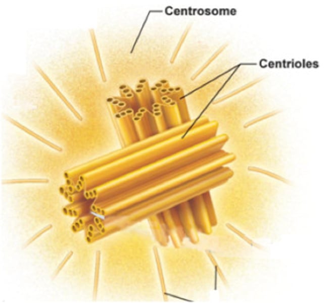
centrioles
Cell organelle that is found in ANIMAL CELLS only; although they are formed by the centrosomes, these don't really do much in the way of cell division, as they are typically used for cilia and flagella, the locomotive organelles
Why is Interphase significantly longer than the M-Stage?
this is because in order to divide, the DNA in chromatin must condense to be chromosomes, whose genes are unable to be expressed - this means no protein synthesis can occur, so the M-Stage must be held to a short amount of time so that the cell can stay alive
G1 Stage
in the G1 stage, the cell is recovering from its last division in time for the S Stage, where DNA is replicated, by growing the metabolic organelles (mitochondria and ribosomes), growing its size, and stockpiling materials
- only has ORIGINAL copy of DNA; all chromosomes have a singular chromatid
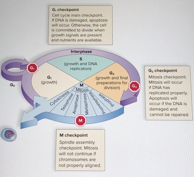
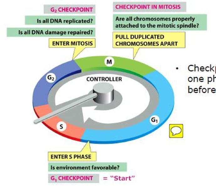
Cell Cycle Checkpoints
mechanisms that monitor the preparedness of a eukaryotic cell to advance through the various cell cycle stages; these are regulated by cyclin proteins that fluctuate in abundance
occur within the G1, G2, and M Stages
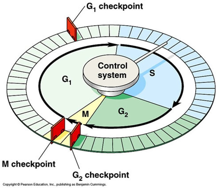
G1 Checkpoint
- main checkpoint of the Cell Cycle
- in addition to checking the cell's current size, available nutrients, and growth factors, it also checks for DNA damage
- if no DNA is found to be damaged, the cell can proceed onto later stages of the cell cycle and becomes committed to division
- if the DNA is found to be damaged, apoptosis will occur to the cell to kill itself off before the damaged DNA is spread
- if it lacks the cell lacks the nutrients/size to divide, it will be stopped at this checkpoint and ejected into G0 phase (does not receive go-ahead signal)
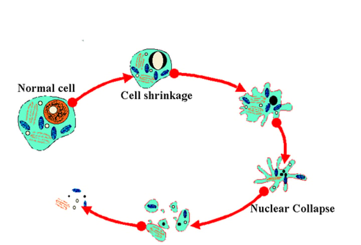
apoptosis
- "programmed cell death"
- cell becomes more spherical in shape, which causes the nucleus to fragment and plasma membrane to blister
- white blood cells/neighboring cells then kill off the fragmented cell by engulfing it
- occurs when DNA is damaged beyond repair
- uses a family of enzymes of caspases to go into apoptosis; vesicles border off the cell undergoing apoptosis so that they do not spread to other cells
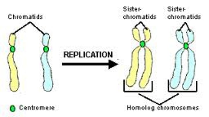
S Stage
- where DNA Synthesis/Replication occurs
- coming from G1, the chromosome consists of just one DNA double helix molecule (chromatid), but at the end of the S Stage, two identical DNA double helices are made (sister chromatids) - doubles chromatin/histone proteins
- the sister chromatids stay together until mitosis' anaphase
G2 Stage
- here, the cell continues to grow and create proteins, such as those that help with division like microtubules
- microtubules will form the mitotic spindle that will later distribute the sister chromatids accordingly in mitosis
- will have ORIGINAL and COPY of DNA (comes after S Stage); all chromosomes have two chromatids
G2 Checkpoint
- in addition to rechecking for cell size, the G2 Checkpoint, which occurs after the S Stage, checks to make sure DNA has completely finished replicating
- if it has, the cell can proceed to the M-Stage
- if not and the DNA is still reparable, the DNA will be repaired by the p53 protein
- if not and the DNA is NOT reparable, the cell will be forced to go into apoptosis
TRUE/FALSE: No matter the context, apoptosis should always be seen as a negative.
FALSE: Apoptosis and cell division, in unison, maintain homeostasis and are perfectly normal in balanced amounts - back and forth of reduction and growth.
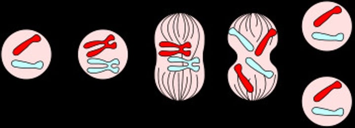
Mitotic Stage (M-Stage)
- characterized by two forms of division, mitosis and cytokinesis
- mitosis consists of the PPMAT stages and is nuclear division
- cytokinesis is the division of the cytoplasm/organelles between daughter cells
Prophase
- the first P in PPMAT
- chromatin BEGINS to condense into chromosomes
- nuclear envelope still intact
- nucleolus disappears
- centrosomes (& centrioles*) begin to migrate towards poles with the formation of spindle microtubules (mitotic spindle)
- still has as many chromatids as it did in G2 phase
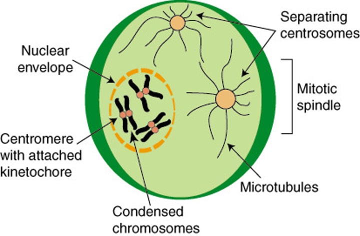
mitotic spindle
a structure made of microtubules that controls chromosome movement during mitosis
- they attach to the kinetochore of the sister chromatids, and they allow for dynein to walk the sister chromatid towards the poles in Anaphase
Prometaphase
- the second P in PPMAT
- nuclear envelope explodes/fragments
- nonkinetochore microtubules and kinetochore microtubules from the centrosomes begin to attach to their respective positions
- the jerky tug of war to align chromosomes BEGINS in Prometaphase
- still has just as many chromatids as it did in G2 phase
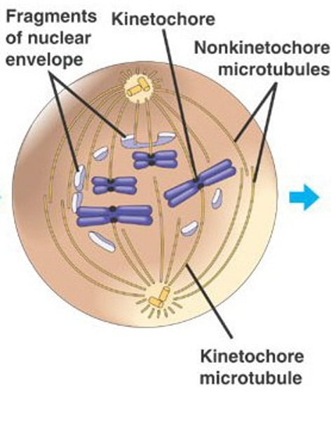
What is the point of the nonkinetochore fibers/microtubules?
- they start to pull apart from each other to elongate the cell, making the daughter nuclei farther apart for proper division
- overlapping occurs at the metaphase plate
- HOWEVER, this doesn't occur in plant cells because of the cell wall and how plant cells are already elongated
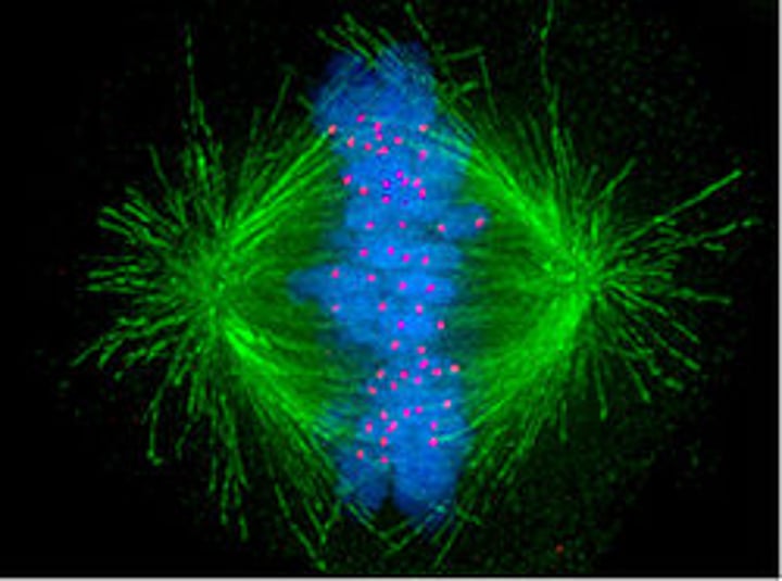
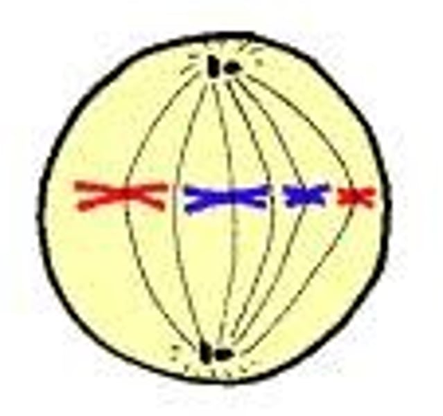
Metaphase
- the M in PPMAT
- chromosomes are MOST HIGHLY condensed at this stage
- chromosomes are aligned in the Middle along the Metaphase plate
- the spindle is across the whole of the cell; it is fully formed
- considered the longest stage of Mitosis
- still has just as many chromatids as it did in G2 phase
Why is Metaphase the longest stage of Mitosis?
- this is because of the M-Stage Checkpoint that takes place during Metaphase (between Metaphase going into Anaphase)
- must make sure of proper attachment with spindle and alignment along metaphase plate to proceed
M-Stage Checkpoint
- found between Metaphase and Anaphase
- checks to see if chromosomes are properly aligned along the metaphase plate AND if the chromosomes are properly attached to the mitotic spindle
- these two requirements must be met before the cell proceeds to Anaphase
- if a drug arrests the spindle fibers from properly forming and attaching, the cell will be arrested by this checkpoint
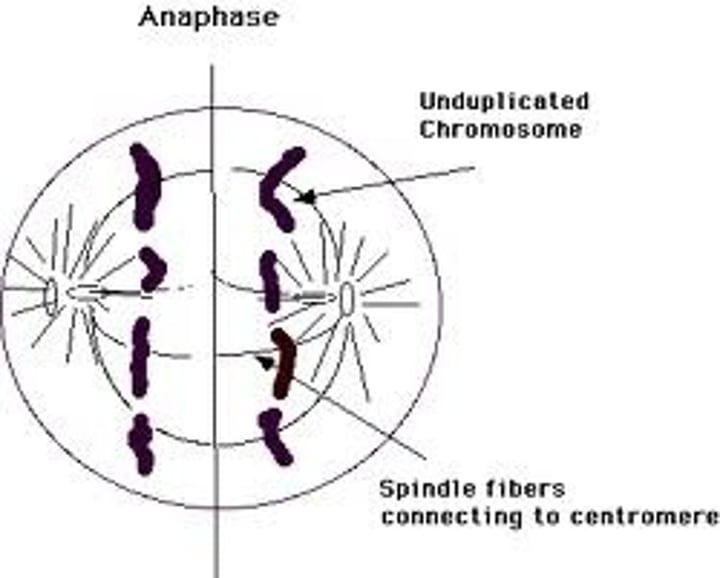
Anaphase
- the A in PPMAT
- chromosomes' cohesin proteins are split Apart by the separase enzyme, effectively splitting the sister chromatids from each other (which are now individual chromosomes)
- the dynein protein, phosphorylated by MPF, walks sister chromatids to the poles of the cell
- kinetochore microtubules disassemble behind the chromatids as they work their way towards the poles (microtubules do not shorten at pole ends)
- still has just as many chromatids as in G2 phase
Telophase
- the T in PPMAT
- near-opposite of Prophase
- The End of Mitosis, the daughter nuclei envelopes begin to form
- chromosomes begin to uncondense into chromatin
- nucleolus reappears
- has HALF as many chromatids as in G2 phase (the two daughter cells have as many chromatids as the original parent cell did, fulfilling the definition of mitosis)
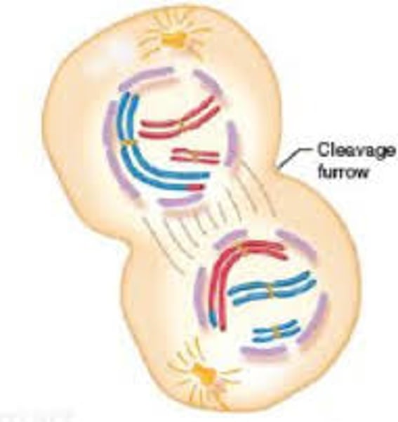
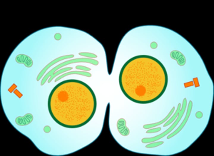
Cytokinesis
- the physical division/cleavage of the parent cell into their respective daughter cells
- is marked by the formation of a cell plate in plant cells or cleavage furrow in animal cells
- does NOT have anything to do with nuclei
- although there isn't an actual system telling which organelles to go where, organelles should be split evenly by the cells (duplicated during Interphase)
TRUE/FALSE: It is accurate to say that Telophase of Mitosis and Cytokinesis occur concurrently.
TRUE; Although this varies from textbook to textbook, it is generally agreed upon that a lot of Cytokinesis occurs during Telophase. In fact, Cytokinesis is considered to begin during Telophase. However, the daughter nuclei forming are considered to be a part of Telophase exclusively, and the physical cleavage of the cytoplasm/cell is considered to be a part of Cytokinesis exclusively.
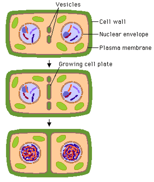
cell plate
- in plant cells
- because of the rigid cell wall, plant cells cannot elongate themselves and cleave in the middle
- instead, a new cell wall must be formed via the Golgi apparatus creating vesicles that form the cell plate, which is actually a plasma membrane that will extend out in both directions until it reaches the old plasma membrane of the cell wall
- once it reaches the old plasma membrane, the cell plate fuses with the old membrane and releases molecules to form the new cell wall (later strengthened by cellulose)
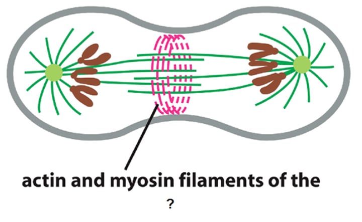
contractile ring
- A thin band of actin and myosin filaments that wraps around the midsection of an animal cell undergoing cytokinesis
- it contracts to pinch the cell inward (think of pinching water balloon in middle)
- although this cannot be visibly seen, evidence of the contractile ring is the cleavage furrow, which is the actual indentations into the cell
cyclin
- a family of proteins that regulates the cell cycle in eukaryotic cells
- fitting with the name, cyclin proteins fluctuate in concentration according to a cycle: generally, [cyclin] is nonexistent during G1/S Stages and [cyclin] increases up to a peak during G2 Stage into M-Stage before a precipituous drop in concentration during Metaphase
- it acts as an allosteric activator for CDKs, an enzyme crucial to cell division
![<p>- a family of proteins that regulates the cell cycle in eukaryotic cells</p><p>- fitting with the name, cyclin proteins fluctuate in concentration according to a cycle: generally, [cyclin] is nonexistent during G1/S Stages and [cyclin] increases up to a peak during G2 Stage into M-Stage before a precipituous drop in concentration during Metaphase</p><p>- it acts as an allosteric activator for CDKs, an enzyme crucial to cell division</p>](https://knowt-user-attachments.s3.amazonaws.com/67602408-627d-429c-88a1-5ed7f65c67f7.image/png)
cyclin-dependent kinases (Cdks)
- an enzyme that catalyzes the phosphorylation of a protein (causing said protein to be active or inactive) but is only activated by cyclin attaching to its active site
- take in a protein and ATP into their active site
- their concentration never varies, but they remain inactive when [cyclin] is very low or nonexistent
![<p>- an enzyme that catalyzes the phosphorylation of a protein (causing said protein to be active or inactive) but is only activated by cyclin attaching to its active site</p><p>- take in a protein and ATP into their active site</p><p>- their concentration never varies, but they remain inactive when [cyclin] is very low or nonexistent</p>](https://knowt-user-attachments.s3.amazonaws.com/e586a3ee-92fb-49b8-a971-2318e4cd6f99.image/png)
MPF (maturation-promoting factor)
- also called Mitosis Promoting Factor
- a cyclin-Cdk complex that triggers a cell's passage past the G2 checkpoint into the M phase
- this complex is crucial to cell division, as it allows for the phosphorylation of certain activities done by proteins, such as the condensation of chromatin into chromosomes during Prophase, the nuclear envelope fragmenting during Prometaphase, and the activation of separase, the enzyme that separates sister chromatids' cohesin proteins
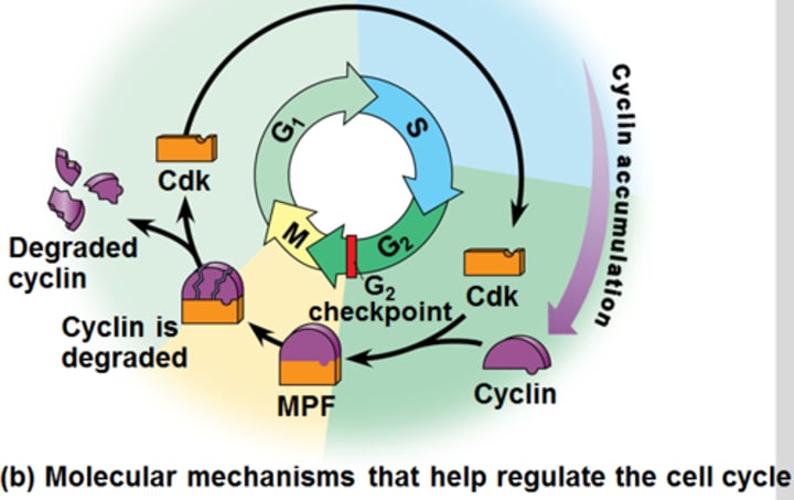
Describe how MPF is regulated and its relation to cell division activity.
REGULATION:
MPF is regulated by [cyclin], as MPF is the complex of the cyclin protein and its associated, specific CDK.
- There is no [cyclin] during G1/S Stages, so there is no premature MPF activity spurring on cell division.
- During G2 Stage and into M-Stage, there starts a rise in [cyclin], and as a result, MPF activity also increases.
- The peak comes at Metaphase and then a precipitous drop occurs due to MPF phosphorylating the enzyme that will catalyze the breakdown of cyclin, meaning MPF is no more going into Anaphase and Telophase.
CELL DIVISION:
- When protein kinase activity increases, after a short period of time, the % of dividing cells also increases.
- This is because protein kinase activity, such as that of MPF, phosphorylates the necessary proteins to change shape, making them become more active or inactive at the right time to spur cell division.
![<p>REGULATION:</p><p>MPF is regulated by [cyclin], as MPF is the complex of the cyclin protein and its associated, specific CDK.</p><p>- There is no [cyclin] during G1/S Stages, so there is no premature MPF activity spurring on cell division.</p><p>- During G2 Stage and into M-Stage, there starts a rise in [cyclin], and as a result, MPF activity also increases.</p><p>- The peak comes at Metaphase and then a precipitous drop occurs due to MPF phosphorylating the enzyme that will catalyze the breakdown of cyclin, meaning MPF is no more going into Anaphase and Telophase.</p><p>CELL DIVISION:</p><p>- When protein kinase activity increases, after a short period of time, the % of dividing cells also increases.</p><p>- This is because protein kinase activity, such as that of MPF, phosphorylates the necessary proteins to change shape, making them become more active or inactive at the right time to spur cell division.</p>](https://knowt-user-attachments.s3.amazonaws.com/38a653aa-0381-4921-ade0-6277b7df1501.image/jpeg)
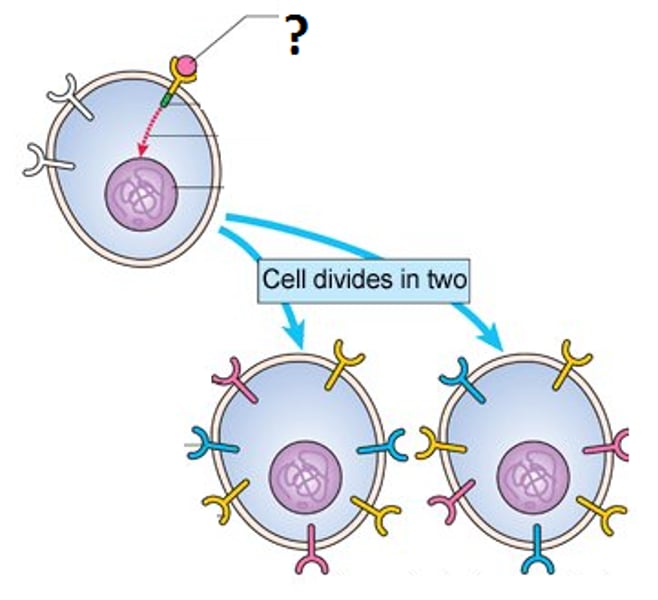
growth factors
- Regulatory proteins that ensure that the events of cell division occur in the proper sequence and at the correct rate
- external signals released by other cells to be received by the cell that wants to divide
- can be a limiting factor in cell division
- example is platelet-derived growth factor (PDGF)
anchorage dependence
- the requirement that to divide, a cell must be attached to a solid surface
- must be attached to some edge or some other surface to divide (such as another cell or container wall)
- external regulation
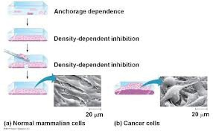
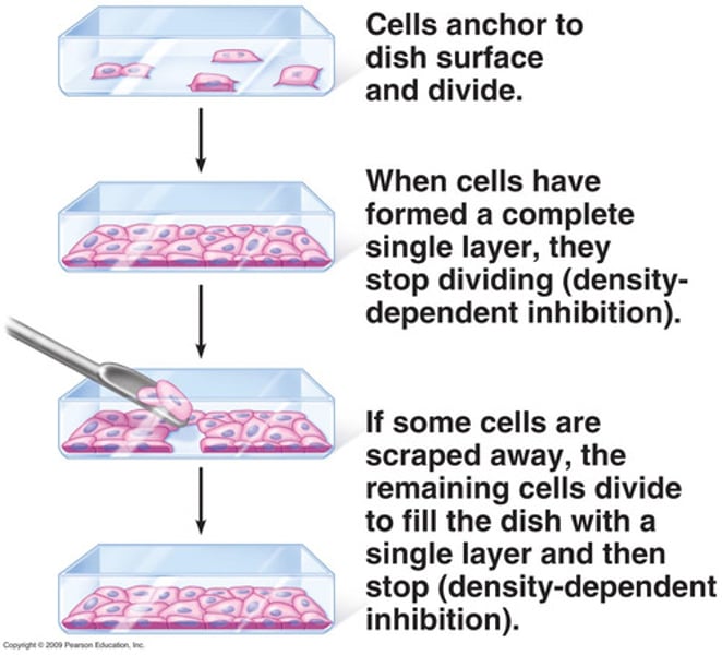
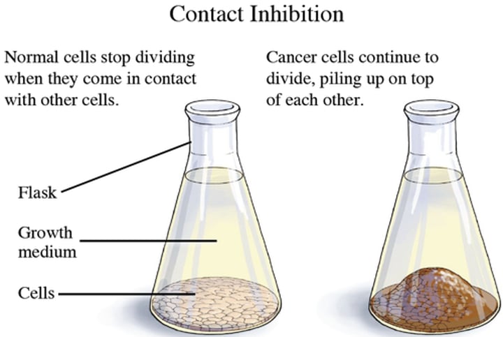
density-dependent inhibition
- aka contact inhibition
- the phenomenon observed in normal animal cells that causes them to stop dividing when they come into contact with one another
- signals are sent between the cells to stop dividing
- example of external regulation
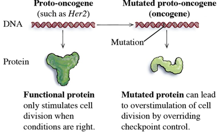
proto-oncogenes
- gene for proteins that stimulate normal cell growth and division
- if mutated, can lead into an oncogene, which is perpetually stimulated to cause the cell to divide an uncontrolled amount of times (with more dividing proteins than they need)
- internal regulation
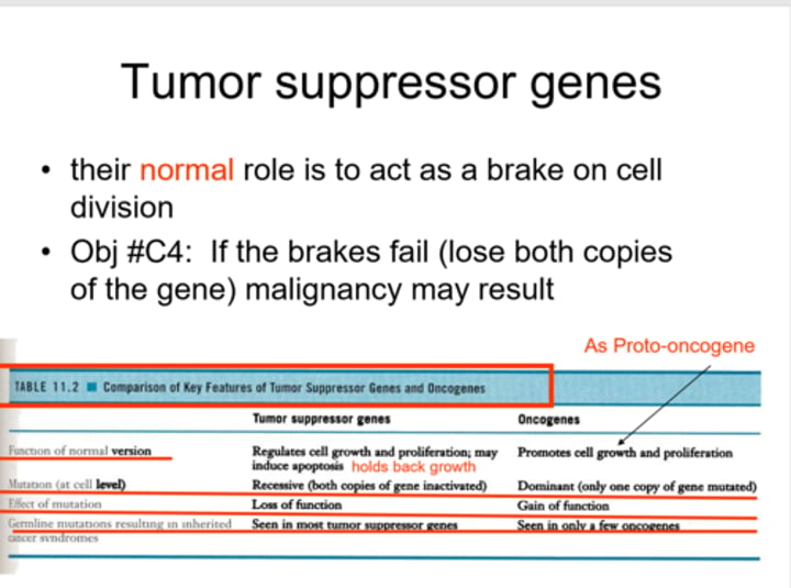
tumor suppressor genes
- A gene whose protein product inhibits cell division, thereby preventing the uncontrolled cell growth that contributes to cancer
- if mutated, can lead to DNA damage and lack of a way to halt cell division (apoptosis is regulated by this gene, so it would halt)
- internal regulation
How do cells become cancerous?
- unchecked cell growth
- mutations in genes can cause cancer by accelerating cell division rates or inhibiting normal controls on the system, such as cell cycle arrest, programmed cell death
- an accumulation of mutations leads to transformation where a normal, functioning cell becomes transformed into a cell where controls are ignored
- they make their own growth factors or create an alternate signal transduction pathway that does not require a growth factor for them to divide
What are the effects of cancerous cells?
- have abnormal nuclei/non-functional DNA, which is allowed to spread like a wildfire to other cells
- angiogenesis - the creation of new blood vessels to transport materials to and from the tumor/cancerous cells at the expense of other tissue
benign tumors
tumors that are surgically removable with little risk of further growth in the body; remain at their original site without causing serious problems

malignant tumors
those tumors that invade surrounding tissues and can metastasize
these impair organ function, and these tumors are those that begin to fall under the definition of "cancer"
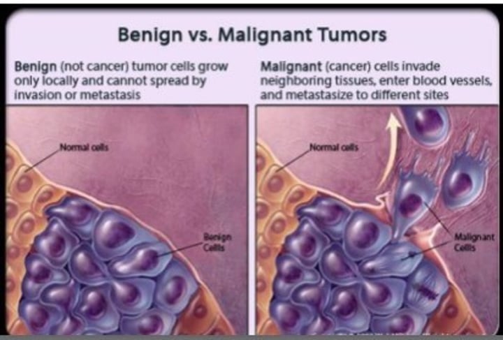
metastatic tumors
Malignant tumors that originate in one organ and spread to another -> spread through the lymph and blood vessels
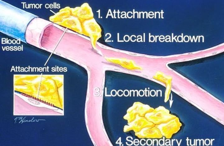
Explain anchorage dependence and contact inhibition in regards to cancerous cells.
AD:
- they will lose anchorage dependence, causing metastasis, the spread of the cancer to other parts of the body through lymphatic or blood vessels, impairing organs from functioning
CI:
- they will lose contact inhibition, creating benign (do not impair surrounding tissue) and malignant (impair surrounding tissue) tumors
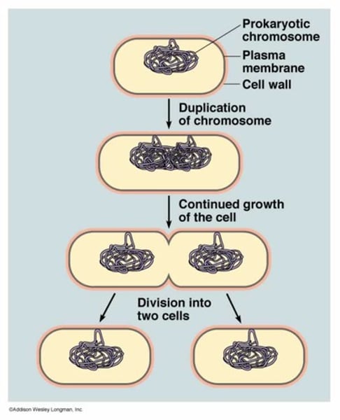
binary fission
type of asexual reproduction in which an organism replicates its DNA and divides in half, producing two identical daughter cells
- the method by which prokaryotic cells divide; so named because it produces identical daughter cells
- in comparison to mitosis: both make identical haploid daughter cells; binary fission is meant for reproduction (mitosis for development, growth, repair except for unicellular eukaryotes); BF has relatively few proteins involved in it
- no mitotic spindle
Describe the process of binary fission.
1. Singular circular chromosome of the cell attaches to a special site of the plasma membrane.
2. This triggers enlargement of cell wall, plasma membrane, and overall volume.
3. Chromosome duplicates.
4. Cell wall and plasma membrane then begin to pinch inward, starting the process by which it divides, which it finishes by elongating itself.
5. New cell wall made after pulling apart daughter cells; chromosomes were pulled apart and cytoplasm was split evenly.