Integumentary System & Wound Healing: Pathophysio
1/71
There's no tags or description
Looks like no tags are added yet.
Name | Mastery | Learn | Test | Matching | Spaced |
|---|
No study sessions yet.
72 Terms
skin basics
- largest organ of the body
- makes up 15-20% of body weight
- differs anatomically and physiologically in different parts of body
skins primary function
protector of underlying structures from external injury and harmful substances
other skin functions
insulator, holds organs together, controls temperature, absorb UV radiation, metabolize vitamin D, synthesize lipids in oil glands
acts in the insulator for skin
adipose tissues and the blood vessels
primary layers of skin
epidermis, dermis, hypodermis
epidermis
superficial layer
dermis
middle layer
hypodermis (subcutaneous tissue)
deepest layer
epidermis composition
keratinized stratified squamous epithelium
epidermis key features
-protective shield of the body
-renews self every 28-30 days
-varies in thickness (usually about as thick as sheet of paper)
-outer most layer composed of dead cells packed with keratin and lipids
keratin
insoluble protein (also makes up nails and hair); gives structural strength to skin
lipids
-protect against water loss and penetration
-provides flexibility for keratin
layer of skin you can touch
epidermis
accessory structures of epidermis
-hair shaft surrounded by epithelial cells down to the hair root
-shaft has nerve endings that help with light touch
-collection of BVs
-sebaceous glands which produce sebum (lipids)
-sweat ducts (gland is in dermis)
dermis composition
-mesh like network composed of collagen and elastin, blood and lymph vessels, and specialized cells (main ones are fibroblasts)
-cells are surrounded by a gel-like substance: ground substance
dermis functions
-fibrous network provides structure and resilience to the skin
-BVs in the dermis help in thermoregulation of the body by constricting or dilating to conserve or release heat
-BV's in the dermis also aid in immune function and provide oxygen and nutrients to the lower layers of the epidermis
ground substance role
maintain hydration and moisture levels within the skin
made by fibroblasts
collagen
hypodermis function
-fat tissue that provides insulation and padding; insults body from cold temperatures and provides shock absorption
-stores nutrients and energy
hypodermis key features
-deepest section of skin
-thickest in the buttocks, palms of the hands, and soles of feet
-blood vessels and nerves for the skin pass through this layer
benefits of treating the skin
increases circulation and healing, as the blood vessels and nerves for skin pass through the hypodermis
skin layer where fascial trains are found
hypodermis
allows muscles in the body to work as a unit
fascial trains
attaches muscle to other muscle
fascia
melanin
-produced by melanocytes
-gives color to skin, hair, iris
-provides protection against ultraviolet radiation
-production stimulated by UV and hormones
scarring
areas of fibrous tissue that replaces skin after injury
fibrous tissue being laid down after injury is abnormal. (T/F)
false
atrophic scar
scar that takes on a sunken appearance
hypertrophic scar
body overproduces collagen, causing the scar to be red in appearance and raised about surrounding skin
keloid scar
scars that extend beyond the wound area and become hard and thickened
stretch marks
form of scarring caused when the skin stretches rapidly, due to growth spurt or weight gain
required when scars are adhesive- deep and stiff to the point of hindering mobility
-deep tissue mobilization
-skin rolling
instrument assisted short treatment times mobilization (IASTM)
-for relatively short treatment times
-deep or specific treatments; more aggressive form of scar tissue therapy
scleroderma
-disease that involves the hardening and tightening of skin and connective tissues
-thickening and inflammation over joints is a classic sign
-symptoms vary depending on systems affected
-affects women > men
-age of onset 30-50
-no cure
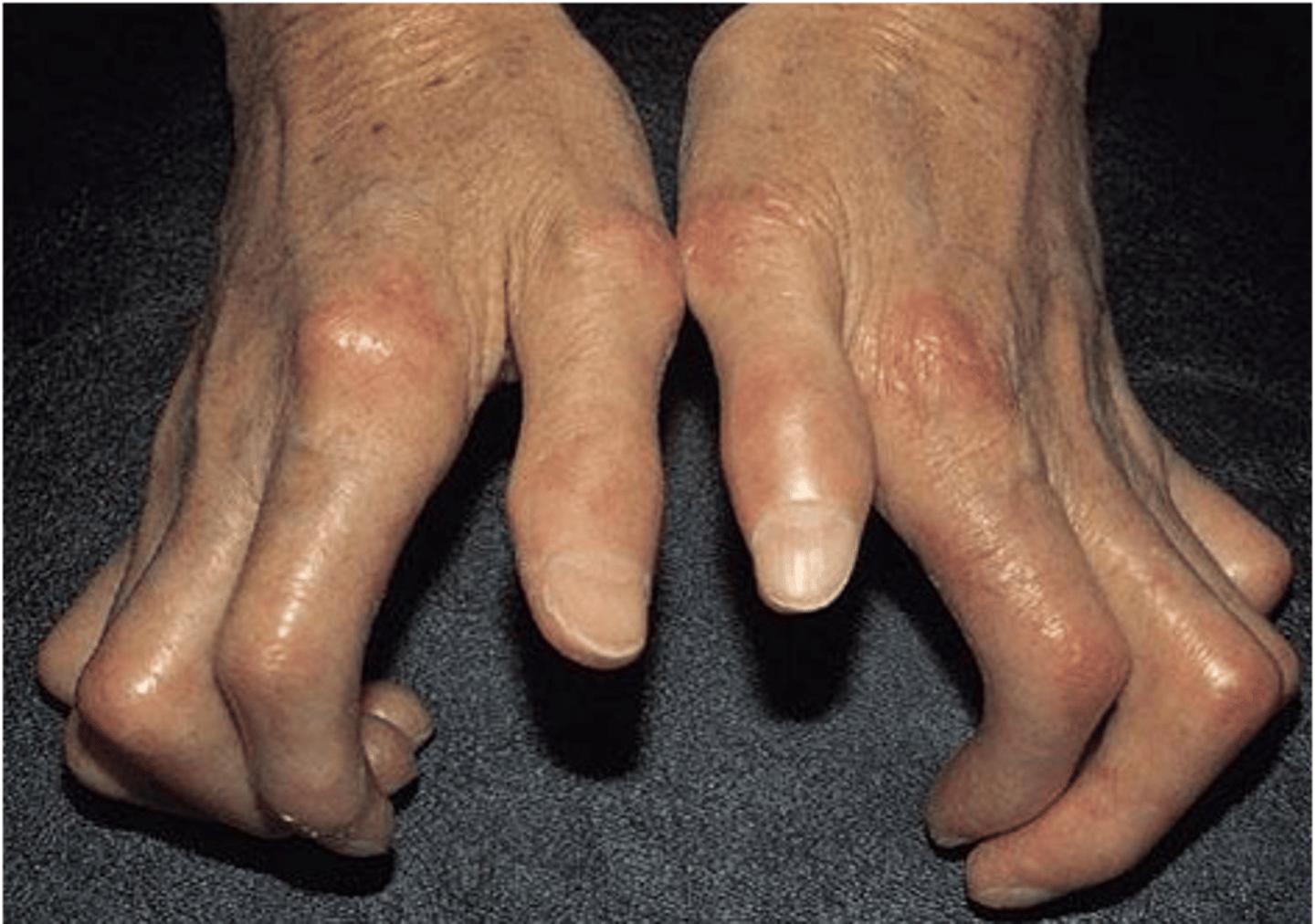
structures affected by scleroderma
blood vessels, internal organs, and the digestive tract
wounds
-disruption of normal anatomical structure
-disruption of function
-pathological process
-may begin internally or externally
internal wounds
disease process
external wounds
trauma
debridement
removal of foreign material and dead or damaged tissue from a wound
process of wound healing
-wound closure
-restoration of function to the damaged tissue
wound closure occurs via
-removal and resorption of devitalized tissue
-regeneration of replacement tissue
phases of wound healing: soft tissue
1. hemostasis
2. inflammation
3. proliferation
4. remodeling
hemostasis
-begins as soon as tissue destruction occurs
-normally lasts about 30 minutes
-platelets aggregate at injury site and release enzymes that trigger clotting process
-endothelial cells release prostacyclin
-prevents excessive bleeding, edema, and further tissue damage
inflammation
-normally last 3-7 days
-begins with invasion of neutrophils that lyse and clear away nonviable cellular components
-platelets and neutrophils release growth factors
-eosinophils phagocytose devitalized tissue
-mast cells and basophils release histamine to increase vascular permeability to attract monocytes
-monocytes become macrophages
-lymphocytes produce antibodies for immune respons
proliferation- granulation
-characterized by granulation tissue
-macrophages are orchestrators of transition from inflammation to proliferation
-fibroblasts produce collagen--> connective tissue; also produce growth factors
-endothelial cells produce new capillaries and transport oxygen and nutrients to the wound
-macrophages continue the phagocytic process and also secrete growth factors
-myofibroblasts produce fibronectin, collagen, GAGs, enzymes, encourage wound contraction
proliferation-epithelialization
-platelets and fibroblasts secrete fibronectin
-platelets, macrophages, and other cells release growth factors
-keratinocytes initiate wound coverage at the edges
-epithelial cells cover surface of wound
-basal cells bond dermal and epidermal cells
growth factors
-released by platelets, macrophages, neutrophils
-aka cytokines, interleukins, colony simulating factors
-composed of proteins (polypeptides)
-occur in all of these stages of tissue repair
-serve as the messengers between cells to attract them to the injury site
remodeling
-fibroblasts produce type III collagen (disorganized) that is lysed and replaced with type I (organized)
-epithelial cells, macrophages, fibroblasts, leukocytes secrete collagenase to lyse excess collagen
-collagen refines and realigns to accommodate function of the affected area
-skin returns to patient's original color by the migration of melanocytes toward the surface area of the dermis
phase of remodeling that goes wrong, causes scarring
failure of cells to secrete collagenase -- needed to lyse/break down excess collagen
length till full maturation of wound healing
365 days after injury
venous ulcers
-caused by long standing venous insufficiency (pooling of blood in the limbs); causes opening of skin
-ulcers typically occur along the medial aspect of the distal portion of the lower leg
-will often be accompanied by skin hyperpigmentation and discoloration, wet
treating venous ulcers
compression therapy, antibiotics, painkillers, and elevating the affected area
arterial ulcers
-caused by arterial insufficiency commonly called ischemic ulcers
-typically seen on the ankle and distal digits
-ulcer will have a "punched out" like appearance, dry due to lack of blood flow; constricting ulcer
neuropathic ulcers
-caused by neuropathy secondary to diabetes
-located on the bottom of the foot and tips of the toes
-often surrounded by calluses
primary intention
-wounds have a small, clean defect that minimizes the risk of infection and requires new blood vessels and keratinocytes to migrate only a small distance
-surgical incisions, paper cuts and small cutaneous wounds
secondary intention
-healing of a wound in which the wound edges cannot be approximated
-secondary closure requires a granulation tissue matrix to be built to fill the wound defect
-requires more time and energy than primary wound closure, and creates more scar tissue
how majority of wounds close
by secondary wound closure
delayed primary intention
-delayed primary closure is a combination of healing by primary and secondary intention
-intentional method by the wound care specialist to reduce the risk of infection
-in delayed primary closure, wound is first cleaned and observed for a few days to ensure no infection is apparent before it is surgically closed
examples of traumatic injuries such as dog bites or lacerations
delayed primary intention
factors inhibiting wound healing assessed via
Braden scale
6 subscales of braden scale
1. sensory perception
2. moisture
3. activity
4. mobility
5. nutritional status
6. friction/shear
severe risk on the braden scale
total score less than or equal to 9
high risk on the braden scale
total score 10-12
moderate risk on the braden scale
total score 13-14
mild risk on the braden scale
total score 15-18
systemic factors inhibiting wound healing
-chronic diseases (diabetes, peripheral vascular disease, venous insufficency)
-protein energy malnutrition
-cancer
-aging
-stress
-smoking
-obesity
-hypothermia
local factors inhibiting wound healing
bioburden and foreign bodies
bioburden
caused by inflammatory state secondary to infection
foreign bodies
necrotic tissue
clinician induced factors inhibiting wound healing
-medications
-radiation
-improper patient positioning
-inappropriate dressings
-cyotoxic topical agents (i.e. hydrogen peroxide)
best treatment for pressure areas that may cause wounds/ulcer
-prevention
-positioning is important to avoid too much time spent on common pressure areas
wound prevention and PT
-PT plays a critical role in wound prevention
-patient and family education is key
-setting up the BEST positioning
-refer to wheelchair clinic to help with positioning