Circulation
1/131
Earn XP
Description and Tags
Name | Mastery | Learn | Test | Matching | Spaced | Call with Kai |
|---|
No analytics yet
Send a link to your students to track their progress
132 Terms
what is hypertension?
high blood pressure where the force of blood against artery walls is too high
causes: overexcitement, narrowed arteries, or high blood volume.
myocardium
thickest layer of muscle of the heart, does most of the mechanical work
contracts to pump blood throughout the body
mesentery artery, vein
artery: Supplies oxygenated blood to the intestines (small and large)
vein: Drains nutrient-rich, deoxygenated blood from the intestines and delivers it to the hepatic portal vein
hepatic portal vein
carries nutrient-rich, oxygen poor blood from the digestive organs (e.g. intestines, stomach) to the liver for processing.
compare the blood in the renal artery to the renal vein
renal artery: blood high in wastes
renal vein: blood low in waste
compare the blood in the mesenteric artery to the mesenteric vein
mesenteric artery: blood low in nutrients
mesenteric vein: blood high in nutrients (the mesenteric vein has more nutrients because it collects them from the intestines after absorption.)
iliac artery and vein
artery: carries oxygenated blood from the aorta to the pelvis and legs.
vein: Returns deoxygenated blood from the pelvis and legs to the inferior vena cava
frq 6a: Compare the chemical composition of blood inthe right atrium of a fetus to the blood in the right atrium of an adult. Provide reasons for your answer.
The blood in the right atrium of a fetus contains a mixture of oxygenated and deoxygenated blood. This is because oxygenated blood from the placenta enters the fetus through the umbilical vein and largely bypasses the liver via the ductus venosus, flowing into the inferior vena cava. There, it mixes with deoxygenated blood from the fetus’s body before entering the right atrium.
the right atrium of an adult receives only deoxygenated blood from the body through the superior and inferior vena cava, since adults do not have an umbilical vein supplying oxygenated blood, and all oxygenation occurs in the lungs.
frq 6b : Detail three key differences between fetal and adult circulation, and how they change at birth.
Fetal circulation includes special structures that allow blood to bypass the liver and lungs, which are not fully functional before birth. Three major differences are:
ductus venosus: allows oxygenated blood from the placenta to bypass the liver and flow directly into the inferior vena cava. After birth, when the umbilical cord is cut, the ductus venosus closes and becomes a ligament.
Foramen Ovale – This is a hole between the right and left atria that allows blood to bypass the lungs by flowing directly from the right atrium to the left atrium. When the baby takes its first breath, pressure in the left atrium increases, causing the foramen ovale to close permanently.
Ductus Arteriosus – This vessel connects the pulmonary artery to the aorta, letting most blood bypass the fluid-filled lungs. After birth, the ductus arteriosus closes and becomes the ligamentum arteriosum due to changes in oxygen levels and pressure.
In addition, deoxygenated blood is carried to the placenta via the umbilical arteries, and oxygenated blood returns via the umbilical vein — both of which cease function after birth and become ligaments.
frq 3: describe what the graph is illustrating in terms of blood velocity, pressure, and cross sectional area in each of the major types of blood vessels (label)
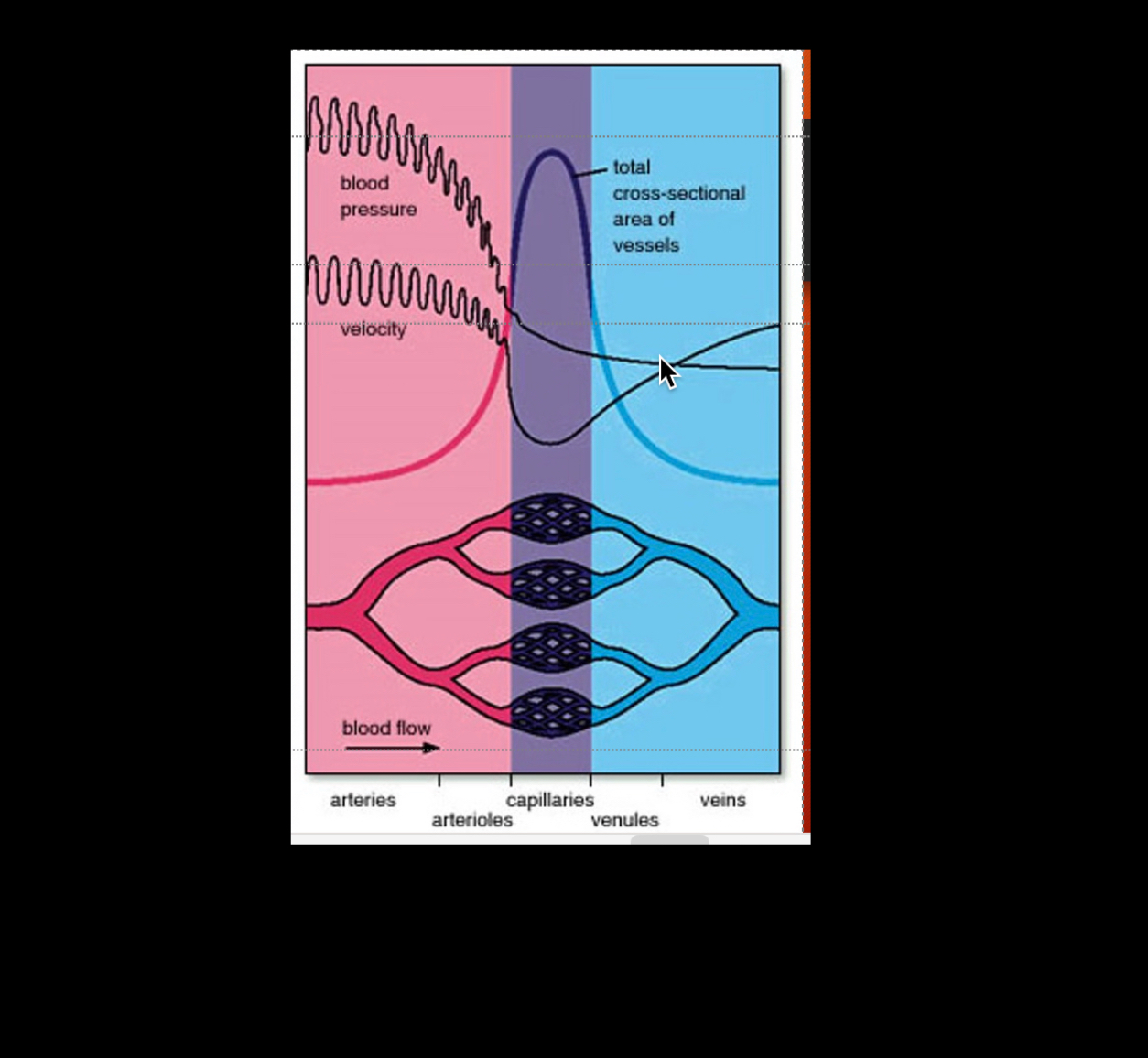
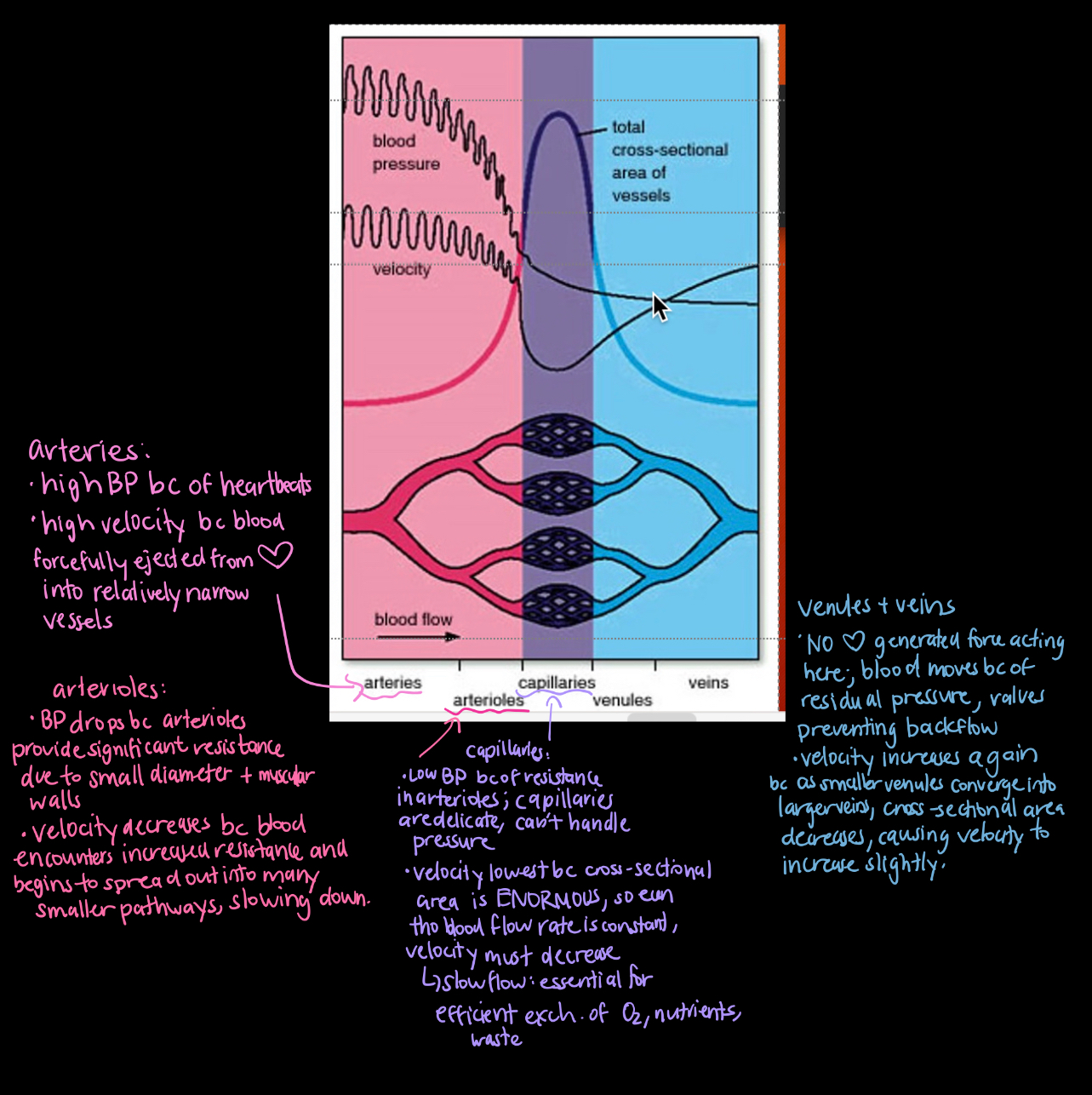
(labled on image)
how many capillary beds does a red blood cell pass through during one full circulation, and why?
a red blood cell passes through two capillary beds during one full circulation
systemic capillary bed, where it delivers oxygen to body tissues
pulmonary capillary bed, where it picks up oxygen in the lungs
hypotension
low blood pressure, vessels dialate
how does overexcitement lead to hypertension
leads to higher heart rate, increased bloodflow and constricted blood vessels = increased blood pressure
excess salt in diet can increase blood pressure because..
blood vessels constrict, extra H2O moved into blood, increased blood volume
atheroscelerosis
accumulation of fatty deposits in veins
thrombus
stationary clot
embolus
moving clot
stroke
embolus that moves to a vessel in the brain and blocks it
heart attack
portion of heart dies due to lack of oxygen
jugular vein
major blood vessels in the neck that drain deoxygenated bloo dfrom the head/neck back into the heart
pulmonary circulation
Carries oxygenated blood from the left side of the heart to the body, delivers oxygen/nutrients, and returns deoxygenated blood to the right atrium.
systemric circulation
Carries oxygenated blood from the left side of the heart to the body, delivers oxygen/nutrients, and returns deoxygenated blood to the right atrium
describe how a tumour would form in the brain, and the route a cancerous cell would follow if it metastasized from the brain to the liver
mutations in the dna cause cells in the brain to divide uncontrollably → these mutations can disable tumour suppressor genes/activate oncogenes, leading to rapid growth + formation of a mass.
cancerous cells can enter the cerebral veins, draining into the internal jugular vein
travel through the superior vena cava → heart → pulmonary circulation. passes through the lungs → enters systemic circulation via the aorta
reaches the liver through the hepatic artery (a branch of the aorta), where they may form secondary tumours and spread to other organs
describe the process the body undergoes to destroy foreign invaders with specific reference to the structure/function of the lymphatic system
The antigen is picked up by tissue fluid, enters lymphatic capillaries, becoming lymph. Lymph transported thru lymphatic vessels to lymph nodes. Lymph nodes filter lymph, contain imune cells (B/T cells, macrophages)
B cells recognize the antigen and, with help from helper T cells, produce antibodies specific to it, neutralizing it or causing agglutination (clumping), making it easier to destroy.
Cytotoxic T-cells/macrophages destroy infected/marked cells direclty.
Filtered lymph w/ waste and destroyed invaders drains into the lymphatic ducts (right lymphatic duct or leftthoracic duct): returns fluid to subclavian veins and back into the circulatory system
angina
radiating pain caused by blockage of coronary artery
process of angioplasty
invasive, very thin wire inserrted into blood vessel, small balloon at end inflated to break up blockage
varicose veins
visible, protruding veins below surface of skin, caused by "leaky" valves in veins
phlebitis
inflammation of a vein (usually in legs), can sometimes lead to thrombosis (blood clot) that can travel to other parts of the body
pulmonary embolism
blockage in a pulmonary artery (in the lungs), usually caused by a blood clot.
- It reduces blood flow to lung tissue
heart transplants
results from congestive heart failure, completely remove old organ and replace with new organ from donor. use of medication to prevent heart rejection
arteries
blood vessels that bring oxygen rich blood to body parts, branch into smaller arterioles. carry blood away from heart (exception: pulmonary artery)
capillaries
the smallest blood vessels that exchange gases and nutrients at the cellular level. They are distributed among all body tissues, and link arteries and veins.
have high cross sectional area bc there are millions of them in the body, their combined total area (when you add up all of them) is very large.
This high cross-sectional area spreads out the blood flow.
describe the pathway that leads away from capillaries
capillaries ⇒ venules ⇒ veins
veins
thin walled, elastic, valve-containing blood vessels that bring oxygen poor blood TO the heart (exception: pulmonary vein)
correct order of blood vessels
arteries, arterioles, capillaries, venules, veins
pulmonary arteries
bring oxygen low blood from heart to the lungs
coronary arteries
blood vessels on the surface of the heart muscle that supply oxygen-rich blood to the heart muscle (myocardium) istself. theyhelp remove waste, and if they are blocked it can result in heart attacks
pulmonary veins
bring oxygenated blood from lungs to the heart
pulmonary circuit
carries deoxygenated blood from the right ventricle to the lungs for gas exchange and returns oxygenated blood to the left atrium
systemic circuit
vessels that circulate blood to body tissues that aren't part of the pulmonary tissues
cardiac cycle
complete sequence of one heartbeat: atrial contraction, ventricular contraction, and relaxation
systole
heart muscle CONTRACTION
diastole
heart muscle RELAXATION
cause of first part of heartbeat
vibrations of the heart when atrioventricular (tricuspid and bicuspid ) valves close
second part of our heartbeat is caused by
vibration of the semilunar valves closing
starting at the right atrium, describe the flow of blood through the heart to the lungs and to the body
right atrium, travels through tricuspid valve, right ventricle, travles through the pulmonary semilunar valve, pulmonary artery, pulmonary veins, left atirum, travels through the bicuspid valve, left ventricle, aorta, dosral aorta
Right atrium
Upper right heart chamber that receives deoxygenated blood from the body.
movement through tricuspid valve
Blood flows from right atrium to right ventricle.
Right ventricle
Lower right heart chamber that pumps blood to the lungs.
the pulmonary semilunar valve
Blood exits right ventricle into the pulmonary artery.
Left atrium
Upper left heart chamber that receives oxygenated blood from lungs.
the bicuspid valve
Blood flows from left atrium to left ventricle.
Left ventricle
Lower left heart chamber that pumps oxygenated blood to the body.
Aorta
Largest artery that carries oxygenated blood from heart to body.
Dorsal aorta
Continuation of the aorta running along the back, distributing blood to the lower body.
purpose of valves
to prevent backflow of blood from the ventricles back into the atria when the ventricles contract
Septum
muscle tissue dividing the left and right sides of the heart.
interventricular septum is the part of the septum that separates the left and right ventricles (the lower chambers).
There is also an interatrial septum, which separates the left and right atria (the upper chambers).
Superior vena cava
Brings deoxygenated blood from upper body to right atrium.
Inferior vena cava
Brings deoxygenated blood from lower body to right atrium.
Chordae tendonae
tendon-like "strings" that pull down valves to allow flow of blood through the heart
SA node
pacemaker of the heart, intiates heartbeat and sents out excitation pulses to cause atria to contact. found in upper dorsal wall of the right atrium
AV node: what is it, what does itt do
Receives signals from the SA node.
passes the signal to the ventricles via Purkinje fibers, triggering ventricular contraction. found at base of right atrium (near septum)
what happens if the SA node fails to work?
the heart still beats, but irregularly
EKG
a test that records the electrical activity of the heart over time. it has 3 waves: P (contraction of atria), QRS (contraction of ventricles), T (recovery of ventricles)
blood pressure
force of blood pushing against the inside wall of blood vessels, esp. arteries. decreases as it moves further from the heart
basic components of blood
plasma proteins, platelets, red blood cells, white blood cells
blood is composed of what two main groups
formed elements (solid portion of blood) and plasma (the liquid portion)
Plasma
The liquid portion of blood that carries water, nutrients, hormones, and waste; makes up about 55% of blood volume
Formed elements
The solid components of blood: red blood cells, white blood cells, and platelets.
What are plasma proteins and where are they produced
proteins found in blood plasma that help with osmotic pressure, clotting, and immunity.
They are mainly produced by the liver.
platelets
responsible for clotting, made in red bone marrow
red blood cells
transport oxygen, made in red bone marrow - contain hemoglobin
white blood cells
fight infection, made in red bone marrow
blood velocity
high in arteries, drops at capillaries (due to large area/volume), increases speed at veins
blood pressure across the body
highest in arteries, steady decline in capillaries and veins
why is cross sectional area highest in capillaries
Because there are millions of tiny capillaries, their combined area is much greater than that of arteries or veins.
Cross-sectional area
The total combined area of all blood vessels at a certain level of circulation
Capillaries have the highest cross-sectional area → slows blood flow for efficient exchange.
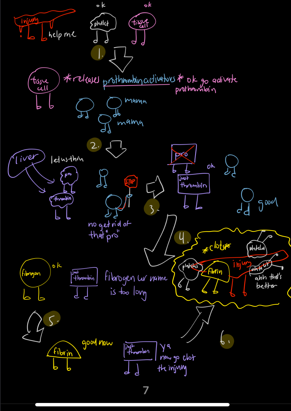
Chemical reactions involving blood clotting
Vessel injury signals platelets & tissue cells.
Tissue cells release prothrombin activators.
Prothrombin (from liver) → thrombin.
Thrombin converts fibrinogen → fibrin.
Fibrin forms the clot at injury site.
Calcium & vitamin K are essential for activating prothrombin and clotting enzymes.
blood proteins
carry hormones, vitamins, lipids in the blood, thickening blood and providing pressure
O₂ and CO₂ transport via hemoglobin
In lungs, hemoglobin binds oxygen to form oxyhemoglobin ⇒ delivers O₂ to tissues ⇒ in tissues, hemoglobin picks up CO₂ (as carbaminohemoglobin (some) or helps convert it to bicarbonate (most)) ⇒ transports CO₂ back to lungs for exhalation.
Hemoglobin
A protein in erythrocytes (red blood cells) that binds oxygen in the lungs and carries it to tissues; also helps transport carbon dioxide back to the lungs.
HDL cholesterol
"good" cholesterol, high density lipoprotein - helps remove excerss cholesterol from blood and artery walls
LDL cholesterol
"bad" cholesterol, low density lipoprotein - can deposit in arteries and cause atherosclerosis
components of the lymphatic system
lymphatic vessels, lymphoid organs, lymph nodes, spleen, thymus gland, red bone marrow
Lymphoid organs: what are they, and what are some main examples?
organs where lymphocytes are produced/activated. Help with immune response against pathogens.
bone marrow (produces all blood cells, b and t cells)
thymus (where t cells mature)
lymph nodes (bean-like structures filtering lymph, house immune cells)
spleen (filters blod, removes old rbc’s, activates lymphocytes
Lymph nodes
Small, bean-shaped structures that filter lymph and house immune cells to fight pathogens.
Spleen
Organ that filters blood, removes old red blood cells, and helps fight infection.
Thymus gland
Gland where T cells mature; active in childhood and shrinks with age.
Red bone marrow
Tissue inside bones where blood cells, including lymphocytes, are produced.
Lacteal
A lymph capillary in the small intestine that absorbs dietary fats.
Subclavian vein
Large vein under the collarbone where lymph reenters the bloodstream.
Lymphatic duct
Major vessel that drains lymph into the subclavian veins (e.g. thoracic duct).
Thoracic duct
The largest lymphatic duct; drains lymph from most of the body into the left subclavian vein.
Lymph vessels
General term for all vessels that carry lymph, including capillaries and ducts. they are structurally like veins, have valves
flow of lymph fluid: right upper body
tissue fliod ⇒ lymphatic capillaries, lymphatic vessels, right lymphatic duct, right subclavian vein, superior vena cava
flow of lymph fluid: lower body and upper left
tissue fluid ⇒ lymphatic capillaries ⇒ lymphatic vessels ⇒ thoracic duct ⇒ left subclavian vein ⇒ superior vena cava
Antibodies
Proteins made by B cells (lymphocytes/white blood cells) that specifically bind to antigens to neutralize or mark them for destruction. There are specific antibodies for each antigen, able to recognize them because of receptor specificity.
How antibodies work
Bind to antigens
Immune response
Antigen detection triggers immune cells (B and T cells)
How does the immune system respond to an antigen using the lymphatic system?
Antigens enter lymph fluid → carried to lymph nodes
Helper T-cells activate B-cells and cytotoxic T-cells
B-cells produce specific antibodies → bind antigens → cause agglutination
T-cells destroy tagged pathogens (some by engulfing)
Memory cells remain → enable faster response if antigen returns