AP II prac (anatomy)
 Call Kai
 Learn
Learn Practice Test
 Spaced Repetition
 Match
Match Flashcards
 Knowt Play
1/98

There's no tags or description
Looks like no tags are added yet.
Name | Mastery | Learn | Test | Matching | Spaced | Call with Kai | Chat |
|---|
No analytics yet
Send a link to your students to track their progress
99 Terms
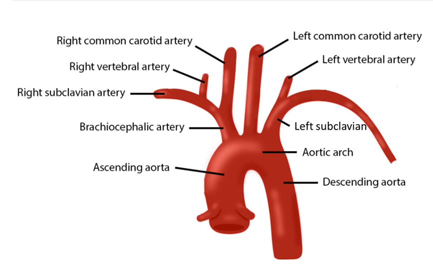
3 branches of aortic arch
brachiocephalic trunk
left common carotid artery
left subclavian artery
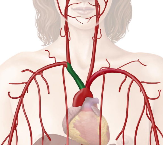
Brachiocephalic trunk
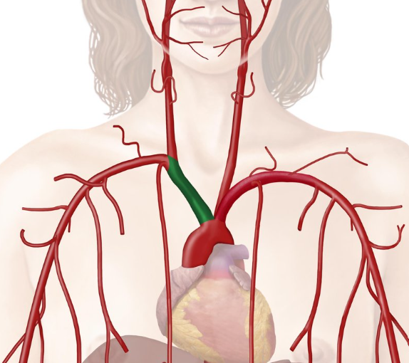
Brachiocephalic trunk’s function:
take blood to the right side of head, neck, upper limb
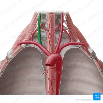
Right common carotid artery
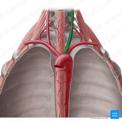
Left common carotid artery
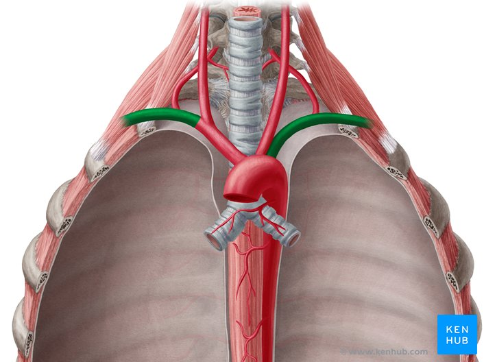
subclavian artery (R/L)
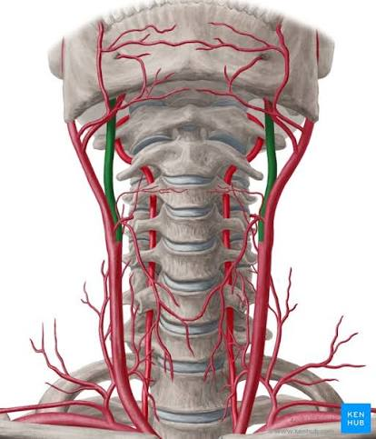
Internal carotid artery (R/L)
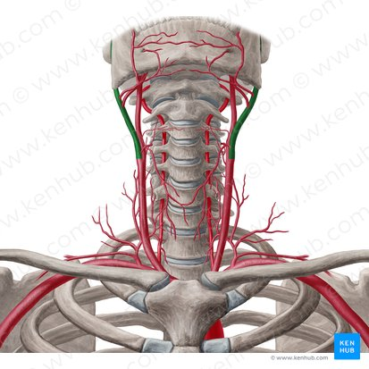
External carotid artery (R/L)

Brachiocephalic vein (R/L)
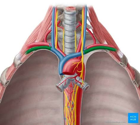
subclavian vein (R/L)
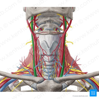
Internal jugular vein (R/L)
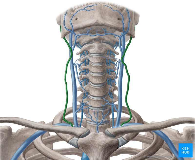
External jugular vein (R/L)

Pulmonary trunk

Pulmonary veins (R/L)

Pulmonary arteries (R/L)
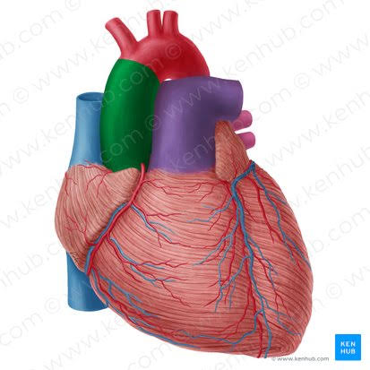
Ascending aorta
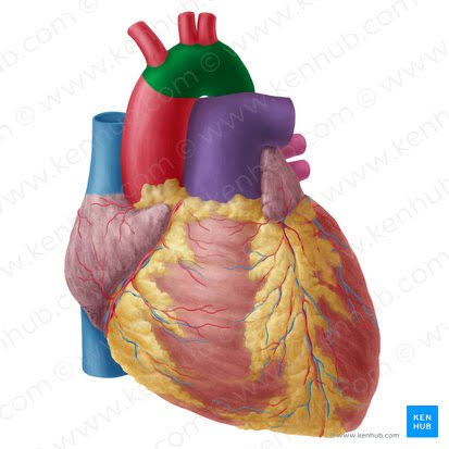
Aortic arch
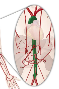
Descending aorta

Superior vena cava

Inferior vena cava
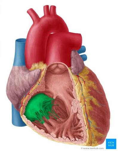
Tricuspid valve
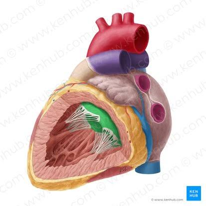
Bicuspid valve
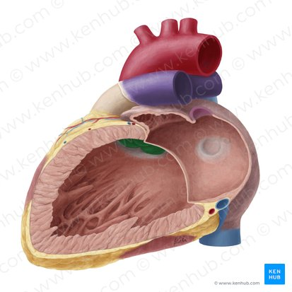
aortic valve
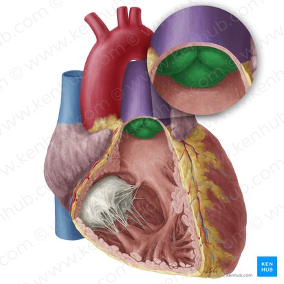
pulmonary valve
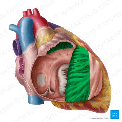
Pectinate muscles
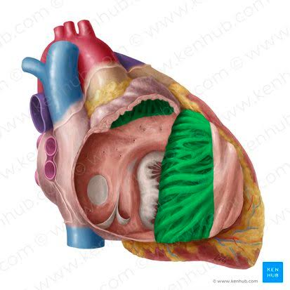
Pectinate muscles’ location
in atrial walls
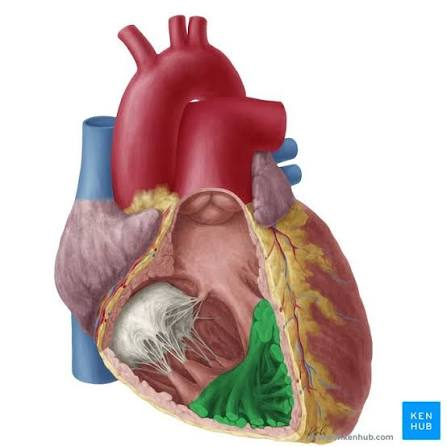
Trabeculae carneae

Trabeculae carneae’s location
in ventricular walls

Papillary muscles
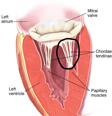
Chordae tendineae

Chordae tendineae’s location
attached to AV vavles
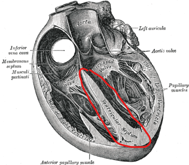
Interventricular septum
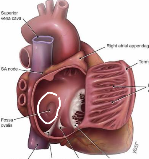
Fossa ovalis
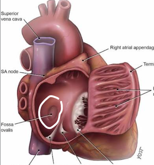
Fossa ovalis is a remnant of:
Foramen ovale
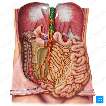
Abdominal aorta

Celiac trunk

Celiac trunk’s 3 branches:
splenic artery
left gastric artery
common hepatic artery

Left gastric artery
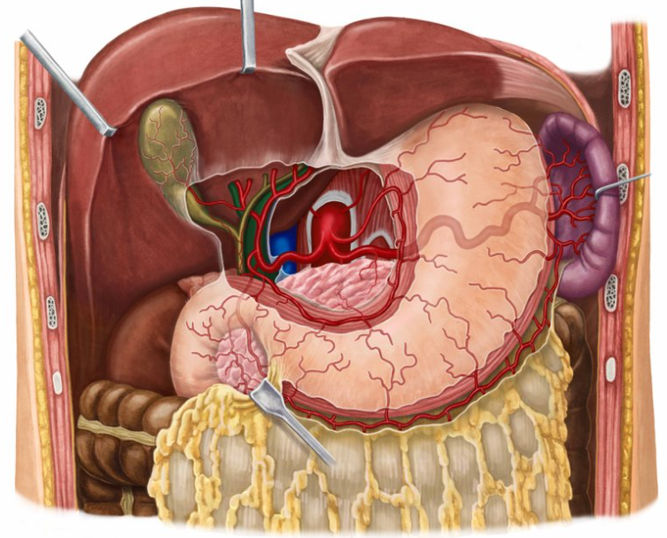
Hepatic portal vein
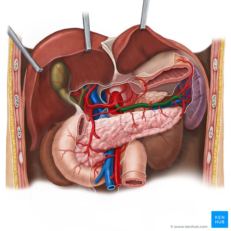
Splenic artery
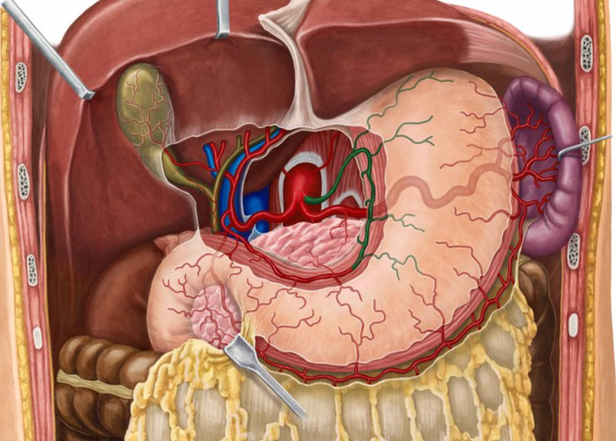
Left gastric artery
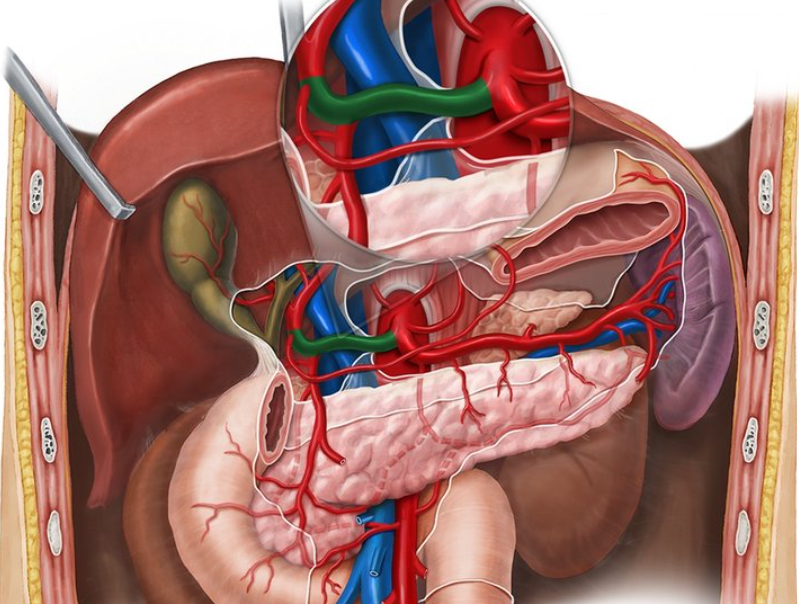
Common hepatic artery

Hepatic artery proper
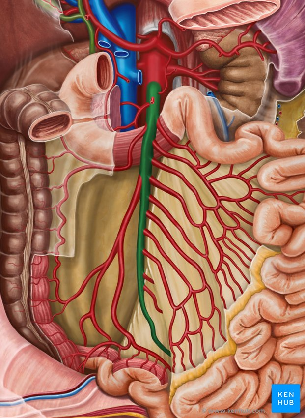
Superior mesenteric artery

Inferior mesenteric artery
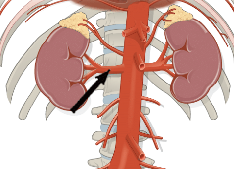
Renal artery (R/L)
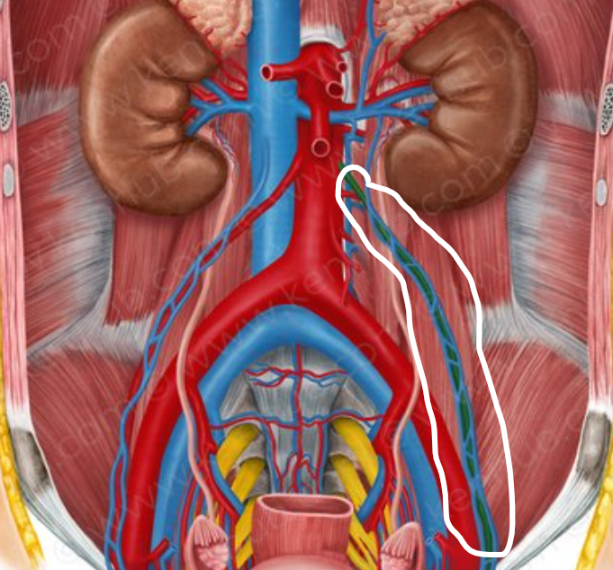
Gonadal artery (R/L)

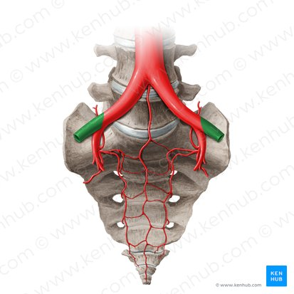
Common iliac artery (L/R)
External iliac artery (L/R)
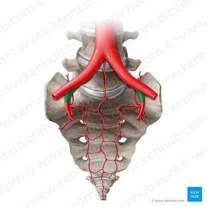
Internal iliac artery (L/R)
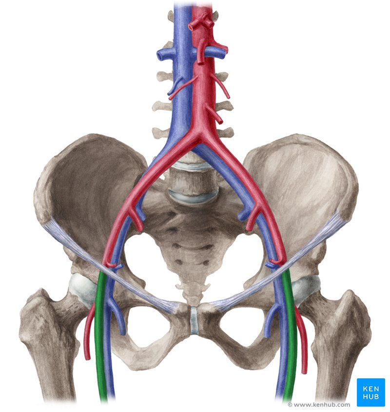
Femoral artery (L/R)
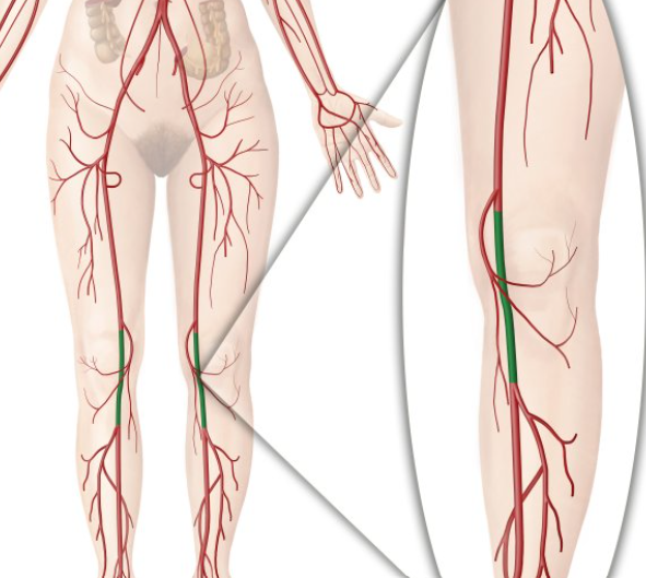
Popliteal artery (L/R)
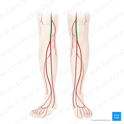
Popliteal artery’s subdivisions:
anterior, posterior tibial artery
fibular artery
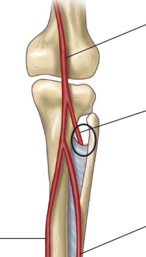
Anterior tibial artery
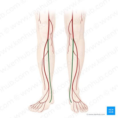
Posterior tibial artery
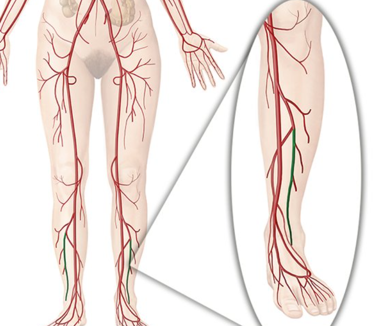
Fibular artery
![<p>Posterior tibial artery is on the […] side</p>](https://assets.knowt.com/user-attachments/74eca77e-982f-404c-ab86-5c53206a9a76.png)
Posterior tibial artery is on the […] side
medial
![<p>Fibular artery is on the […] side</p>](https://assets.knowt.com/user-attachments/f3980990-1433-44a9-babe-8cf7068ff727.png)
Fibular artery is on the […] side
lateral
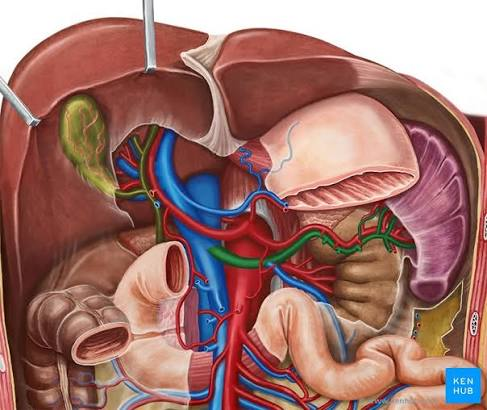
Splenic vein
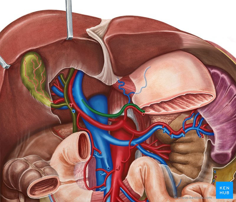
Gastric vein (L/R)
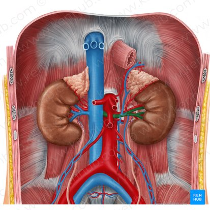
Renal vein (R/L)
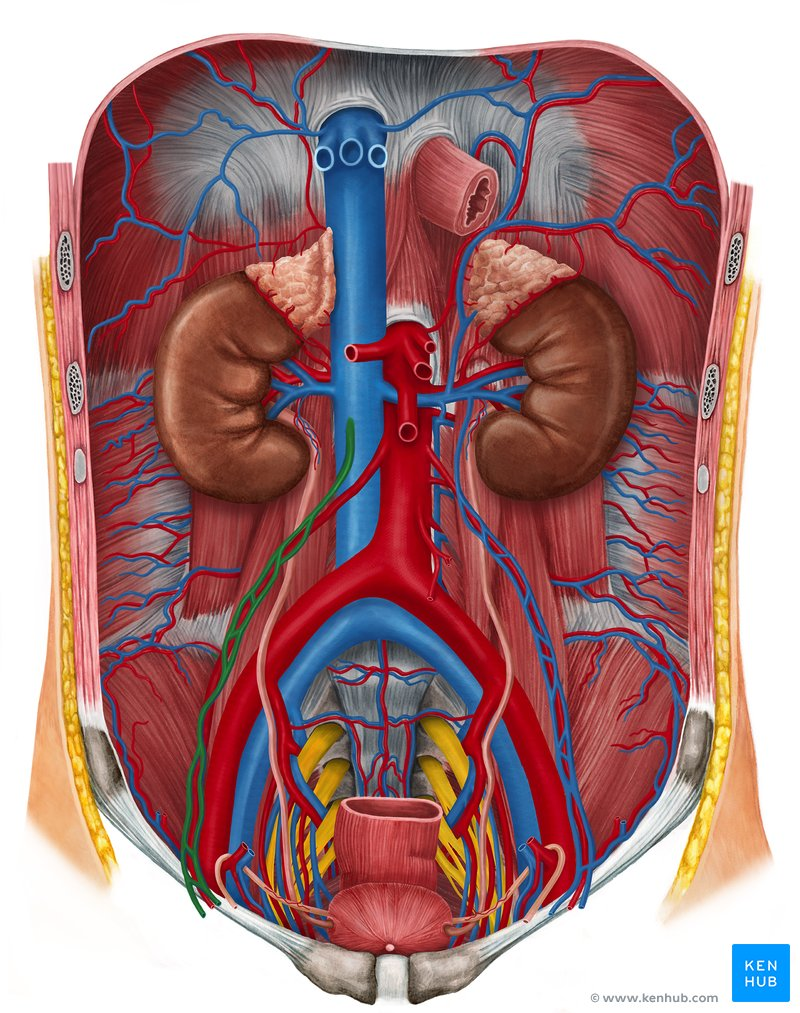
Right gonadal vein

Where does Right gonadal vein drain into?
into Inferior vena cava
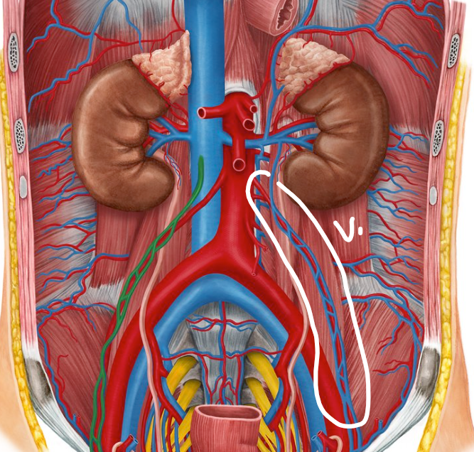
Left gonadal vein
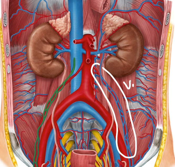
Where does Left gonadal vein drain into?
into Left renal vein

Common iliac vein (R/L)
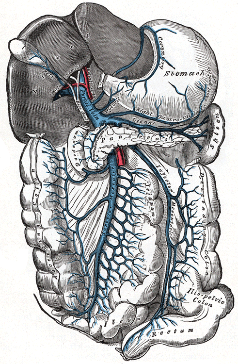
Hepatic portal system (veins) contains:
hepatic portal vein
splenic vein
inferior mesenteric vein
superior mesenteric vein

Hepatic portal vein
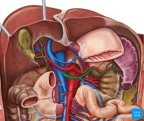
splenic vein

Inferior mesenteric vein
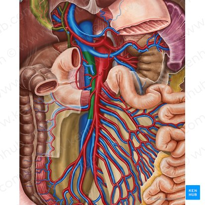
Superior mesenteric vein
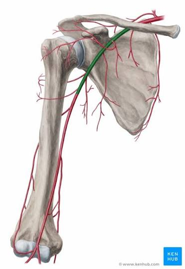
Axillary artery
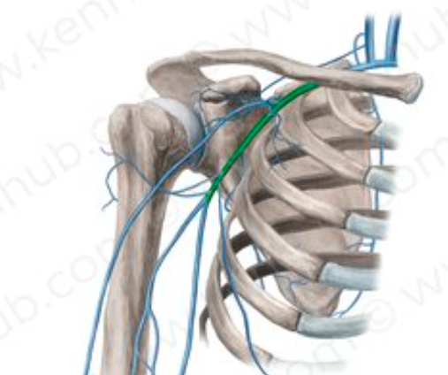
Axillary vein
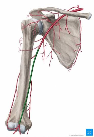
Brachial artery

Brachial vein
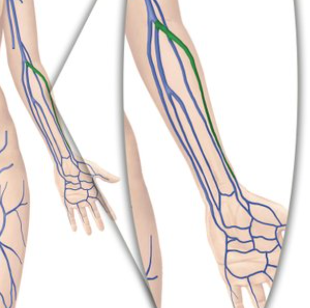
Radial vein (L/R)

Median cubital vein

Cephalic vein
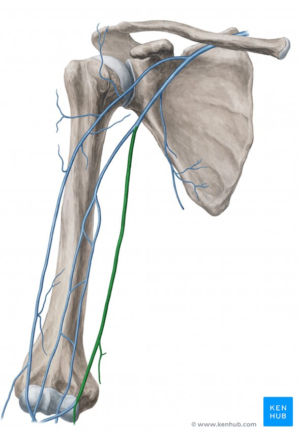
Basilic vein

(medial side)
Great saphenous vein

Where does the Great saphenous vein dran into?
into Femoral vein

(posterior side)
Small saphenous vein
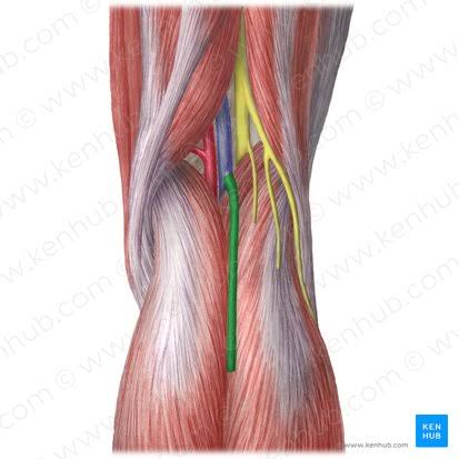
Where does small saphenous vein drain into?
into Popliteal vein
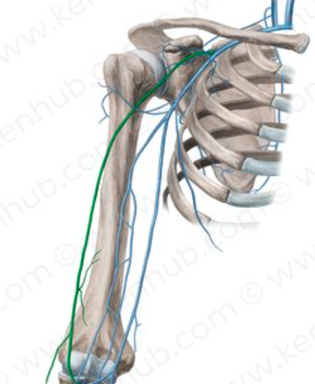
what does ‘Cephalic’ mean?
on the lateral (thumb) side

what does ‘Basilic’ mean?
on the medial (pinky) side
what does ‘cubital’ mean?
in the middle (elbow region)

urinary bladder
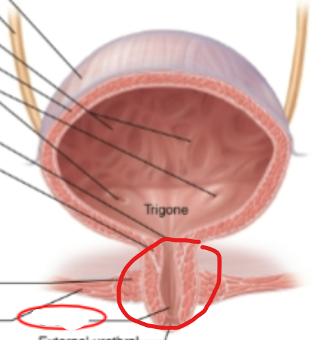
urethra
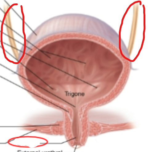
ureter

renal hilum
what is the anatomical location of the kidneys?
in retroperitoneum
- posterior abdominal wall
what muscle lines the urinary bladder?
detrusor muscle

renal cortex
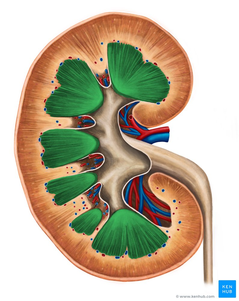
renal medulla
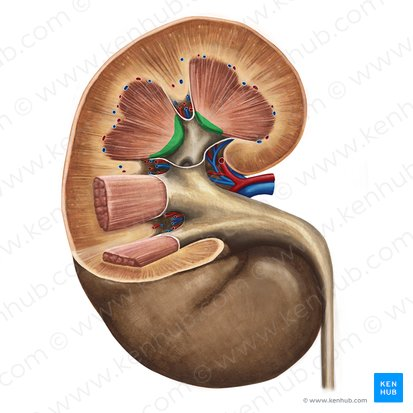
renal pyramid of medulla

minor calyx

major calyx
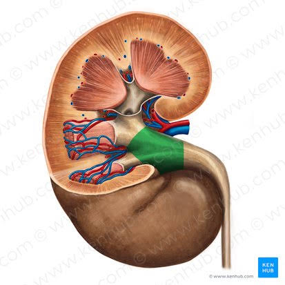
renal pelvis