Leukocytes
 Call Kai
 Learn
Learn Practice Test
 Spaced Repetition
 Match
Match Flashcards
 Knowt Play
1/216

There's no tags or description
Looks like no tags are added yet.
Name | Mastery | Learn | Test | Matching | Spaced | Call with Kai | Chat |
|---|
No analytics yet
Send a link to your students to track their progress
217 Terms
what are the 4 main types of neutrophilia (Count Abnormalities)
Physiologic
Glucocorticoid associated
Acute inflammatory
Chronic inflammatory
what 2 types of neutrophilia can occur in healthy animals (Count Abnormalities)
Physiologic
Glucocorticoid associated
what 2 types of neutrophilia can occur due to disease (Count Abnormalities)
Acute inflammatory
Chronic inflammatory
what causes Physiologic Neutrophilia (Count Abnormalities)
Occurs as the result of epinephrine and norepinephrine release associated with fear, excitement or vigorous exercise
Glucocorticoid is associated with (Count Abnormalities)
stress
what causes Glucocorticoid (Count Abnormalities)
Caused by the effects of endogenous (cortisol) or exogenous glucocorticoids.
Glucocorticoid results in (4) (Count Abnormalities)
Fewer neutrophils in the Marginated Pool and more in the Circulating Pool.
Fewer neutrophils enter the tissue so they have an increased circulating lifespan.
The older neutrophils may become hypersegmented.
This also results in increased release of segs mostly from the storage pool.
Acute Inflammatory Neutrophilia causes an increased release of (3) (Count Abnormalities)
Increased release of segmented and band neutrophils
Increased release from the storage pool, which takes a few hours after the onset of inflammation.
Release of cells from the maturing pool occurs after the depletion of the storage pool.
what causes Acute Inflammatory Neutrophilia (Count Abnormalities)
infection
what causes Chronic Inflammatory Neutrophilia (Count Abnormalities)
occurs because the release of neutrophils from the marrow exceeds the migration of neutrophils into the tissues.
what are the 2 main types of neutropenia (Count Abnormalities)
Inflammatory
Decreased production
what causes Inflammatory Neutropenia (Count Abnormalities)
when margination and emigration of neutrophils into inflamed tissue exceeds the release from the bone marrow
overwhelming bacterial infections
some viral infections
endotoxins
inflammatory states in cattle
Decreased Production Neutropenia occurs when (Count Abnormalities)
due to destruction of stem of blast cells in the bone marrow.
Ineffective Production Neutropenia occurs when (Count Abnormalities)
occurs when neutrophil precursors are damaged and die before they can be released.
what causes Decreased or Ineffective Production Neutropenia (6) (Count Abnormalities)
Toxoplasmosis
Parvovirus
Felv
Neoplastic (abnormal growth)
toxic
Myelofibrotic (abnormal stem cell production)
what are the 4 main types of Lymphocytosis (4) (Count Abnormalities)
“Lymphocytosis” of young animals:
Physiologic
Chronic inflammatory
Neoplastic
what causes Physiologic Lymphocytosis (Count Abnormalities)
Due to the effects of the catecholamines, causing lymphocytes to move from the marginated to the circulating pool.
what causes Chronic Inflammatory Lymphocytosis (Count Abnormalities)
Results from increased lymphopoiesis in response to chronic antigenic or cytokine simulations.
define Hyperplastic (Count Abnormalities)
enlargement of a part due to an abnormal numerical increase of its cells.
what causes Neoplastic Lymphocytosis (2) (Count Abnormalities)
Due to the neoplastic proliferation of lymphoid cells in the lymph nodes, bone marrow and other tissues.
Extreme lymphocytosis occurs with many lymphocytes having abnormal, immature morphology.
what are the 2 types of Lymphopenia (Count Abnormalities)
Glucocorticoid – Associated (Stress)
Acute Inflammatory
what causes Glucocorticoid – Associated (Stress) Lymphopenia (Count Abnormalities)
Due to endogenous or exogenous glucocorticoids
Movement of lymphocytes from the circulating pool into the tissues and or bone marrow and lymph nodes.
Chronically elevated glucocorticoid levels cause (2) (Count Abnormalities)
lymphoid hypoplasia
decreased production.
define Hypoplasia (Count Abnormalities)
abnormal deficiency of cells or structural elements.
what causes Acute Inflammatory Lymphopenia (Count Abnormalities)
Due to changes in lymphocyte kinetics stimulated by inflammatory mediators
define Mediators (Count Abnormalities)
A substance or structure that mediates a specific response in a bodily tissue
Lymphoma can be classified in one of two ways: (Count Abnormalities)
Anatomical Location
Extent of the disease
what are the 4 forms of Lymphoma (Count Abnormalities)
Multicentric
Mediastinal
Alimentary
Extranodal
define Multicentric lymphoma (Count Abnormalities)
generalized enlarged lymph nodes.
define Extranodal lymphoma (Count Abnormalities)
affecting organs and tissues
define Alimentary lymphoma (Count Abnormalities)
palpable abdominal mass
define Mediastinal lymphoma (Count Abnormalities)
enlarged mediastinal lymph nodes
clinical signs of Multicentric lymphoma (3) (Count Abnormalities)
Anorexia
weight loss
lethargy
nothing
clinical signs of Mediastinal lymphoma (8) (Count Abnormalities)
Dyspnea
tachypnea
regurgitation
cough
anorexia
depression
weight loss
pleural effusion
clinical signs of Alimentary lymphoma (3) (Count Abnormalities)
Anorexia
weight loss
v/d
clinical signs of Extranodal lymphoma (4) (Count Abnormalities)
renomegaly
itching
seizures
paralysis
how to diagnose (7) (Count Abnormalities)
enlarged lymph nodes
sent for cytology
Biopsy of mass
possibly bone marrow
Radiographs for mediastinal
radiographs abdominal masses
most diagnostic for diagnosis
how to treat Lymphoma (Count Abnormalities)
chemotherapy
Drug protocols for chemotherapy are typically divided into four phases: (Count Abnormalities)
Induction of Remission
Intensification
Maintenance
Rescue
All chemotherapy drugs have side effects, some include: (Count Abnormalities)
Anorexia
Vomiting
Leukopenia
Renal toxicity
Hemorrhagic cystitis
There are only two important causes of Monocytosis (Count Abnormalities)
Glucocorticoid
Inflammatory
define Mastocytemia (Count Abnormalities)
A mast cell is found in the peripheral blood
The parameters that must be examined in order to determine which of these four patterns a particular leukogram fits are: (5) (Count Abnormalities)
Total WBC numbers
Number of neutrophils
Age of neutrophils
Number of lymphocytes
Whether a monocytosis is seen
signs of a left shift (6) (Count Abnormalities)
immature neutrophils (bands) are present.
A lot of band cells present.
Mostly seen with acute inflammatory responses
Immature neutrophils exceed the number of mature neutrophils
the leukocyte count is low or normal.
animals condition is deteriorating.
when does left shift occur (Count Abnormalities)
Occurs when tissue demands are high.
signs of right shift (3) (Count Abnormalities)
hypersegmented neutrophils
Leukocytosis due to neutrophilia with the appearance of immature neutrophils.
Increased concentration of old neutrophils
what does right shift and left shift refer to (Count Abnormalities)
Refers to the change in numbers and ages of neutrophils.
— are the most numerous leukocyte in the blood and bone marrow
neutrophils
— and — are the most numerous in the entire body.
Lymphocytes and macrophages
function of neutrophil
to locate and phagocytose pathogenic microorganisms.
— are the first cells to arrive at the site of infection.
neutrophils
what are the 4 steps in migration sequence of neutrophils
Margination
Diapedesis
Chemotaxis
Migration
define the Margination step in neutrophil migration sequence
Approximately 50% of the neutrophil population in the blood vessels are adhered to the blood vessel walls (marginal pool)
define the diapedesis step in neutrophil migration sequence
Neutrophils that are in the marginating pool squeeze between the endothelial cells of the blood vessel wall and enter the tissues
define the chemotaxis step in neutrophil migration sequence
Neutrophils perform diapedesis due to the presence of chemotaxis. Inflammatory chemicals (chemotaxins) are released from other neutrophils and cells in the presence of microorganisms in tissues that draw the marginating neutrophils to the site
define the migration step in neutrophil migration sequence
Neutrophils follow the chemotaxins to the highest concentration (the source)
what are the 2 blood pools
The Circulating Pool
The Marginating/Marginal Pool
define circulating pool
Within the lumen of the blood vessel
define Marginating/Marginal Pool
Line the walls of the endothelium of blood vessels
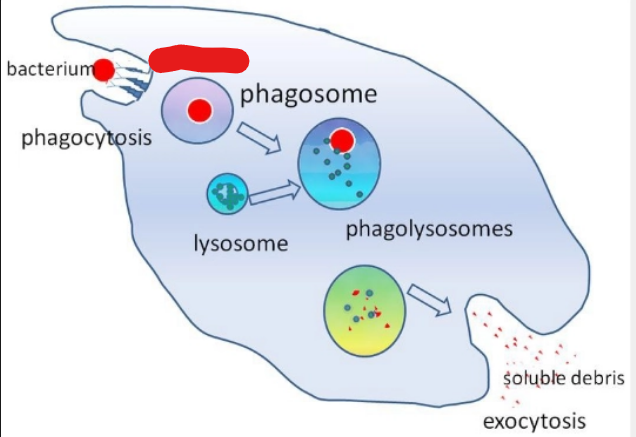
what is it
receptors
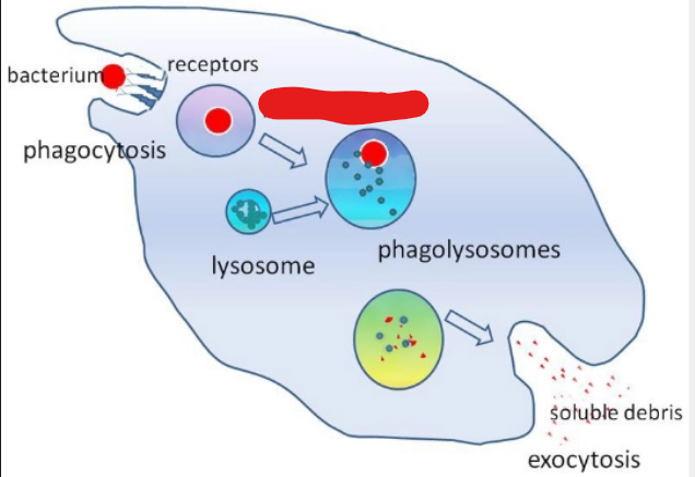
what is it
phagosome
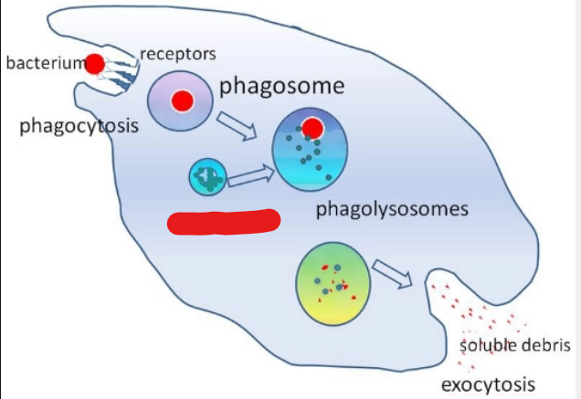
what is it
lysosome
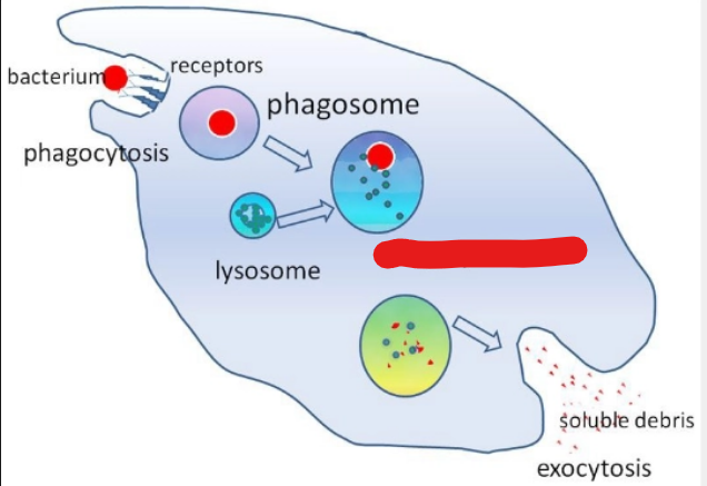
what is it
phagolysosomes
define Immune Adherence
Neutrophil recognizes the microbe by binding to its cell surface
define Endocytosis
The microbe is engulfed by the neutrophil and becomes a phagosome.
define Lysosome Fusion
The neutrophilic granules are lysosomes which attach to the walls of the phagosome and discharge their contents into the vacuole (lysosomal enzymes).
how does leukocyte Killing and Digestion occur (2)
Lysosomal enzymes released into the phagosome from the lysosomes kill the microbe and break down the remains of the microbe
Useful components from the microbe are absorbed into the cytoplasm and used by the neutrophil.
define exocytosis
Waste products in the phagosome are eliminated from the cell.
— will continue killing microbes until they run out of granules, then they die.
Neutrophils
Dead neutrophils form —
pus
steps of opsonization (2)
Encapsulated microorganisms become coated with plasma proteins (antibodies) that are called opsonin
Once the microorganisms are coated in the opsonin the neutrophils can recognize them as microorganisms and perform phagocytosis
why is opsonization needed
Some microorganisms are more challenging for neutrophils to phagocytose as they can “hide” within a capsule
define Mononuclear Phagocyte System
the bodies population of macrophages and monocytes.
Macrophages have many functions outside of the immune response including (3)
Catabolism of other dead tissue cells.
Removal of toxins, activated coagulation factors and immune complexes from the blood.
Catabolism of rbcs and iron metabolism.
define catabolism
to break down.
what are the 3 steps of phagocytosis
a bacterium engulfed by a macrophage is encased in a vacuole
lysosome fuse with the vacuole and digest the bacterium
antigens from digested bacterium are presented with MHC ll on the cell surface
— produce monokines
Macrophages
Macrophages can become activated by — secreted by Helper T cells
lymphokines
monokines
chemical substances which enhance other cells in the immune process and act as pyrogens in the inflammatory process.
APC stands for
Antigen Presenting Cells
Antigen Presenting Cells are Essential for the activation of (2)
Cell-Mediated Immunity (Memory cells)
Specific Immunity Response.
what does CMI stand for
Cell-Mediated Immunity
CMI is mediated by
T-lymphocytes
CMI Involves cells attacking cells and is directed against — pathogens and cells such as cancer cells and foreign tissue transplants
intracellular
T-cells can only bind to a foreign antigen and become activated if
the foreign antigen (FAg) is presented on the surface of an Antigen Presenting Cell.
Antigen Presenting Cells are
macrophages that ingest the foreign cell, process the FAg and then present it on their cells surface.
The activated T cell begins dividing rapidly and the resulting cells differentiate into 5 types of T cells:
Helper T-cells
Killer T-cells
Suppressor T-cells
Memory T-cells
Natural Killer Cells (NK cells)
The main function of Helper T cells is to produce chemical mediators called
Lymphokines
if lymphokines touches macrophage it’ll get an upgrade and … (4)
move fast
eat things faster
also attract B lymphocytes to help
bind process macrophages which signals multiplication of other T cells
function of supressor T-cells (4)
Regulate the immune response
producing lymphokines that inhibit T-cell proliferation
reduce Helper and Killer T-cell functioning
prevent B-cells from transforming to plasma cells.
function of memory T-cells (2)
recognize the FAg that originally stimulated their production
will stimulate a larger, quicker response if the FAg appears again.
NK cells stand for
Natural Killer Cells
NK cells are aka
null cells
how are NK cells identified
large granular lymphocytes
what does LGL stand for
large granular lymphocytes
define NK cells
cells kill target cells that ARE NOT sensitized by antibody or complement.
What does AMI stand for
antibody - mediated Immunity
AMI is mediated by
B-lymphocytes
An activated B cell begins dividing, resulting in two populations of cells:
Memory B cells
Plasma cells
Each Memory B-cell carries the same specific antigen receptor as the activated
B-cell.