ASCI 438: Systemic Physio
1/199
There's no tags or description
Looks like no tags are added yet.
Name | Mastery | Learn | Test | Matching | Spaced |
|---|
No study sessions yet.
200 Terms
Which tissues are “excitable”?
Neural and Muscle tissues
Signal transduction
refers to the process by which incoming signals are conveyed into the target cell where they are transformed into the dictated cellular response
Neurons send signals to what other cells?
Other neurons
Muscles
Glands
Resting Membrane Potential
The membrane potential in a cell at rest is negative, not neutral
-70mV
Why is the neuron interior negatively charged relative to the exterior?
3 influences
Na+ K+ pumps on the cell membrane pump 3 Na+ out of the cell for every 2 K+ into the cell
Large negatively charged protein molecules exist inside the cell
K+ leak channels are more active than Na+ leak channels
Na+ K+ ATPase
Moves 3 Na+ out of the cell and 2 K+ into the cell with each cycle
Over time this yields more positive charges outside the cell relative to inside the cell
Requires Energy
Leak Channels
Also play a role in maintenance of resting potential
Concurrently with the ions being moved by the pumps, Na+ and K+ are also passively leaking across the membrane through their specific leak channels
These leak channels are always open
The membranes pumps purpose is:
to set up a gradient that the Na+ and K+ can Flow down
Once the gradient is in place, ions will flow down their gradient across the membrane once a channel opens
In addition to the leak channels, the ions will also flow through transiently opened gated channels
Threshold Potential
-55 mV
4 Types of Gated Channels
Voltage Gated
Chemically Gated
Mechanically Gated
Thermally Gated
A flow of positive ions into the ICF —— the cell
depolarizes (makes less negative)
A flow of positive ions into the ECF —— the cell after depolarization
Depolarizes
From resting state, a flow of positive ions out of the cell into the ECF—- the cell
hyper polarizes
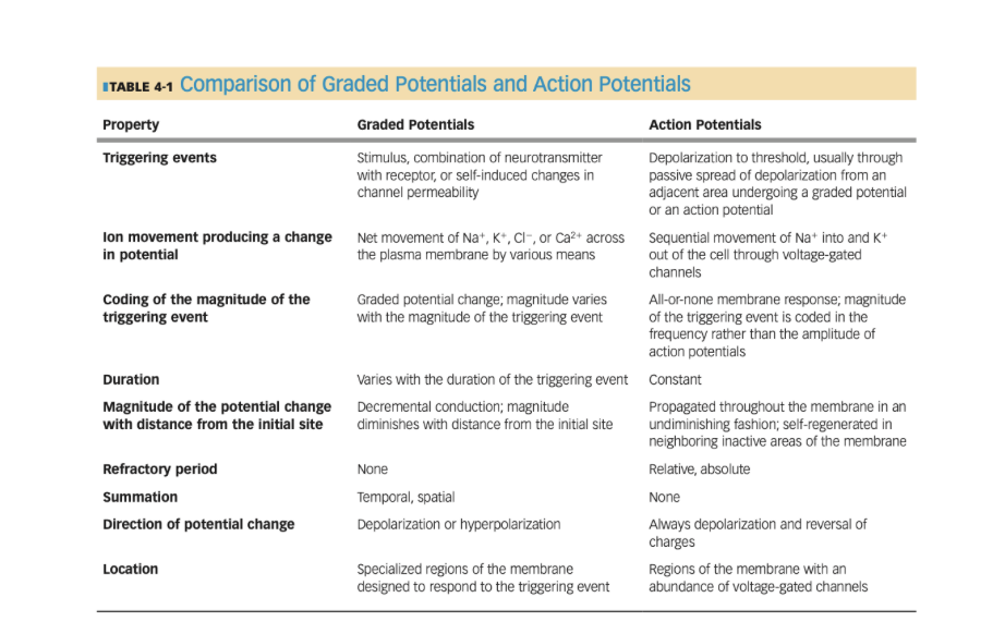
Graded Potentials vs. Action Potentials
Graded Potential: Short distance decremental signal
different sizes
propagate in both directions
Action Potential: long distance non decremental signal
same sizes (all or none principle)
travel in only one direction in the axon
Graded potentials
Short distance signals
Decremental
Spreads in both directions away from the site of initiation
The stronger the triggering event, the more ion channels will be opened and the larger the resulting graded potential (but they then still dwindle down)
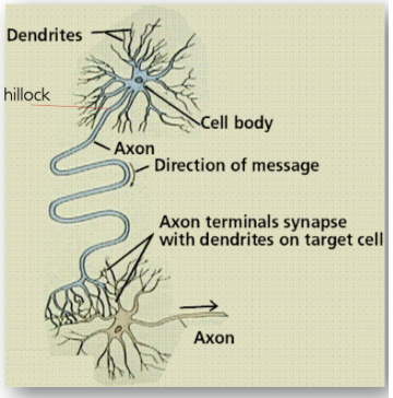
Neuron Anatomy
Cell Body
Has dendrites (“receiving zone”)
The cell membrane covering the cell body and dendrites is where graded potentials arise
Axon
Axon Hillock ('“trigger zone”)
The axon hillock is where action potentials arise
Axon
Terminals
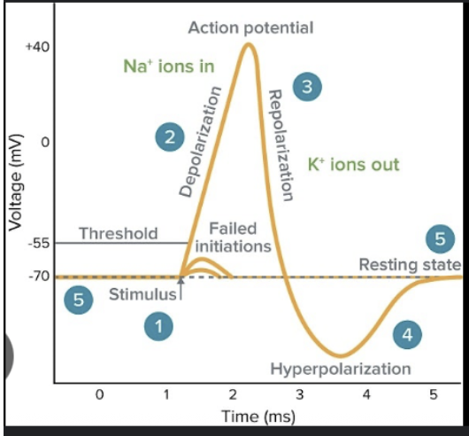
Action Potentials
An action potential happens when the excitable cell membrane is depolarized to threshold potential (-55mV) by a graded potential
At threshold potential, voltage gated Na+ and K+ channels open
The opening of these channels elicits a momentary increase in the permeability of the neuronal membrane to these ions
The ions flow through their channels, causing a transient reversal of membrane potential from –70 to +30 mV
This is an action potential
If enough Na+ enters locally from a graded potential to take the neuron from –70 to –55 in the area of the hillock, the neurons threshold voltage is reached
At threshold voltage, the voltage gated Na+ channels open, allowing an immediate influx of Na+
As the Na+ enters, the membrane potential becomes increasingly more positive. This is the upstroke of the action potential
After a millisecond, the voltage gated Na+ channels close and the voltage gated K+ channels, which began opening slowly at threshold become fully open. K+ flows out of the neuron
As K+ leaves, the exit of positive charge causes the downstroke of the action potential
The k+ channels close slowly, so that K+ continues to flow out and the membrane potential briefly becomes hyperpolarized
The Na+/K+ pumps which are always active, help return the ions to their respective spaces across the membrane and reestablish resting potential
The action potential causes a brief reversal of membrane potential in that it starts at –70mV, goes up to around +30mV and then returns to –70mV
This is the nerve impulse that travels along at hundreds of meters per second
Action Potentials
Long distance signals
Fast moving, large changes in membrane potential
All or none phenomenon: once threshold reached, a uniform AP will be triggered irrespective of magitude of stimulus
Each AP generated by a neuron is uniform and of maximum magnitude each time
An AP does not diminish in strength or die out after a short distance: they are conducted down entire length of membrane to the nerve terminals
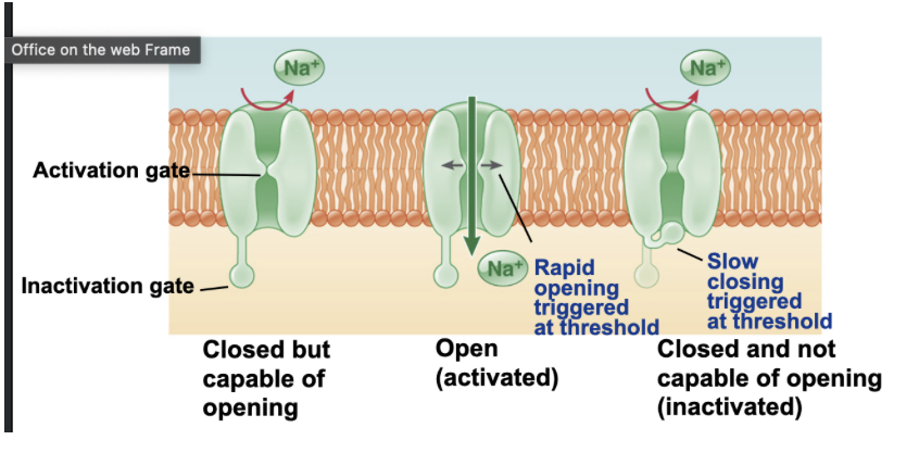
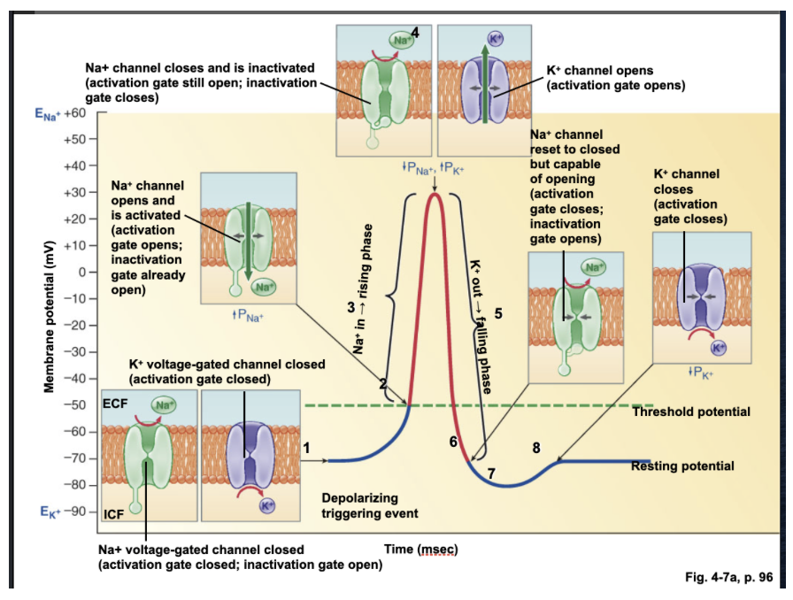
Voltage Gates Na+ Channel Configurations
Closed but capable of opening
Internal Activation gate is closed
Inactivation gate open
Open (activated)
Activation gate open
Inactivation gate open
Closed and not capable of opening (inactivated)
Inactivation gate closed
Activation gate open
Voltage Gated K+ Channel Configuration
The voltage gated K+ channels exist in two configurations
Open
Closed
Refractory Period
the time after a neuron fires an action potential when it cannot fire another. This prevents the neuron from firing too rapidly
The refractory period limits the number of action potentials and ensures AP’s only travel unidirectionally
Absolute Refractory Period
during this time a second stimulus will not elicit a new AP
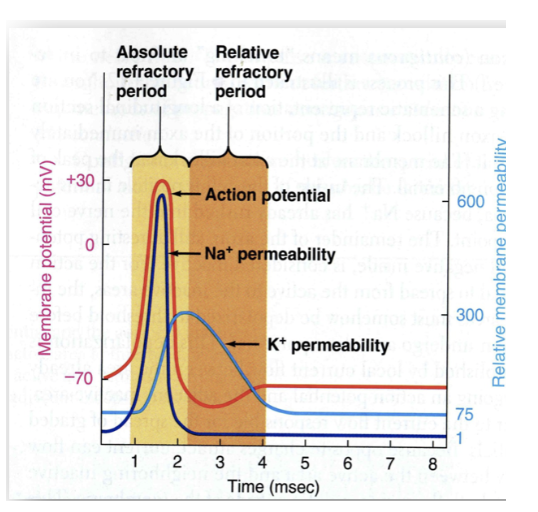
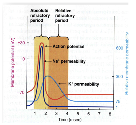
Relative Refractory Period
the interval in which a second AP can be produced but only if the stimulus is considered greater than normal
Action Potential Sequence with Activation Gates ?
At resting potential (-70mV) the Na+ voltage gated channel, the inactivation gate is open, activation gate closed (closed but capable of opening)
Voltage Gated potassium channel at –70 is closed
At threshold (-55 mV) both Na+ gates are open, sodium comes flooding in due to concentration gradient created by Sodium Potassium ATPase pump
At +30 mV, Na+ gates activation gate is open but inactivation gate is closed (closed)
K+ channel opens
Potassium will flood out (going down its concentration gradient) (Repolarize, get more negative)
Na+ channel resets to closed but capable of opening at negative 70 again
Where are voltage gated Na+ and K+ channels concentrated
At the Nodes of Ranvier (Schwaan Cell)
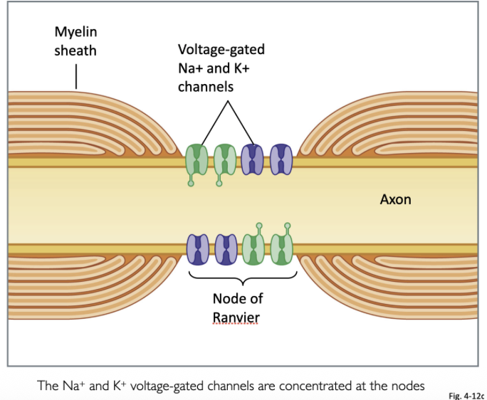
Cells that create myelination in peripheral nervous system
Schwaan Cells
cells that create myelination in central nervous system
oligodendrocytes
Synapse
What happens when a traveling electrical signal (action potential) reaches the nerve terminal at the end of the axon?
It innervates another neuron, a muscle cell or a gland
The tiny spatial gap between nerve ending and target is called a synapse
Events at the Synapse
Arriving Action potential triggers voltage gated Ca2+ channels in presynaptic knob to open
Ca2+ ions rapidly flow into the cell from ECF
Synaptic vesicles containing neurotransmitters fuse with the plasma membrane
Neurotransmitters diffuse across synaptic junction and bind with receptor on postsynaptic membrane
This chemical binding activates chemically gated ion channels
Neuronal Configuration and Events at the Synapse
Voltage gated calcium channels, calcium comes in
Calcium is the cation that facilitates things contracting or moving (actin filaments contract in response to calcium)
Synaptic vesicles (preformed neurotransmitter vesicles) packaged and stores by Golgi
Neurotransmitter vesicles made of same phospholipid bilayer, contract down and fuse, NT is released, goes down and binds to proteins in postsynaptic membrane (receptors are CHEMICALLY gated Na+ channels)
On dendrite they are chemically gated Na+ channels
On axon/ hillock they are voltage gated Na+ channels
In this picture: presynaptic membrane belongs to axon terminal, postsynaptic membrane dendrite
What type of Na+ channels are on the dendrite?
CHEMICALLY gated Na+ channels
What type of Na+ channels are on the axon/hillock
VOLTAGE gated Na+ channels
Excitatory Post-synaptic Potential (EPSP)
If binding of NT (such as ACh) opens Na+ and K+ channels the result is a small depolarization called an excitatory post-synaptic potential (EPSP)
EPSP’s bring the cell closer to threshold
A type of graded potential
Inhibitory Post-Synaptic Potential (IPSP)
If binding of NT (such as GABA) opens either K+ or Cl- channels the result is a small hyperpolarization called an inhibitory post-synaptic potential (IPSP)
IPSP’s polarize the cell further making it less likely to reach threshold
A type of graded potential
Synaptic Summation
the sum of all the EPSP’s and IPSP’s will determine whether the postsynaptic neuron reaches threshold
Temporal Summation
EPSP’s or IPSP’s from a single, repetitively firing presynaptic input occur so rapidly that they add together
Spatial Summation
Adding of EPSP’s or IPSP’s simultaneously from different presynaptic inputs
Grand Post Synaptic Potential
The summation of all inputs is called the grand post synaptic potential (GPSP)
If excitatory inputs dominate, the cell is brought closer to threshold
If inhibitory inputs dominate, the cell is taken farther from threshold
If excitatory and inhibitory activity is balanced, the membrane potential remains close to resting
Summary of events at Posysynaptic membrane
If positive Ion gates open (allowing more Na+ and Ca2+ to enter than K+ to exit) the membrane becomes depolarized, which results in an excitatory post synaptic potential (EPSP)
If the EPSP’s summate such that treshold potential is reached at the hillock, an action potential is generated and will travel down the axon.
If K+ or Chloride Ion (Cl-) gates open (allowing K+ to exit or Cl- to enter) the membrane becomes more polarized which results in an inhibitory postsynaptic potential (IPSP)
If IPSP’s dominate, the hillock region moves away from threshold and the neuron is less likely to generate an action potential
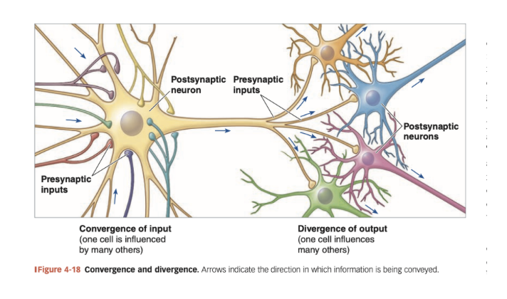
Convergence and Divergence
By converging input, a single cell is influenced by thousands of presynaptic cells
By divergence, branching axon terminals of one neuron affect thousands of postsynaptic cells
At the dendrite, EPSPs and IPSPs can sum in time (temporal summation) and space (spatial summation)
The result is synaptic silence or the grand post synaptic potential, representing the integration of signals from many neurons
Actions of chemical messengers (3 types of receptors)
Opening receptor-channels
Activating receptor-enzymes
Activating an intracellular second messenger via G-protein coupled receptors
GCPR (G-Protein Couples Receptors)
Have 7 membrane domains (7 points of structure that passes through membrane)
These receptors are the mechanism by which water-soluble hormones work
Cannot diffuse across the membrane and enter cell so use an elaborate signaling mechanism to transduce the message into the cell even if the signal molecule cannot enter
Note that the G protein is composed of 3 different subunits and that the alpha subunit can dissociate from the beta and gamma subunits once activated by replacing GDP with GTP
These receptors are all about generaing intracellular second messengers
cAMP
IP3 and DAG: increase Ca2+
Which seconf messenger is generated depends on the type of subunits contained in the G protein connected to the receptor
GCPR’s subunits and monomers
The G protein exists as 3 monomers: Alpha Beta and Gamma
The alpha subunit exists in multiple forms : αS, αi, αq/11, and α12/13
We will concern ourselves with αS and αq/11
If a GPCR’s alpha subunit is an αS it will lead to adenylyl activation and cAMP production
If the alpha subunit is an αq/11 it will lead to phospholipase C (PLC) activation and IP3 and DAG production
If a GPCR’s alpha subunit is an αS it will lead to————
adenylyl activation and cAMP production
If the alpha subunit is an αq/11 it will lead to——-
phospholipase C (PLC) activation and IP3 and DAG production
GCPR: “Alpha S Subunit”
The effector for a G protein containing an Alpha S subunit is the membrane protein adenylyl cyclase
Activation of AC--> cAMP
CAMP activates PKA
PKA activates other proteins in the cytoplasm that will culminate in whatever cellular response was indicated by the binding of the signal molecule at the cell surface
GCPR: Alpha Q Subunit
The effector for a G protein containing an Alpha Q subunit is the membrane protein phospholipase C
PLC cleaves the membrane phospholipid phosohatidylinositol biphosphate (PIP2) into inositol triphosphate (IP3) and diacylglycerol (DAG)
IP3 goes to its receptors on the ER, where it binds and liberates Ca2+ into the cytosol
DAG remains associated with the cell membrane and activates protein kinase C (PKC)
PKC phosphorylates other cytosolic proteins, activating them and leading to a cellular response
Hyperpolarize a cell
open more potassium channels, or open a chloride or other ion channel
An action potential is from opening what channels
from opening voltage gated Sodium and Potassium Channels
If you open a channel for potassium ———-
it will rush from the inside of the cell to the outside (get more negative)
if you open a channel for sodium ——-
sodium will move from outside cell to inside cell
Where are voltage gated calcium channels found
synaptic knob/axon terminal
Sodium potassium ATPase pumps how many ions in and out
3 Na+ (Sodium) ions out and 2 K+ (Potassium) in
Central Nervous System
Consists of the brain and Spinal Cord
Peripheral Nervous System
Afferent and Efferent divisions
Afferent
Efferent
Somatic
Autonomic
Parasympathetic
Sympathetic
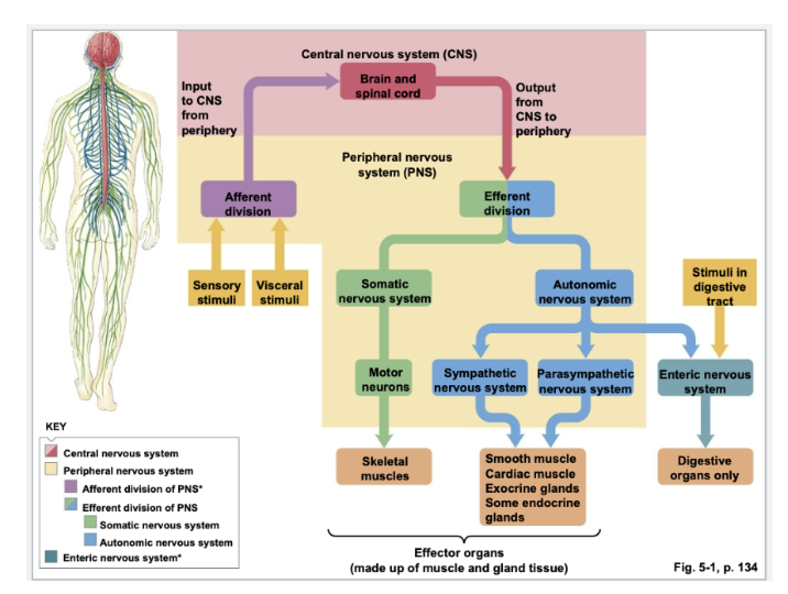
Enteric Nervous System
the nerve network of the digestive tract
Afferent Divisions
Division of PNS
Two afferent divisions carry information into the CNS:
Somatic carries sensory information from skin, skeletal muscles
Visceral carries sensory information from smooth muscle, glands and organs
Efferent Division
Division of PNS
Two efferent divisions carry information out of the CNS:
Somatic nervous system supplies skeletal muscles
Autonomic nervous system innervates smooth muscle, cardiac muscle and glands
Two divisions of the autonomic nervous system
Sympathetic and parasympathetic systems
Divisions of Autonomic Nervous System
Sympathetic and Parasympathetic
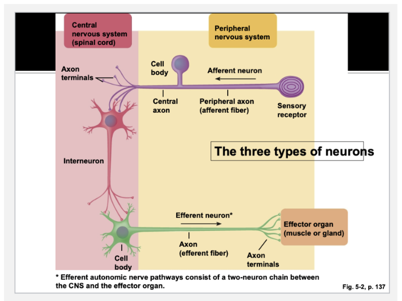
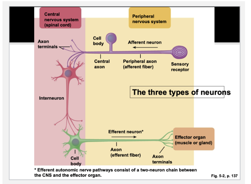
Afferent Neurons
sensory neurons that supply input to the CNS from the internal environment or from the periphery, info about the external environment
SENSORY
Sensory= Ascending = Afferent
Efferent Neurons
carry instructions from CNS to organs, muscles and glands (periphery)
MOTOR
Motor= Descending = Efferent
Interneurons
responsible for integrating afferent information and formulating an efferent response.
These are the most numerous types of neurons
Afferent neurons axon terminals terminate where?
On the dendrites of the Interneuron
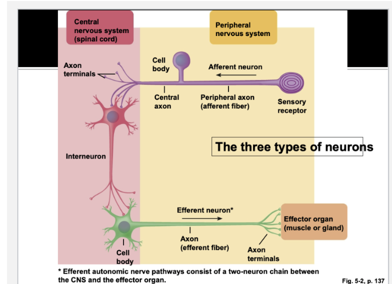
The nerve terminals of the interneuron terminate where?
on the dendrites of the efferent neuron
The nerve terminals of the efferent neurons terminate where?
On the effector organ (muscle, gland or another neuron)
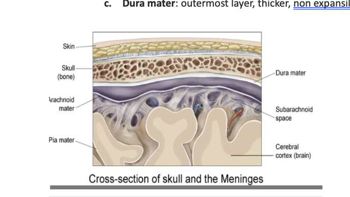
Protection of the CNS
Enclosed by the skull or vertebral column: hard, bony structures
Meninges cover CNS, three layers:
Dura mater
Arachnoid mater
Pia mater
Blood-brain barrier limits access of blood-borne materials into brain tissue
Cerebrospinal fluid (CSF) is the fluid that fills the ventricles and also surrounds and gives hydraulic cushioning to the CNS
Cerebrospinal Fluid (CSF)
is the fluid that fills the ventricles and also surrounds and gives hydraulic cushioning to the CNS
Surrounds and cushions brain and spinal cord
Serves as a shock-absorber to prevent brain from hitting skull
Exchange of materials between neural cells and interstitial fluid surrounding brain
Constant production and flow out of CNS and into venous system
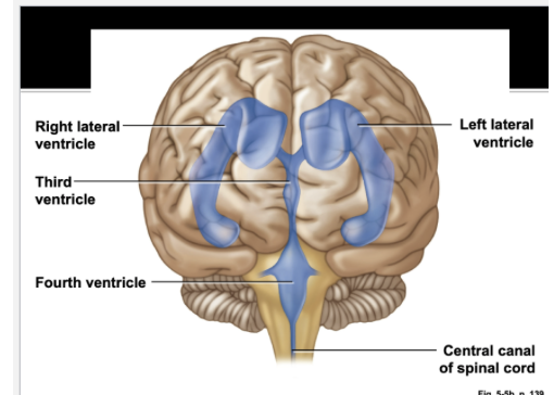
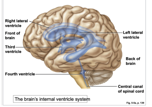
Brain Ventricles
Two Lateral Ventricles
Third and Fourth
(4 total)
Produce CSF
Meninges layers
Dura Mater (outermost)
Arachnoid
Pia Mater (innermost)
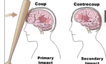
Coup-Contrecoup Injury
Example of one way in which the CSF helps protect the fragile brain tissue
Coup-contrecoup refers to the 2 impact type of closed brain injury that results from traumatic impact
First there is the initial impact and direct crushing injury
A second later comes the secondary impact of the brain against the other side of the skull (when the force knocks the soft brain against the back of the skull)
Closed brain injury without direct trauma: acute acceleration and deceleration
Coup-contrecoup injury can also happen even without direct contact to the head
Whiplash --> there is no direct impact on the head, but the brain still strikes the inner surface of the cranium
In addition, the abrupt acceleration and deceleration causes stretching of the neurons as the brain moves forward and backward inside the cranium: called shear injury
Shear injury damages and destroys neurons and is a significant component of the morbidity associated with closed brain injuries
Skull Interior
The bony skull protects the brain most of the time but can be the source of injury as well
The interior contours of the cranium are not homogenous and smooth
The CSF lies between the brain and cranium, and helps cushion the tender neural tissue from the hard surfaces of the surrounding cranium
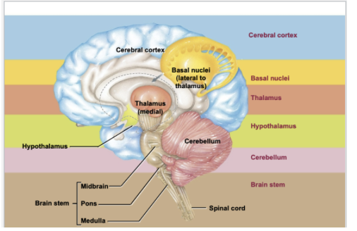
Brain Anatomy Overview
The major parts of the brain from the most complex to most primitive level:
Cerebral cortex
Basal nuclei
Thalamus
Hypothalamus
Cerebellum
Brain Stem
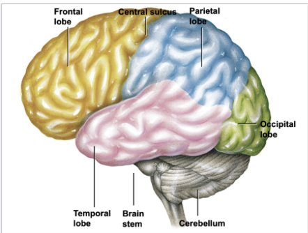
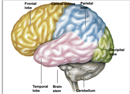
Cerebral Cortex Lobes
Frontal Lobe
Parietal Lobe
Temporal Lobe
Occipital Lobe
Lobes of the Cerebrum
Hemispheres are divided into four major lobes (each has left and right)
Occipital
Temporal
Parietal
Frontal
Occipital Lobe
Houses the visual cortex
Interpretation of visual stimuli and inout
Temporal Lobe
houses the auditory cortex
Auditory processing
Interpretation of language and other things we hear
Parietal Lobe
responsible for reception and perception of somatosensory input (touch,pain, temperature)
Processing of sensory tactile information
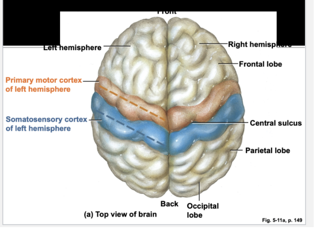
The anterior most gyrus in the parietal lobe is the somatosensory cortex

Frontal lobe
responsible for voluntary motor movement
Responsible for cognition, reasoning, higher language composition: poetry, music
The posterior most part of the frontal lobe is the motor cortex where voluntary movement is initiated
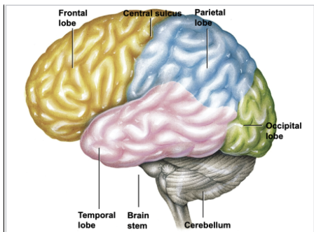
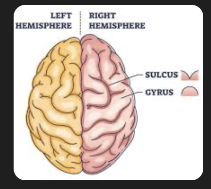
Gyri / Gyrus
Elevated ridges on the brain
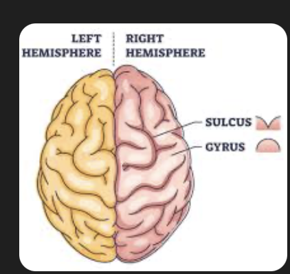
Sulci/ Sulcus
depressions or grooves on the brain
Somatosensory Cortex
Located in the front portion of each parietal lobe just behind the central sulcus (the rostral most gyrus just caudal to the central sulcus)
Site for initial processing and perception of both somesthetic and proprioceptive input
Somesthetic Sensations are from the surface of the body (touch, pressure, heat, cold and pain
Proprioception is the awareness of body position
The somatosensory cortex receives ascending (Afferent) sensory input from the opposite side of the body
Specific regions of the somatosensory cortex receive input from specific areas of the body
The distribution map of sensation in this area is called a sensory homunculus
The size of each body part in the homunculus is proportional to the level of sensory perception associated with each body part
Fingers, lips and the tongue are highly innervated and highly sensitive, command more surface area on the sensory homunculus
Proprioception
is the awareness of body position
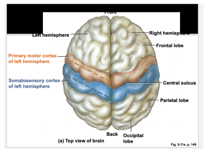
Primary Motor Cortex
Located in posterior portion of frontal lobes, just in front of central sulcus
Controls voluntary movement by skeletal muscles
Motor cortex on each side of the brain primarily controls muscles on the opposite side of body
Basal Nuclei
Consists of several masses of gray matter located deep within white matter
Primary Functions:
Inhibiting muscle tone throughout the body
Selecting, maintaining purposeful motor activity
Suppressing unwanted patterns of movement
Coordinates slow, sustained contractions
Lesions of injuries involving the Basal Nuclei result in unwanted motor activity or body rigidity.
Basal Nuclei Lesions
Putamen
Lesions here cause chorea—involuntary flicking motions of hands, face, shoulders
Globus pallidus
Lesions here cause athetosis- writhing motions of the hands, arms, neck, face
Substantia nigra
Lesions here cause rigidity and tremor—Parkinsons Disease
Spinal Chord White Matter tracts
White matter is organized into nerve tracts—bundles of nerve fibers with a similar function
Each tracts begin or ends within a particular area of the brain
Ascending tracks transmit afferent (sensory) input
Descending tracts relay efferent (motor) input
White matter is myelinated
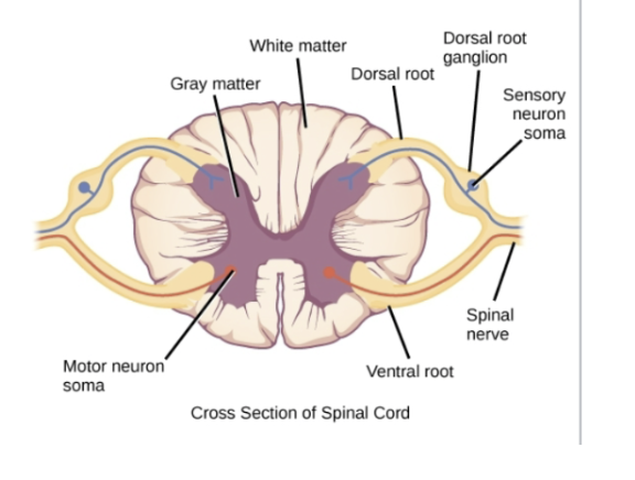
Brain and Spinal Chord White and Grey Matter
In brain grey matter outside/ in periphery white matter more internal
In spinal cord white matter is outside/more superficial and grey matter more internal
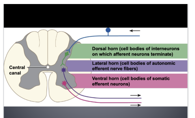
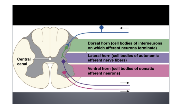
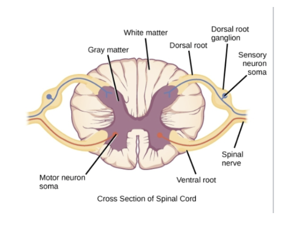
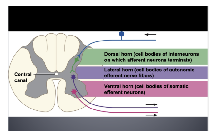
Gray Matter
The gray matter is divided into three horns
Dorsal horn
Contains cell bodies of interneurons on which afferent neurons terminate
Ventral horn
Contains cell bodies or efferent motor neurons supplying skeletal muscles (somatic efferent)
Lateral horn
Contains cell bodies of autonomic efferent nerve fibers (sympathetic or parasympathetic)
Grey matter is non-myelinated (cell bodies)
Dorsal Horn (grey matter)
Contains cell bodies of interneurons on which afferent neurons terminate
Ventral horn (grey matter)
Contains cell bodies or efferent motor neurons supplying skeletal muscles (somatic efferent)
Lateral Horn
Contains cell bodies of autonomic efferent nerve fibers (sympathetic or parasympathetic)
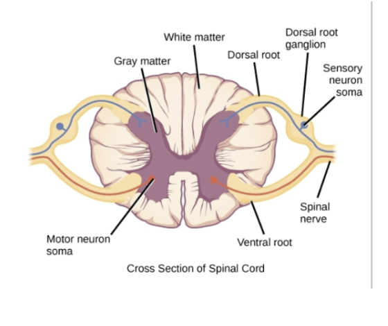
Anatomic Orientation to Spinal Nerves
Afferents enter dorsally (dorsal root carries incoming afferents)
Dorsal Root Ganglion
Efferent enter ventrally (ventral root carries efferent)
No Ventral Root
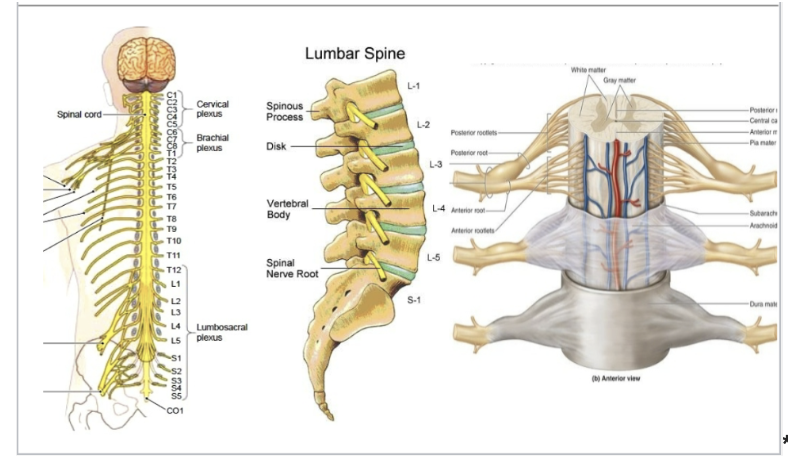
Spinal Nerves
Any given spinal nerve contains both afferent and efferent neurons
Just like the cranial nerves, spinal nerves can carry both sensory and motor neurons
Both Cranial nerves and spinal nerves are peripheral nerves!
The nerves exit the spinal cord in the intervertebral spaces
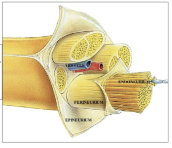
Nerve vs Neuron
A nerve is a bundle of neurons
Coverings of a Neuron
Epineurium
Perineurium
Endoneurium
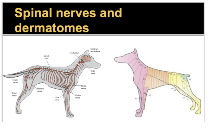
Dermatomes
Each spinal nerve innervates a slice of the body corresponding to where it comes off = dermatome
Each slice of the body is innervated by a single spinal nerve = its dermatome
“ a sensory unit of the skin that is innervated by a single spinal nerve”
Useful diagnostically: if a patient can't feel a pinpoint at a given body site, you can map what nerve is injured and where the injury is
Afferent neurons carry action potentials from —- to blank ——
Carry action potentials from the periphery to the CNS (brain and spinal cord)
What is in the dorsal horn of the grey matter?
Cell bodies of the interneurons and afferent neurons are synapsing on them
What is in the ventral horn of grey matter
Cell bodies of efferent motor neurons going to skeletal muscle
What is in the lateral horn of grey matter
Cell bodies of autonomic efferent neurons (parasympathetic and sympathetic) going to smooth muscles and glands
Thalamus
Serves as “relay station” and synaptic integrating center for sensory input
Helps direct attention to stimuli of interest
Reinforces voluntary motor actions initiated by motor cortex
Capable of crude awareness of sensations but cannot distinguish their location or intensity: relaying the stimuli coming up the spinal cord to the appropriate higher centers is how the specificity of experience occurs