Joint Types, Ligaments & Range of Motion by Region
 Call Kai
 Learn
Learn Practice Test
 Spaced Repetition
 Match
Match Flashcards
 Knowt Play
1/60

Earn XP
Description and Tags
Musculoskeletal Test 1 - Back, Spine & Abdominal Wall
Name | Mastery | Learn | Test | Matching | Spaced | Call with Kai | Chat |
|---|
No analytics yet
Send a link to your students to track their progress
61 Terms
How many joints are in between each vertebra and what do they do?
Two joints between every pair of vertebrae, one disc, and a set of ligaments running the length of the column:
those are what turn 33 separate bones into a structure that moves in a controlled way
Two Kinds of Joint in One Column
Between the bodies
Between the arches
Between the bodies
Secondary cartilaginous joints — symphyses.
The intervertebral disc is the joint.
C2 down to the lumbosacral junction.
Built for load transfer with a little movement at each level.
Between the arches
Synovial joints — the zygapophysial, or facet, joints, between the articular processes.
Also C2 to S1.
Built for guided movement; they decide which direction a region can travel.
The exception:
the craniovertebral joints at the top have no disc at all
— none between the occiput and C1, or between C1 and C2.
Zygapophysial (Facet) Joints → What kind of joint
Synovial.
The articular surfaces are covered in hyaline cartilage, and each joint is enclosed by a capsule containing synovial fluid
Zygapophysial (Facet) Joints → What movement they allow
Gliding between the articular processes.
A single level gives very little; thirty-odd levels together give the column its range
Zygapophysial (Facet) Joints - Why they matter clinically
They are true synovial joints, so they are subject to the same
degenerative changes as any other — which is half of the cervical
spondylosis story from Presentation 2
What Determines Range of Motion (5)
1) Intervertebral disc
Its composition and its height. A taller, well-hydrated disc allows more movement.
2) Facet joint morphology
The plane the articular surfaces sit in — the previous slide.
3) Resistance from muscles & ligaments
Passive tension in the soft tissue crossing the joint.
4) Whether the vertebra carries a rib
Costal articulations tie the thoracic vertebrae down.
5) Surrounding soft tissue
Bulk of tissue around the column limits travel at the extremes
The Intervertebral Disc (3)
Anulus fibrosus
Nucleus pulposus
Where and how numbered
Anulus fibrosus
Concentric fibrocartilaginous rings
forming the outer wall.
Nucleus pulposus
Gelatinous, high water content,
sitting slightly posterior to centre.
Its shape and position shift with
movement
Where and how numbered
C2 to the lumbosacral junction.
Each disc is numbered for the
vertebral body immediately above
it.
The Intervertebral Disc (Nucleus)
Off-centre nucleus
Ligaments: Three Groups
Occiput to cervical vertebrae
Between vertebrae
Spine & sacrum to ilium
The continuity rule:
Occiput to cervical vertebrae
.
Several group 1 ligaments simply change name as they run down the column —
the anterior atlanto-occipital membrane becomes the anterior longitudinal ligament,
the tectorial membrane becomes the posterior longitudinal ligament
the ligamentum nuchae becomes the supraspinous ligament.
Craniocervical Ligaments: External (3)
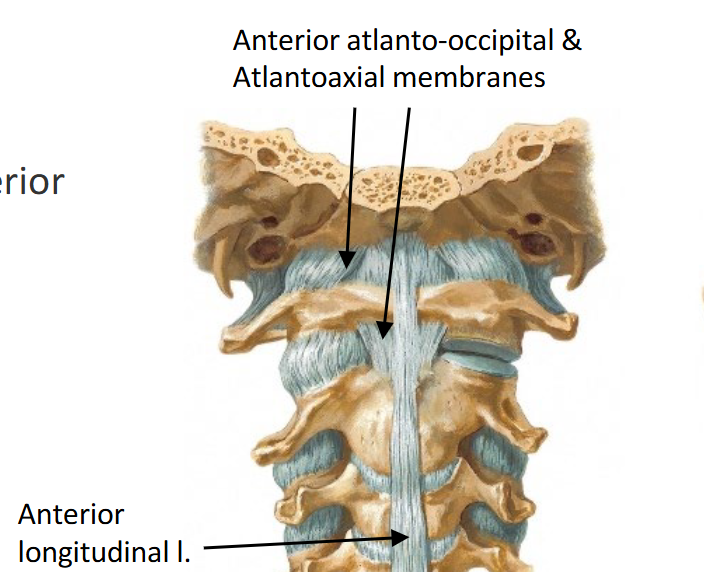
Anterior
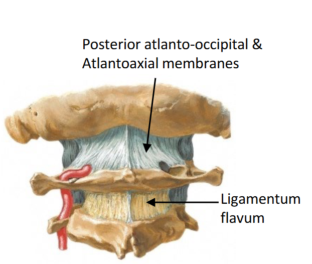
Posterior
Ligamentum nuchae
Anterior (Craniocervical Ligaments: External)
Anterior atlanto-occipital and atlantoaxial membranes
→ continue below as the anterior longitudinal ligament.
Posterior (Craniocervical Ligaments: External)
Posterior atlanto-occipital and atlantoaxial
membranes — continue below as the
ligamentum flavum
Ligamentum nuchae (Craniocervical Ligaments: External)
A midline sheet from the external occipital
protuberance to the C7 spinous process;
continues as the supraspinous ligament

Craniocervical Ligaments: Internal (3)
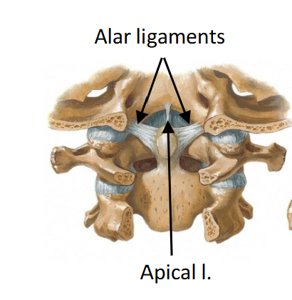
Cruciform ligament
Alar ligaments
Apical ligament
Cruciform ligament
The transverse ligament plus superior and inferior longitudinal bands — together they form a cross.
Transverse band/ligament holds the dens against the anterior arch of C1

Alar ligaments
Paired; dens to the margins of the foramen magnum.
Limit rotation of the head.
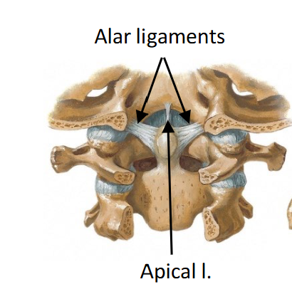
Apical ligament
Tip of the dens to the anterior margin of the foramen magnum
Tectorial membrane
Covering the dens and its ligaments from behind.
It is the upward continuation of the posterior longitudinal ligament
the third of the three continuities

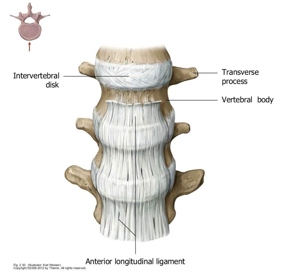
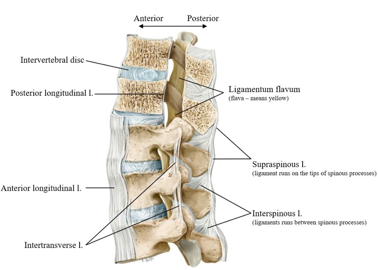
Anterior Longitudinal Ligament (Where it runs)
Down the anterior and lateral surfaces of the vertebral bodies, from the occiput and C2 all the way to the sacrum.
It is firmly attached to the anterior aspect of each intervertebral disc
Anterior Longitudinal Ligament (What it is like)
Broad and strong — the most substantial ligament of the
column, and it gets wider as it descends.
Anterior Longitudinal Ligament (What it limits)
Extension — and it is the only ligament of the vertebral
column that does. Every other one on the next few slides
limits flexion.
Anterior Longitudinal Ligament
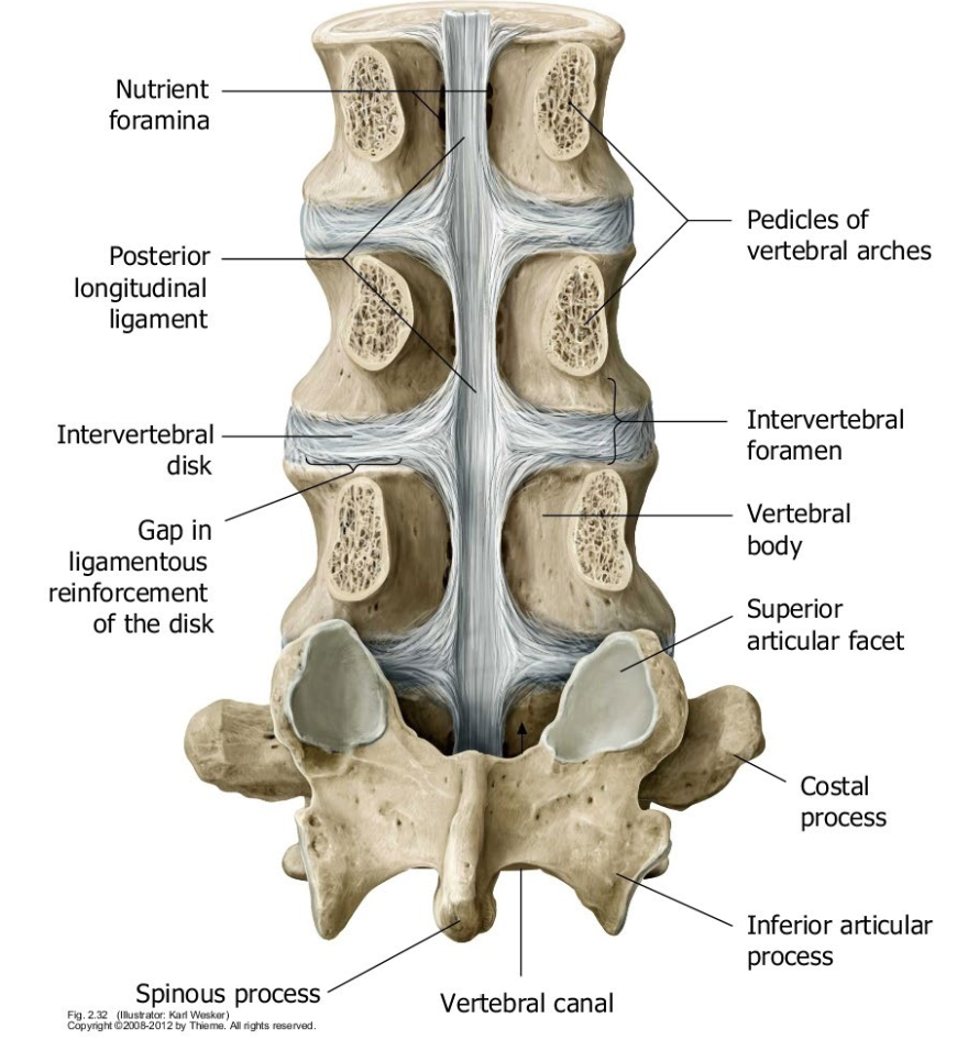
Posterior Longitudinal Ligament (Where it runs)
Down the posterior surface of the vertebral bodies, C2 to the
sacrum — which places it inside the vertebral canal, immediately
in front of the spinal cord.
Continuous above with the tectorial membrane
Posterior Longitudinal Ligament (What it is like)
Narrower and weaker than the anterior longitudinal ligament. It
has lateral expansions where it crosses each disc, so it is broad at
the disc and narrow over the body — giving it a scalloped outline
Posterior Longitudinal Ligament (What it limits)
Flexion — but weakly, given its size.
That scalloped shape is the point. Where the ligament narrows, the disc behind it is least supported.
Posterior Longitudinal Ligament
Ligamentum Flavum Diagram
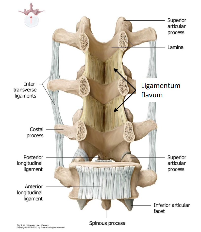
Ligamentum Flavum (Where it runs)
Paired, connecting the lamina of one vertebra to the lamina of
the next. Together the pairs form the posterior wall of the
vertebral canal
Ligamentum Flavum (What it is made of)
High elastin content — which is where the yellow colour and the
name come from. Flavum means yellow.
Ligamentum Flavum (What it limits)
Flexion — it stretches as the laminae separate, then recoils to
resting length without buckling into folds.
Ligamentum Flavum & Sustained Flexion (Why elasticity, and not just strength)
A ligament forming the back wall of the vertebral canal has a design
problem. It must lengthen when you flex, and it must shorten again
when you straighten — but if it shortened by buckling, it would fold
inward, into the canal, toward the cord.
Ligamentum Flavum & Sustained Flexion (Elastin solves it)
Elastic recoil returns the ligament to resting length under its own tension, so it stays taut rather than folding.
The canal keeps its dimensions through the whole range of flexion and extension.
Interspinous, Intertransverse & Supraspinous Location
Interspinous
Between adjacent spinous processes, in the
sagittal plane
Limits: Flexion
Intertransverse
Between adjacent transverse processes, in the frontal plane
Limits: Lateral flexion
Supraspinous
Along the posterior tips of the spinous processes; continuation of the ligamentum nuchae
The supraspinous ligament becomes very thin in the lumbar region — the part of the column where flexion loads are highest
Limits: Flexion
Ligaments: What Each One Limits (6)
Anterior longitudinal
Posterior longitudinal
Ligamentum flavum
Interspinous
Supraspinous
Intertransverse
Anterior longitudinal
Where it runs: Anterior & lateral surfaces of the bodies;
attached to each disc
Extent: Occiput / C2 → sacrum
Limits: EXTENSION — the only one
Posterior longitudinal
Where it runs: Posterior surface of the bodies, inside the
vertebral canal
Extent: C2 → sacrum
Limits: Flexion (weakly)
Ligamentum flavum
Where it runs: Lamina to lamina; posterior wall of the vertebral canal
Extent: C2 → S1
Limits: Flexion
Interspinous
Where it runs: Between spinous processes, sagittal plane
Extent: Whole column
Limits: Flexion
Supraspinous
Where it runs: Along the tips of the spinous processes
Extent: C7 → sacrum
Limits: Flexion
Intertransverse
Where it runs: Between transverse processes, frontal plane
Extent: Whole column
Limits: Lateral flexion
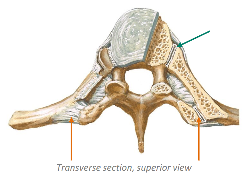
Costovertebral joint
Head of the rib to the superior costal facet of its own vertebra and the inferior costal facet of the vertebra above — with the intervertebral disc between them.
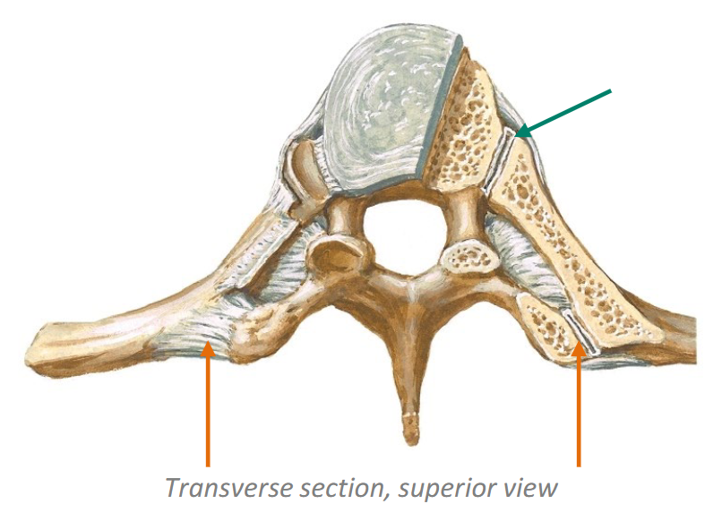
Costotransverse joint
Tubercle of the rib to the transverse process of the vertebra of the same number.
Spine to Pelvis: Lumbosacral & Sacroiliac Ligaments (5)
1) Iliolumbar
2) Sacroiliac (ant., post., interosseous)
3) Sacrospinous
4) Sacrotuberous
5) Sacrococcygeal
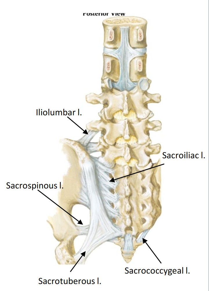
Iliolumbar
Transverse processes of L4 and L5 to the iliac crest
Sacroiliac (ant., post., interosseous)
Sacrum to ilium across the sacroiliac joint
Sacrospinous
Sacrum to the ischial spine
Sacrotuberous
Sacrum to the ischial tuberosity
Sacrococcygeal
Sacrum to the coccyx
Why Discs Herniate Posterolaterally (3)
1) The nucleus already sits posteriorly
2) The anulus is thinnest behind
3) The PLL reinforces the centre, not the corner
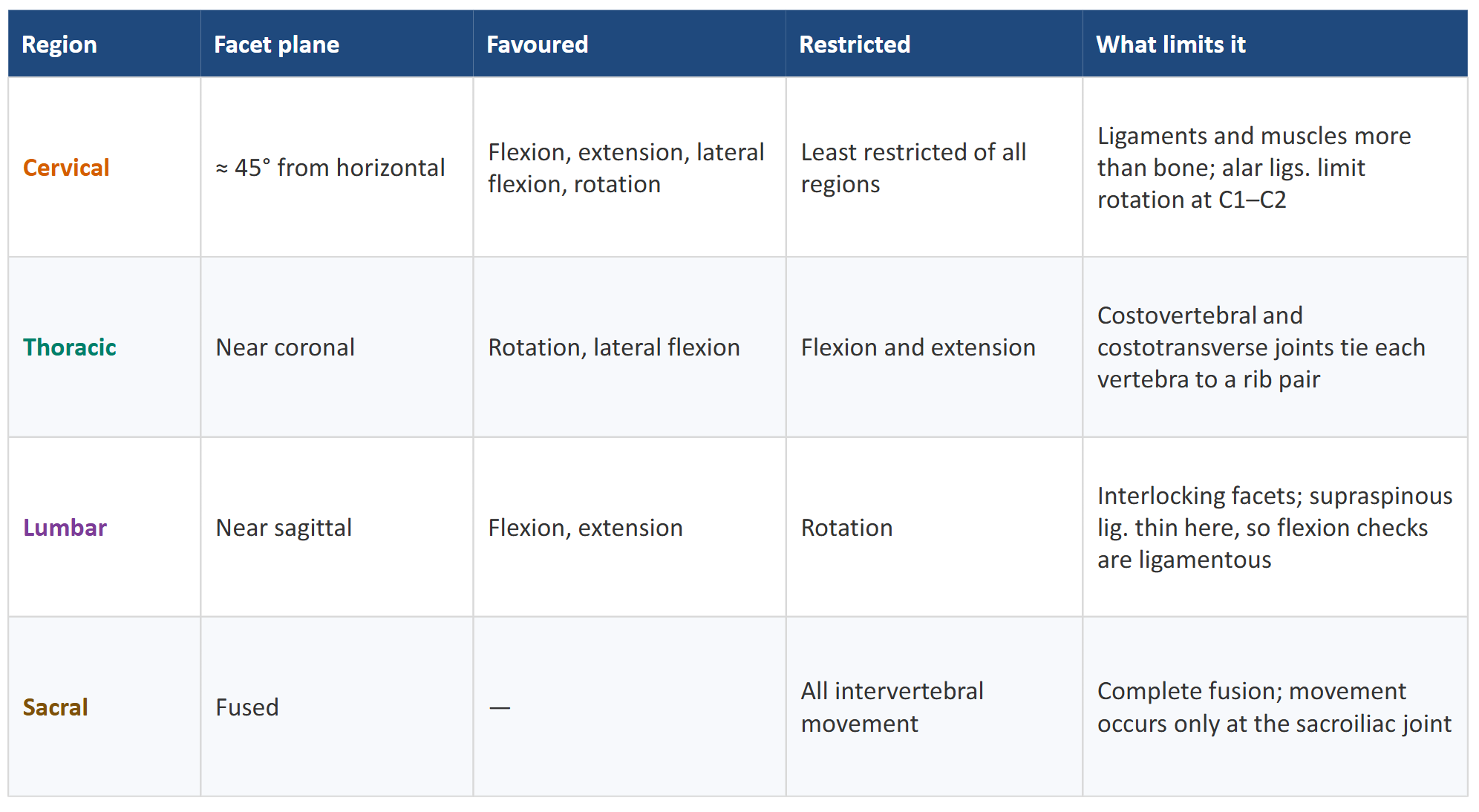
Range of Motion by Region