Micro HSB - Laboratory Major Practicals
1/68
There's no tags or description
Looks like no tags are added yet.
Name | Mastery | Learn | Test | Matching | Spaced |
|---|
No study sessions yet.
69 Terms
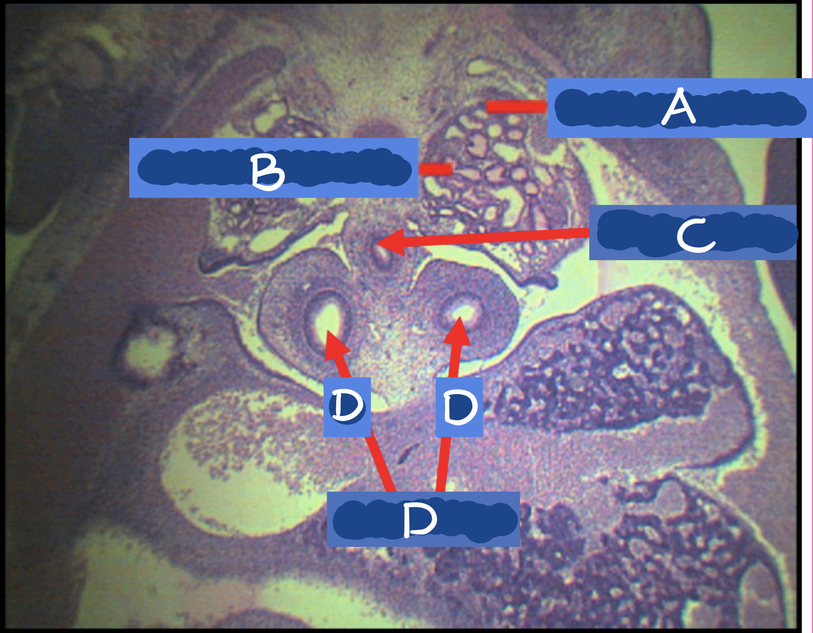
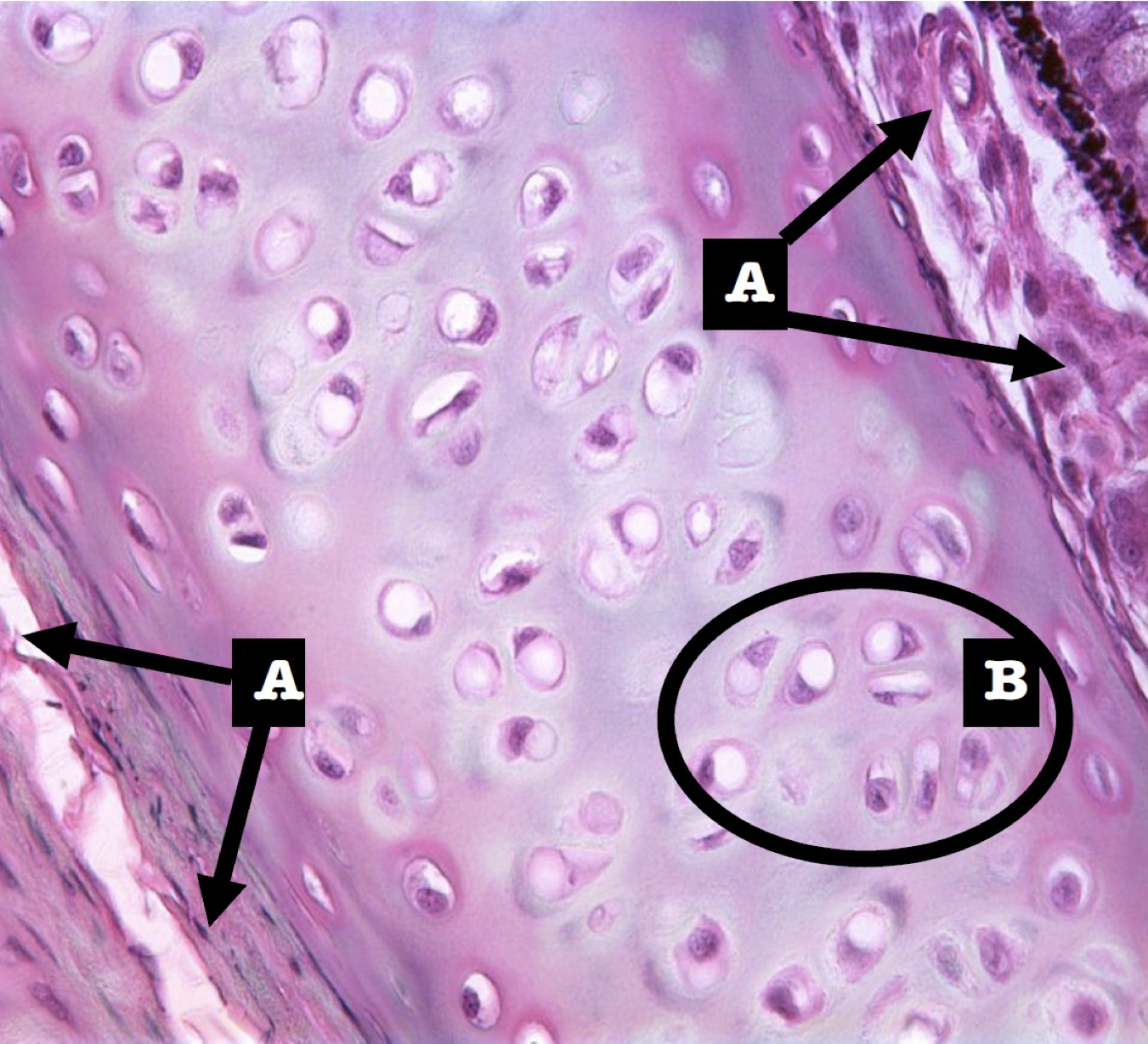
A - pharyngeal arch
B - arytenoid cartilage
C - pharynx
identify the structures

Endoderm
Germ layer derivative for the lining epithelium, larynx, pharynx, bronchi, and lungs
Splanchnic mesoderm
Germ layer derivative of rest of the larynx, connective tissue, muscle, and cartilage
4th and 5th laryngeal arches; cartilages, epiglottis
Respiratory diverticulum came from _________ and will become ________ and _________ area
Lateral plate mesoderm
Germ layer derivative of arytenoid cartilage
4th and 6th pharyngeal arch
Arytenoid cartilage is a derivative of what pharyngeal arch
4th and 5th week
Pharyngeal arch appears in _____ and _____ week of development
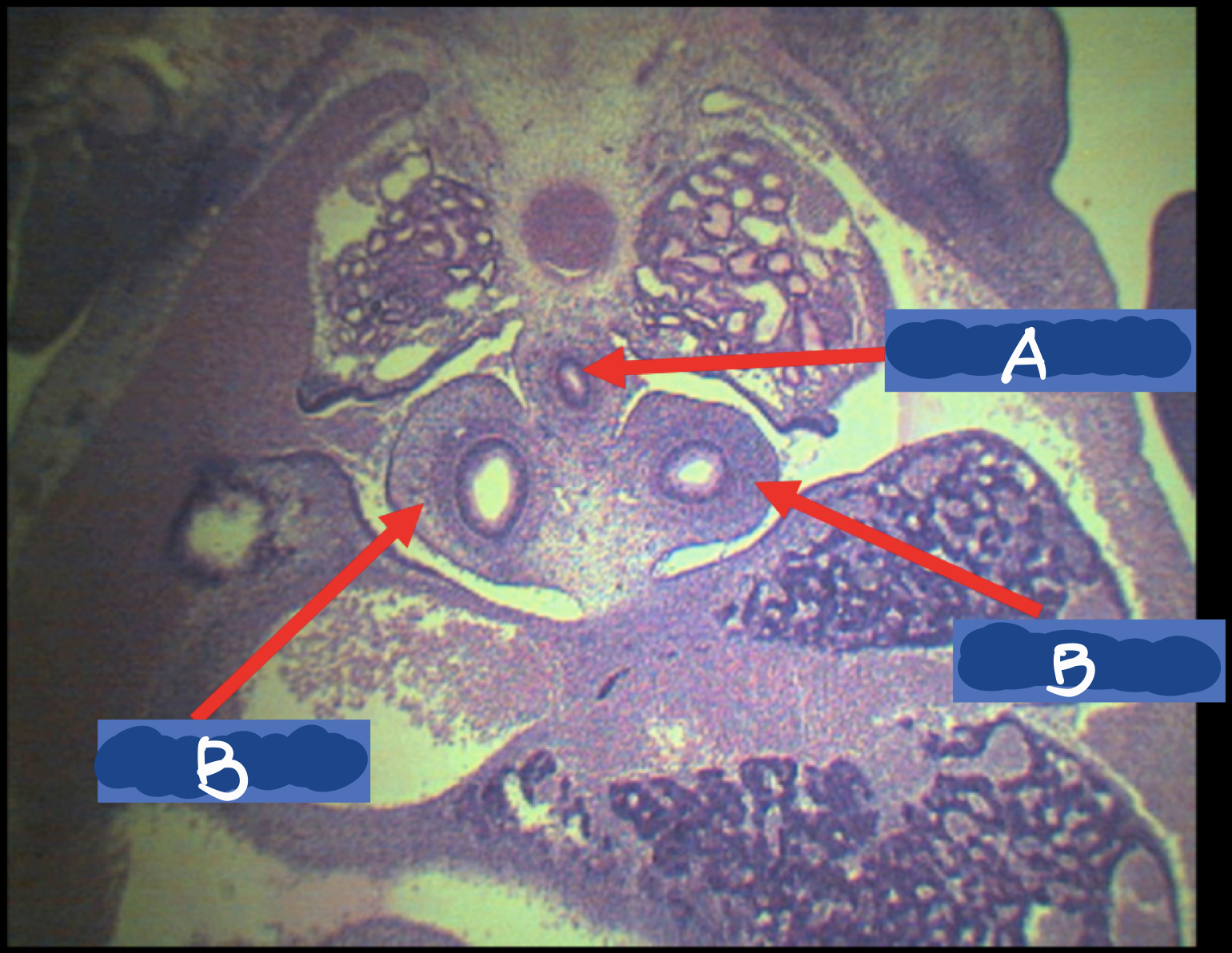
Laryngeal inlet
Identify the structure
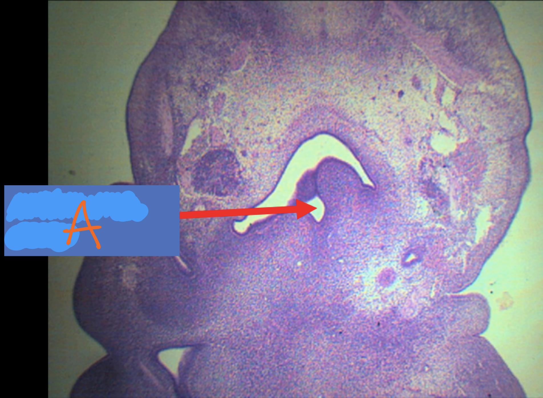
Laryngeal inlet
A sagittal slit that turns into T-shaped opening and is found immediately behind the third median swelling formed by the posterior part of the 4th arch (epiglottis)
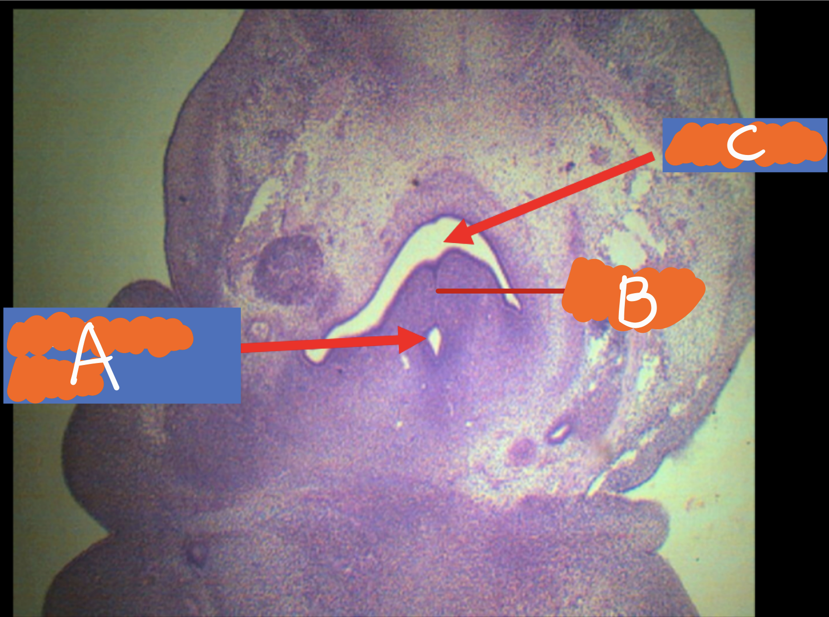
A - laryngeal inlet
B - larynx
C - pharynx
Identify the structures
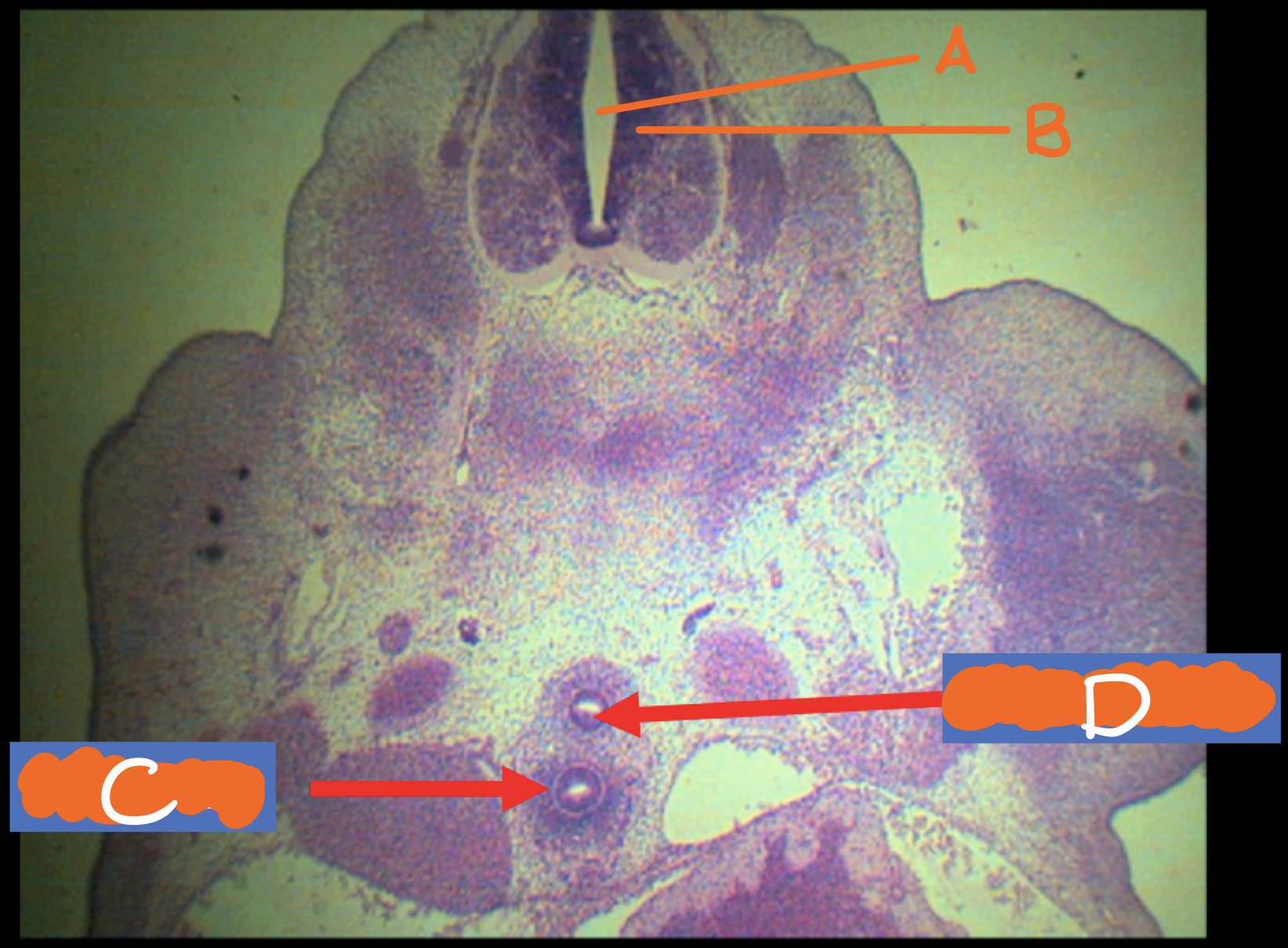
A - neural tube
B - notochord
C - trachea
D - esophagus
Identify the structures
A - mesonephric tubules
B - mesonephric glomeruli
C - esophagus
D - R & L bronchus
Identify the structures
A - esophagus
B - lung bud
Identify the structures
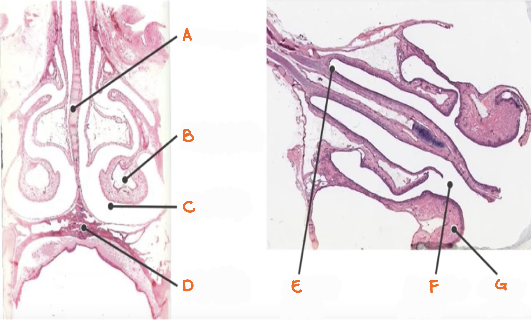
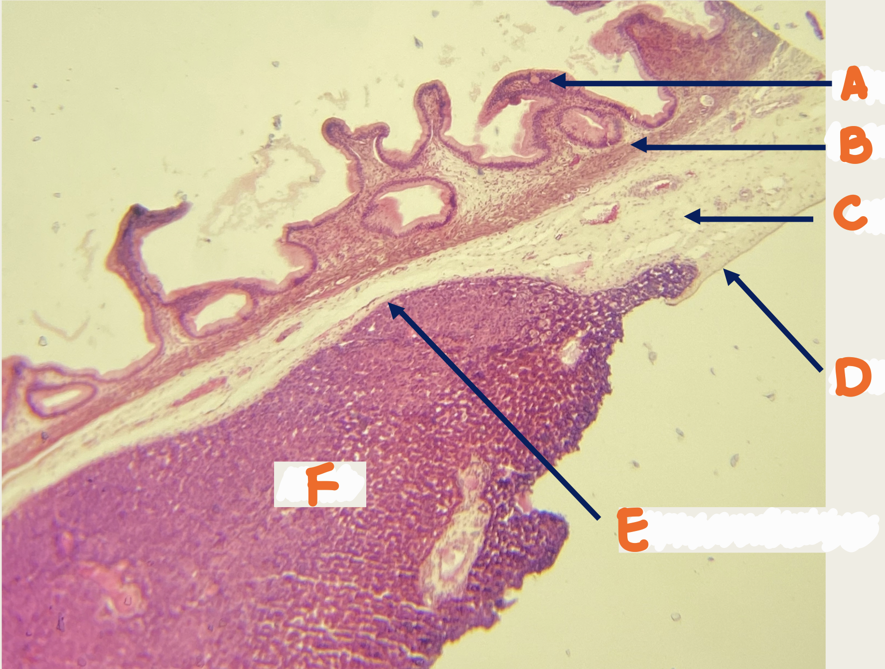
A - nasal septum
B - inferior concha
C - nasal cavity
D - hard palate
E - olfactory region
F - respiratory region
G - inferior concha
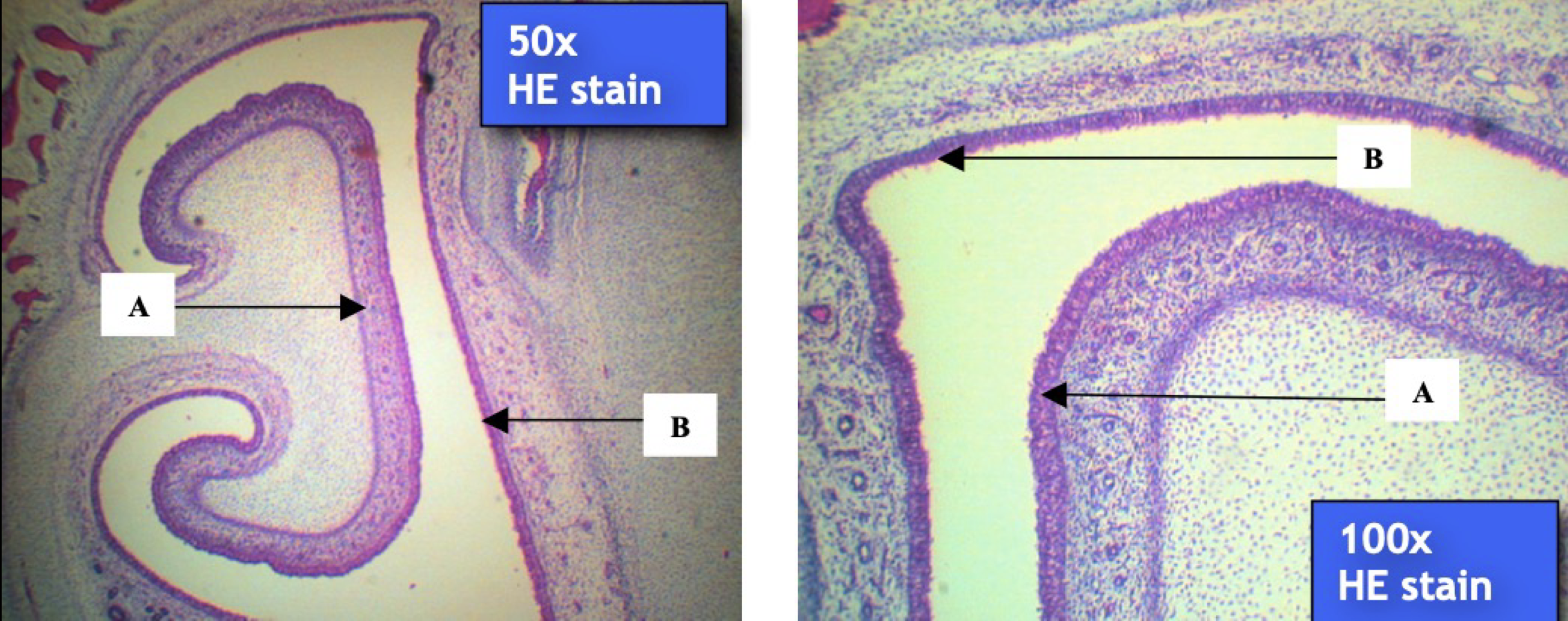
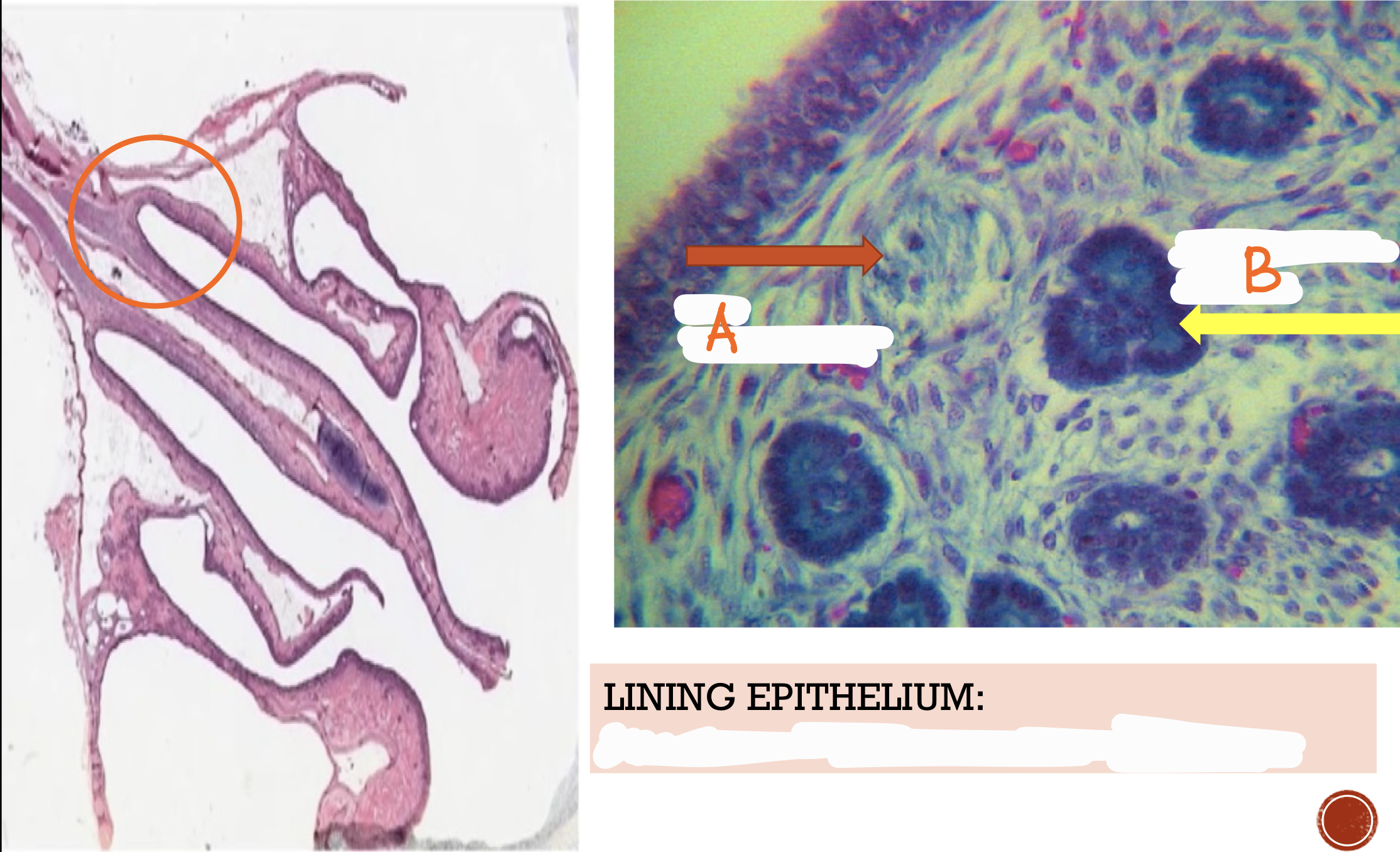
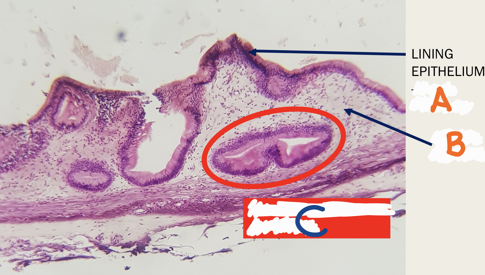
Identify the structure
A - olfactory epithelium
B - respiratory epithelium
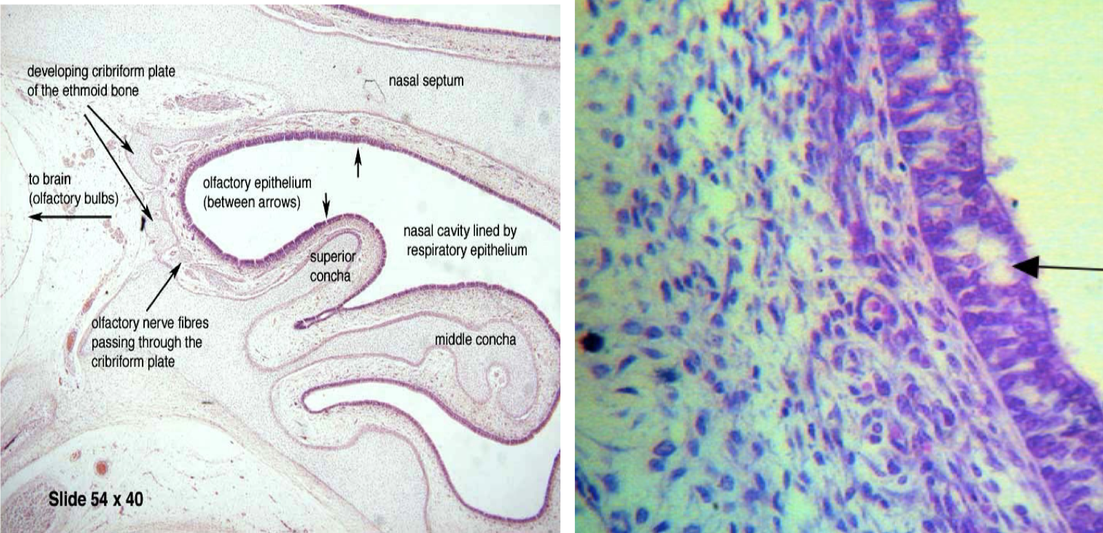
Identify the structures
Encircled - olfactory region
A - fila olfactoria (bundles of axons that make up the olfactory nerve)
B - bowman’s glands
Lining epithelium - pseudostratified ciliated columnar epithelium
Identify the structures and lining epithelium
Superior conchae
This structure covers the _________ conchae

Respiratory region
LE: pseudostratified ciliated columnar epithelium with goblet cells
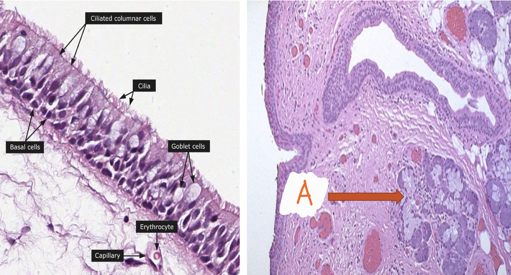
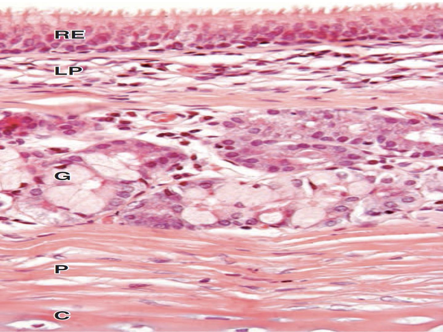
Identify the structure and lining epithelium
Nasopharynx
LE: pseudostratified ciliated columnar epithelium with goblet cells
A: mucus secreting glands
Identify the structure and lining epithelium
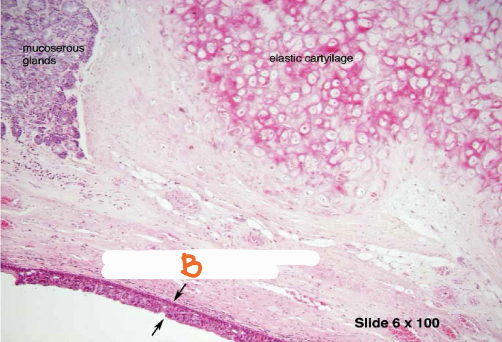
A - pseudostratified ciliated columnar epithelium with goblet cells
B - laryngeal glands
C - laryngeal cartilage
Identify the structures
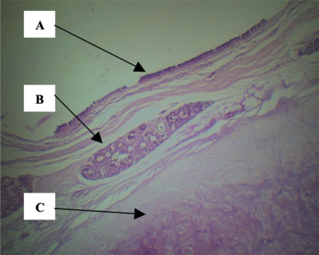
A - Superior epiglottis (lingual portion)
LE: stratified squamous epithelium
Identify the structure and lining epithelium
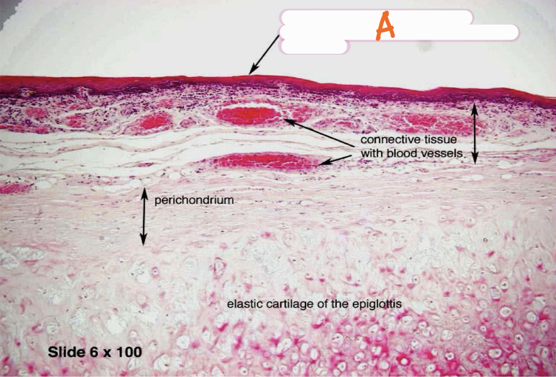
B - Inferior epiglottis (laryngeal portion)
LE: pseudostratified ciliated columnar epithelium with goblet cells
Identify the structure and lining epithelium
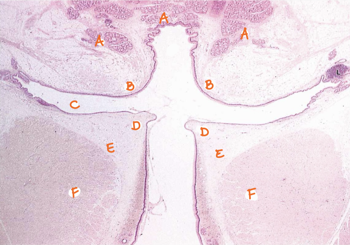
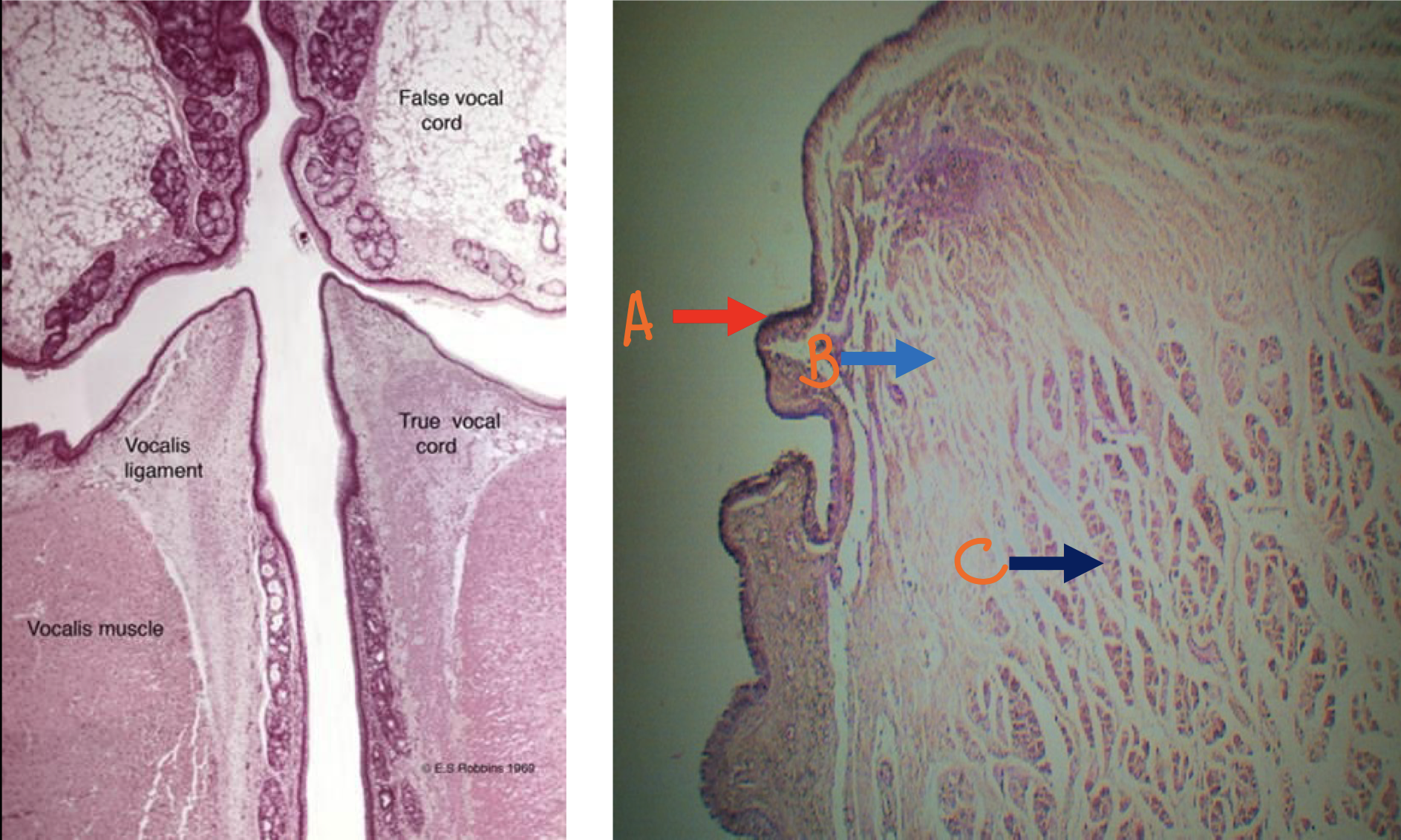
A - seromucous gland
B - vestibular fold
C - ventricle
D - vocal cord
E - vocalis ligament
F - vocalis muscle
Identify the structures
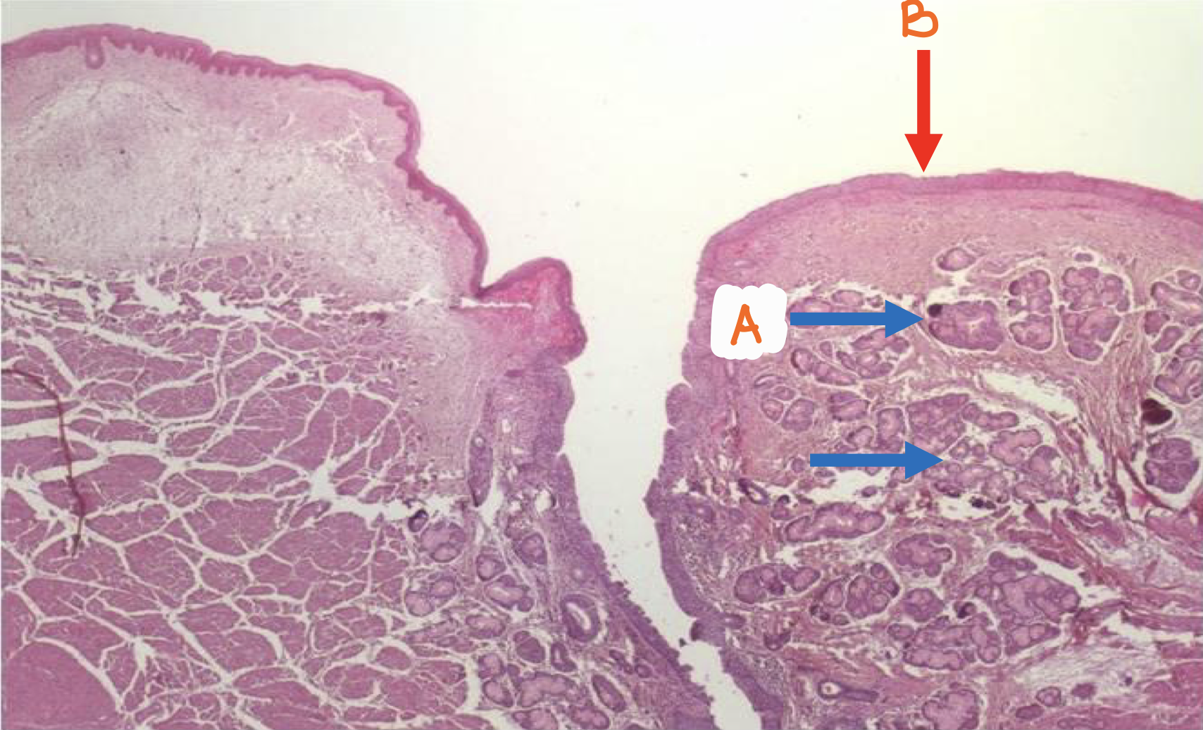
A - seromucous glands
B - false vocal cords / vestibular folds
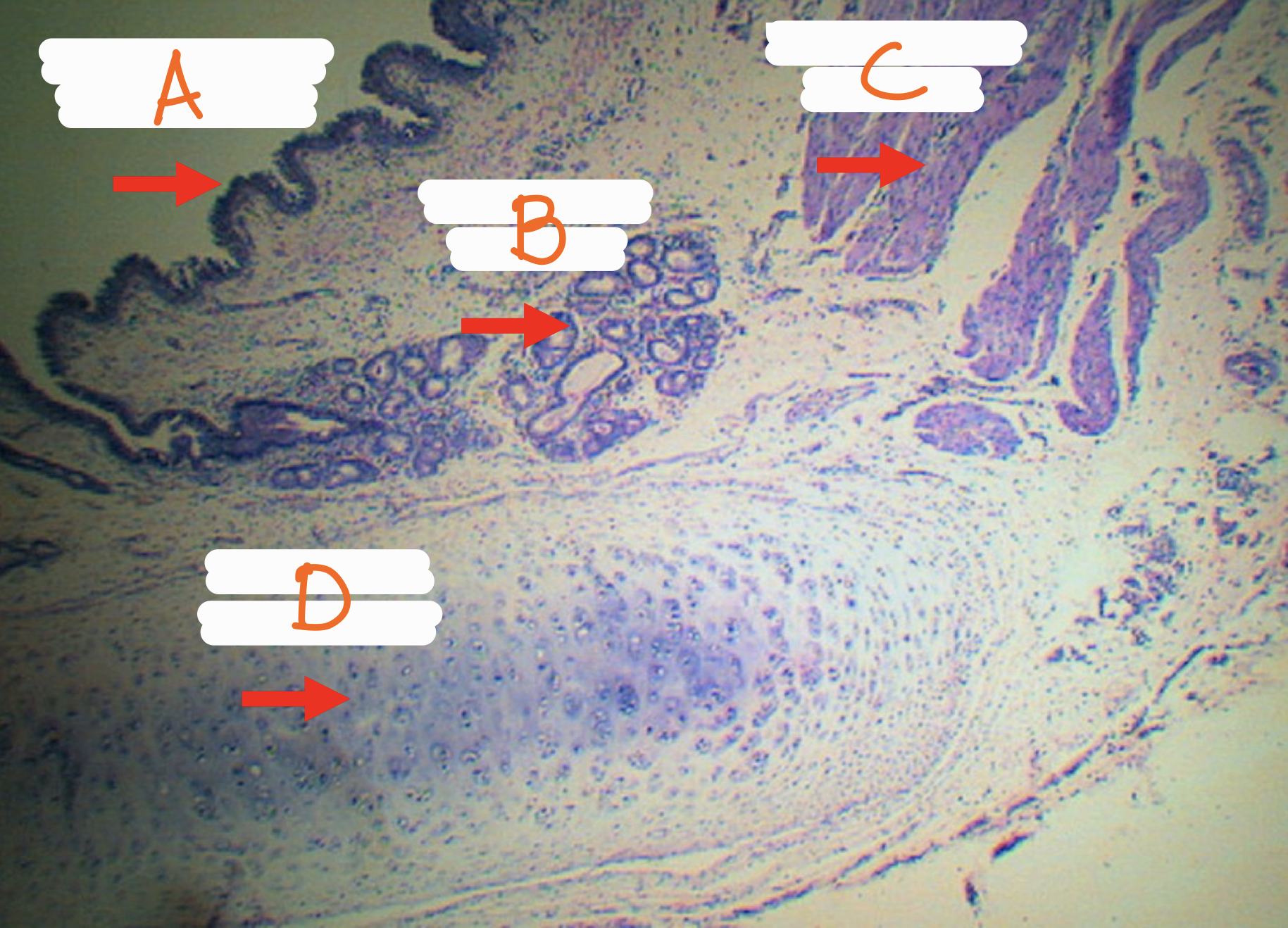
Identify the structure
A - false vocal cords / vestibular folds (pseudostratified ciliated columnar epithelium with goblet cells)
B - true vocal cords / vocal cords (stratified squamous non-keratinized epithelium)
Identify the structure

true vocal cords / vocal cords
A - stratified squamous non-keratinized epithelium
B - elastic fiber bundles
C - vocalis muscle
Identify the structure
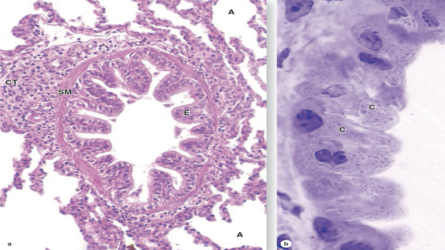
A - pseudostratified ciliated columnar epithelium with goblet cells
B - tracheal muscle
C - trachealis muscle
D - tracheal cartilage
Identify the structures
Trachea
Identify the structure
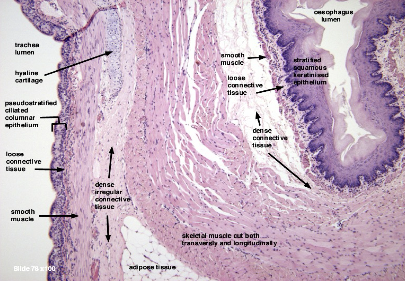
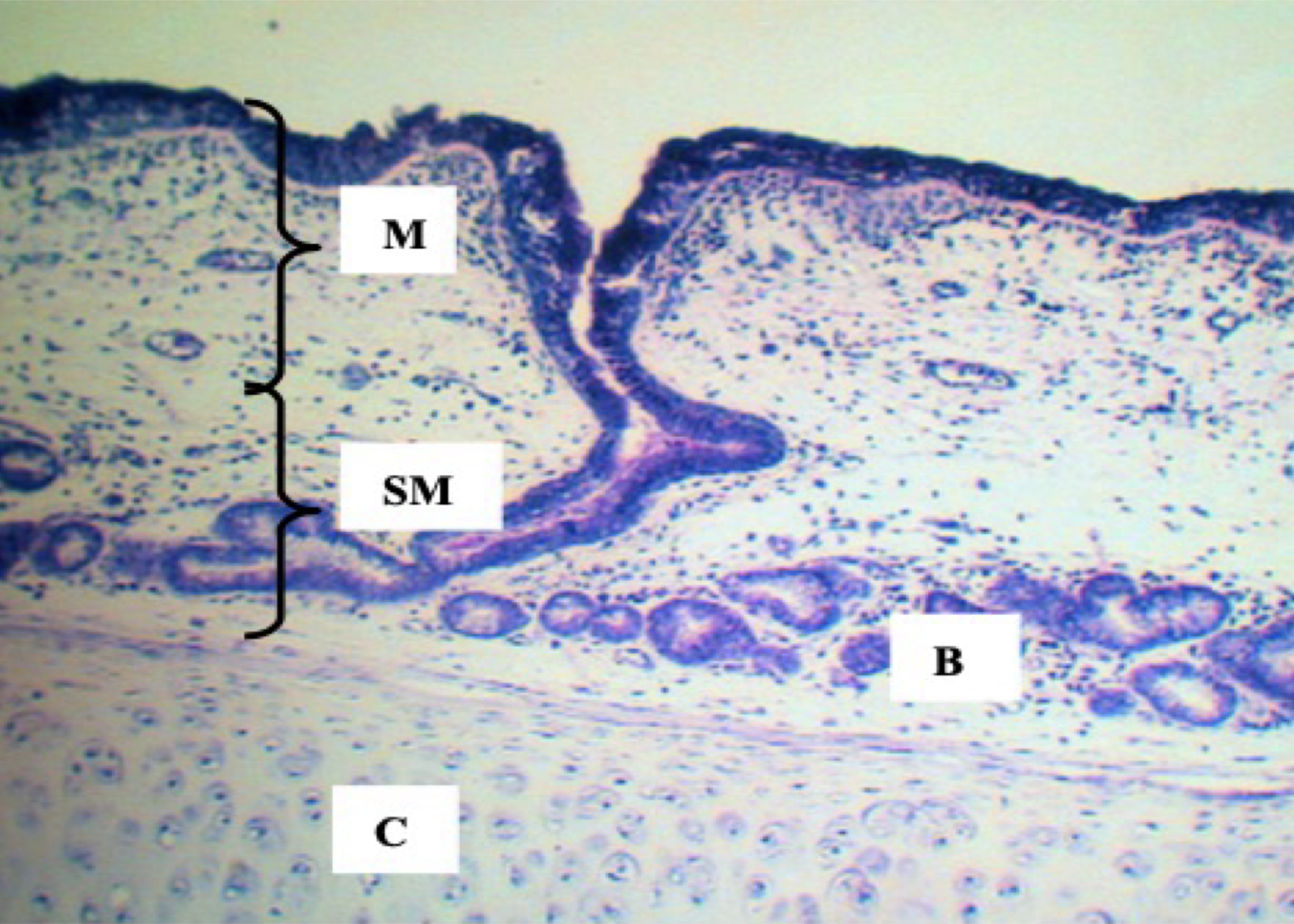
Trachea HPO
M - mucosa
SM - submucosa
B - tracheal glands
C - tracheal cartilage
Identify the structure
Trachea LPO
A - perichondrium (dense irregular CT)
B - chondrocytes
Identify the structure
Trachea
Identify the structure
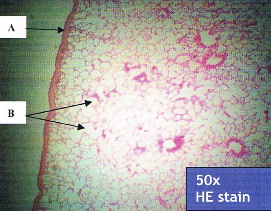
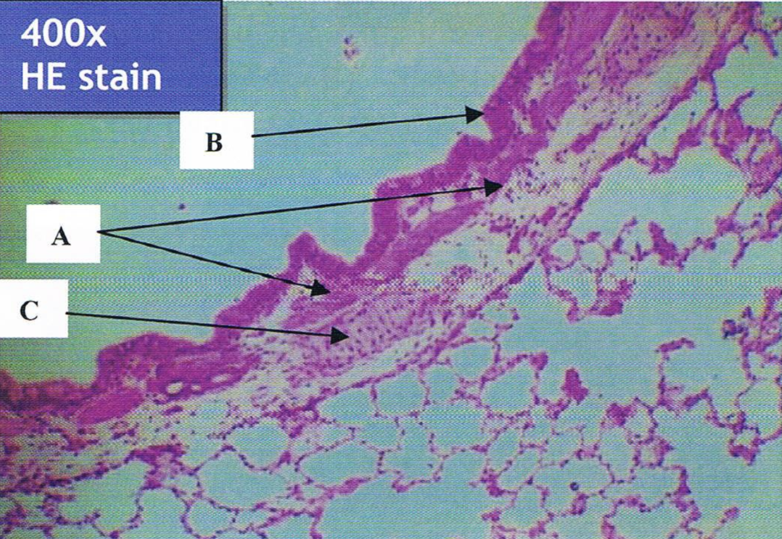
A - visceral pleura (simple squamous epithelium)
B - alveoli
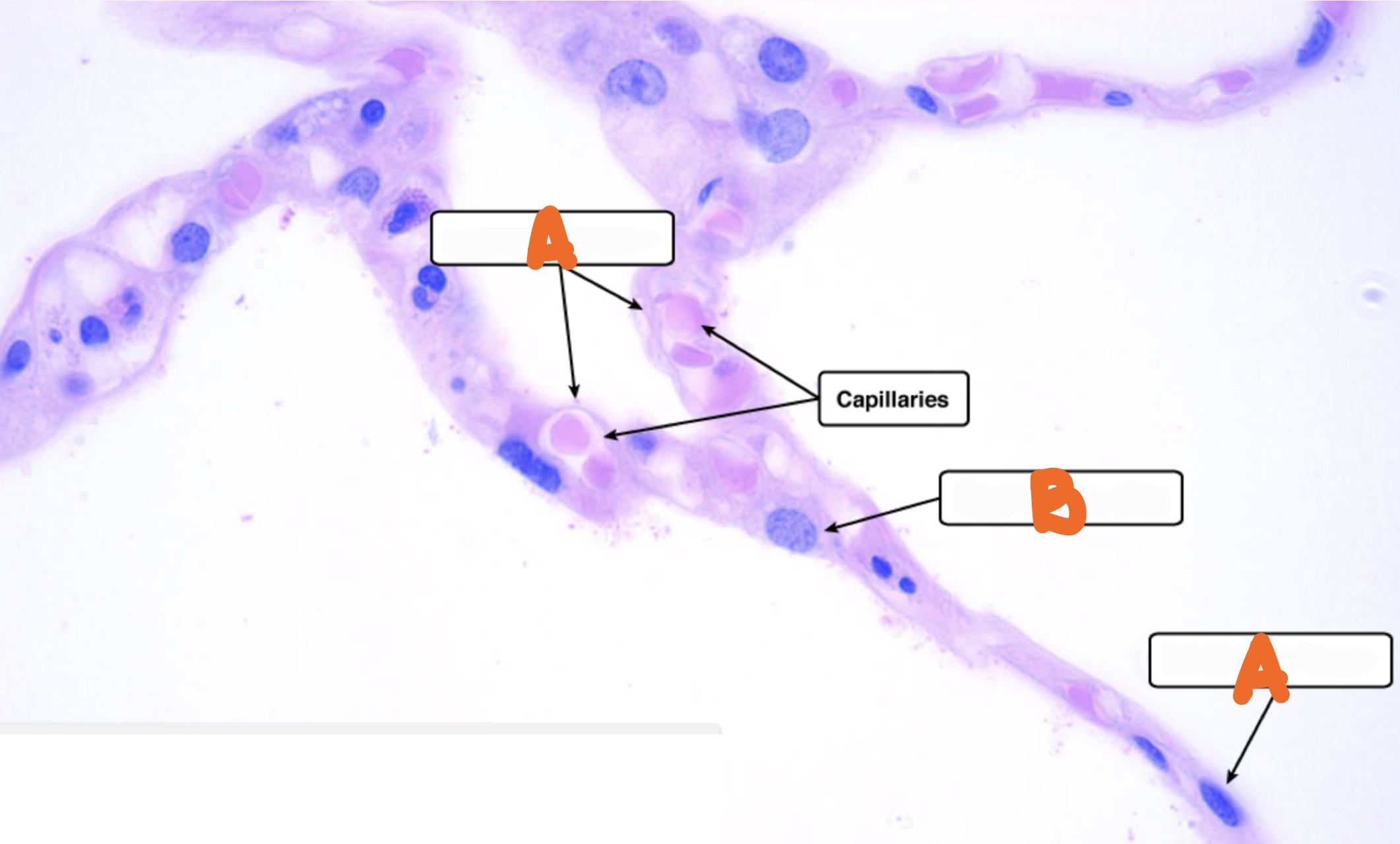
Identify the structure
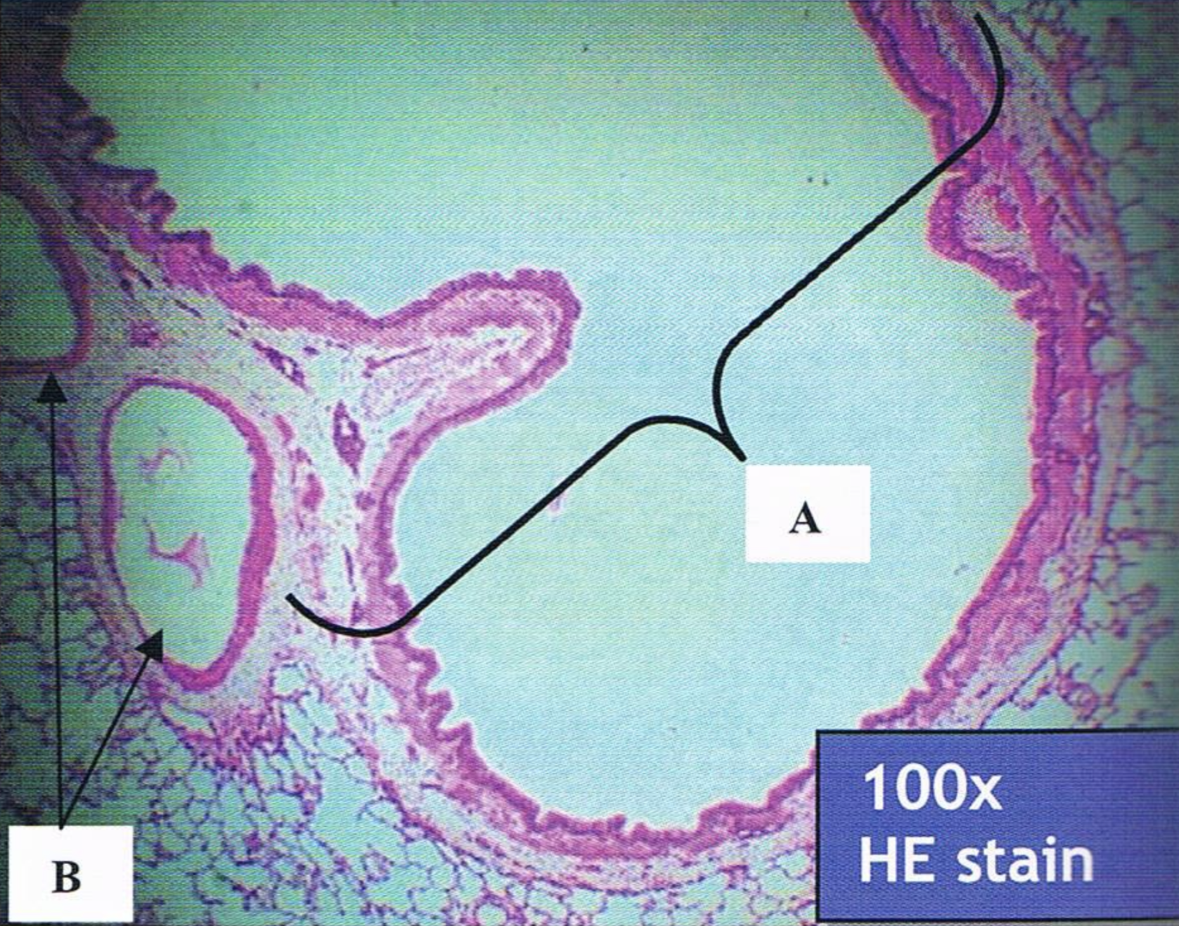
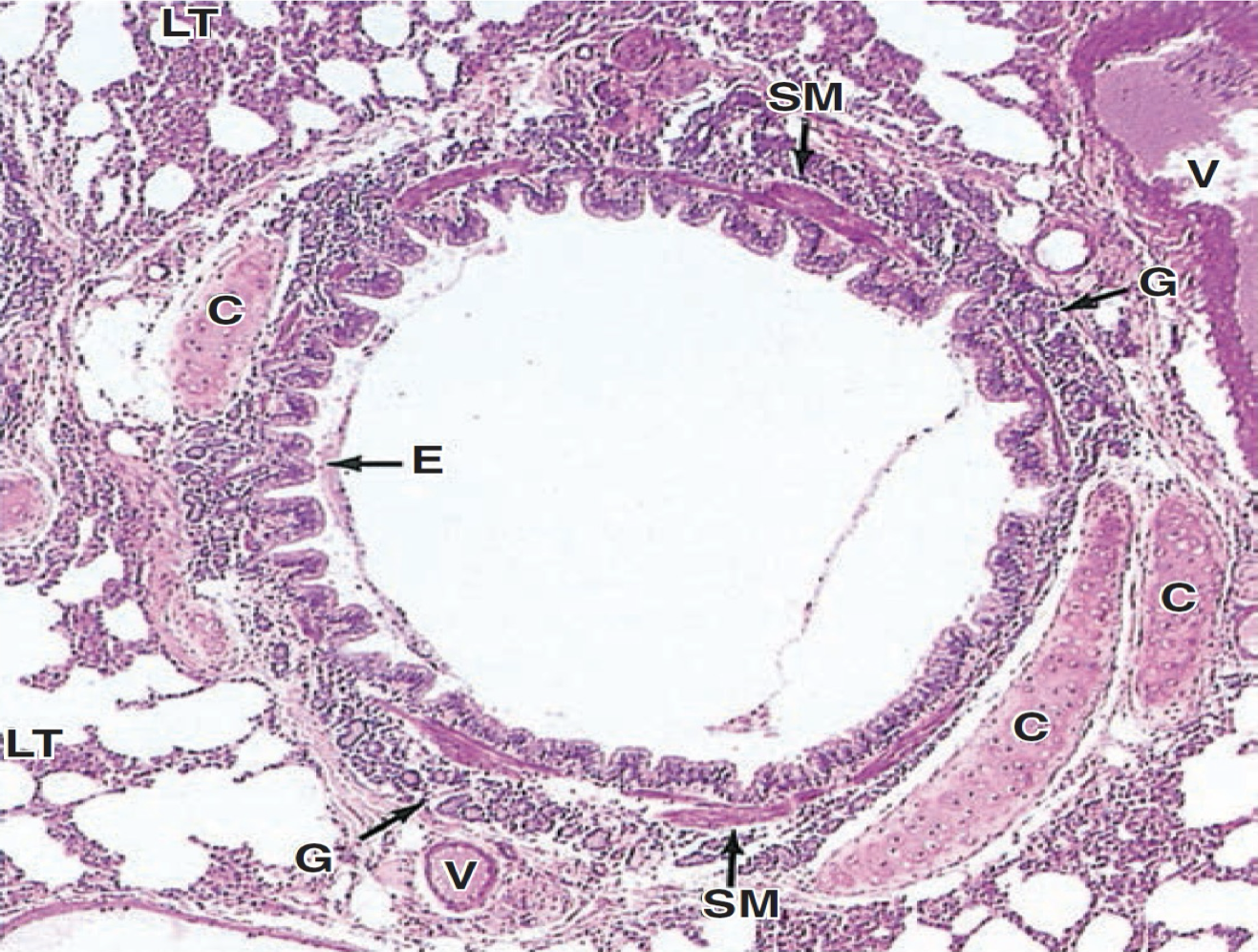
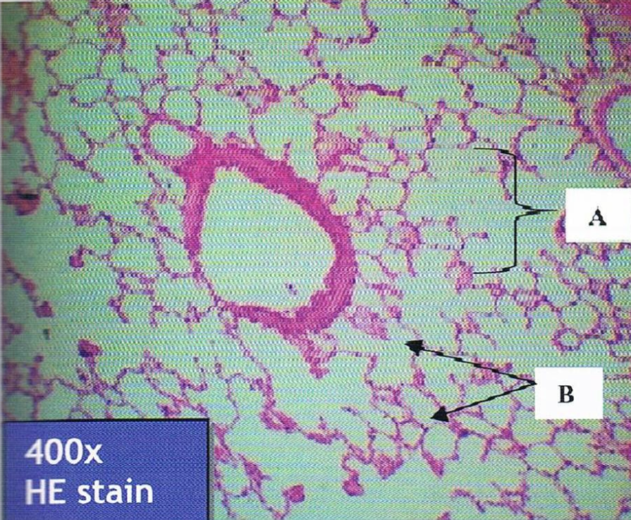
A - intrapulmonary bronchus
B - respiratory vessel
Identify the structure
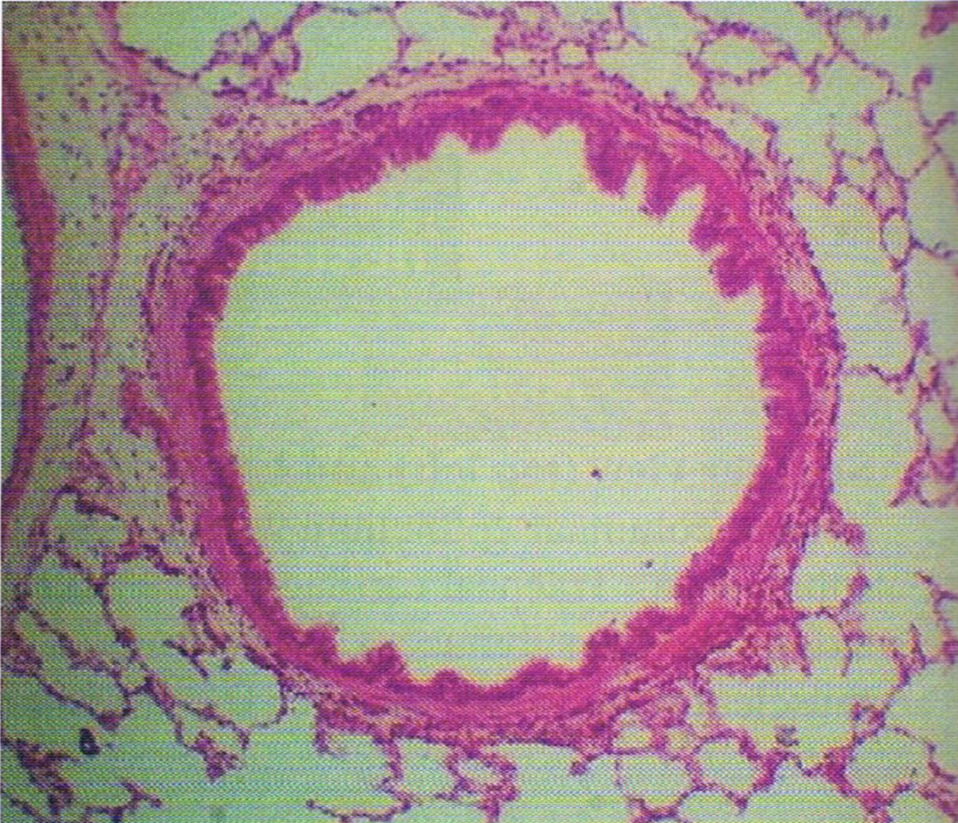
Intrapulmonary bronchus
Identify the structure
Intrapulmonary bronchus LPO
A - smooth muscle
B - pseudostratified ciliated columnar epithelium with goblet cells
C - bronchial cartilage
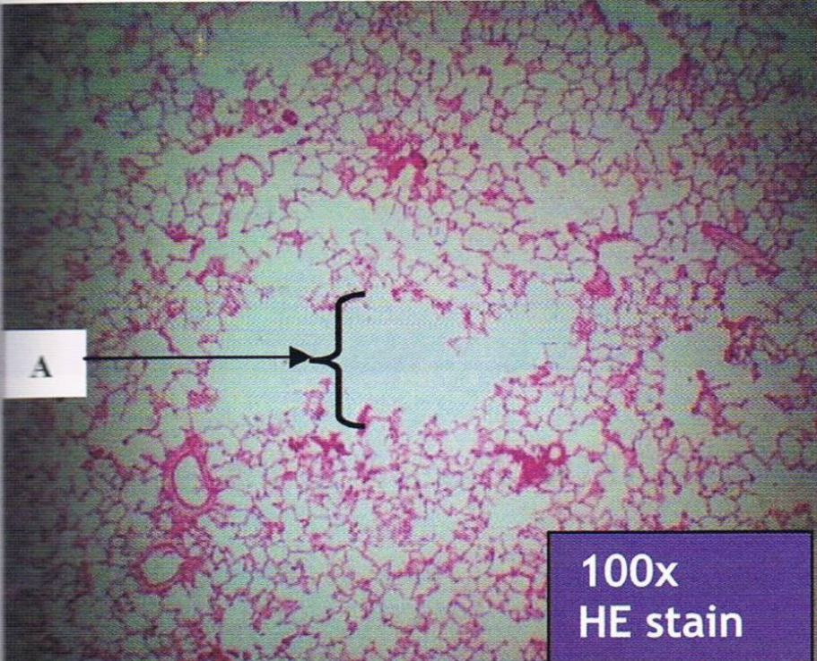
Identify the structure
Bronchioles (no submucosal glands and cartilage)
LE: Simple ciliated columnar and cuboidal epithelium with few goblet cells
Identify the structure and LE
Terminal bronchiole
Identify the structure
Terminal bronchiole
LE: Simple ciliated cuboidal epithelium with club cells
A - alveolar sac
B - alveolar duc
Identify the structure and LE
Respiratory bronchiole
LE: Simple ciliated cuboidal epithelium with club cells and scattered alveoli
Identify the structure and LE
A - alveolar duct (fewer smooth muscle and more surrounded by alveoli)
LE - simple squamous epithelium
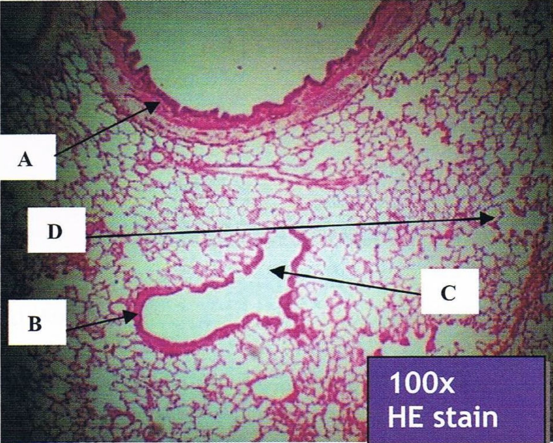
Identify the structure
A - intrapulmonary bronchiole
B - terminal bronchiole
C - respiratory bronchiole
D - alveolar duct
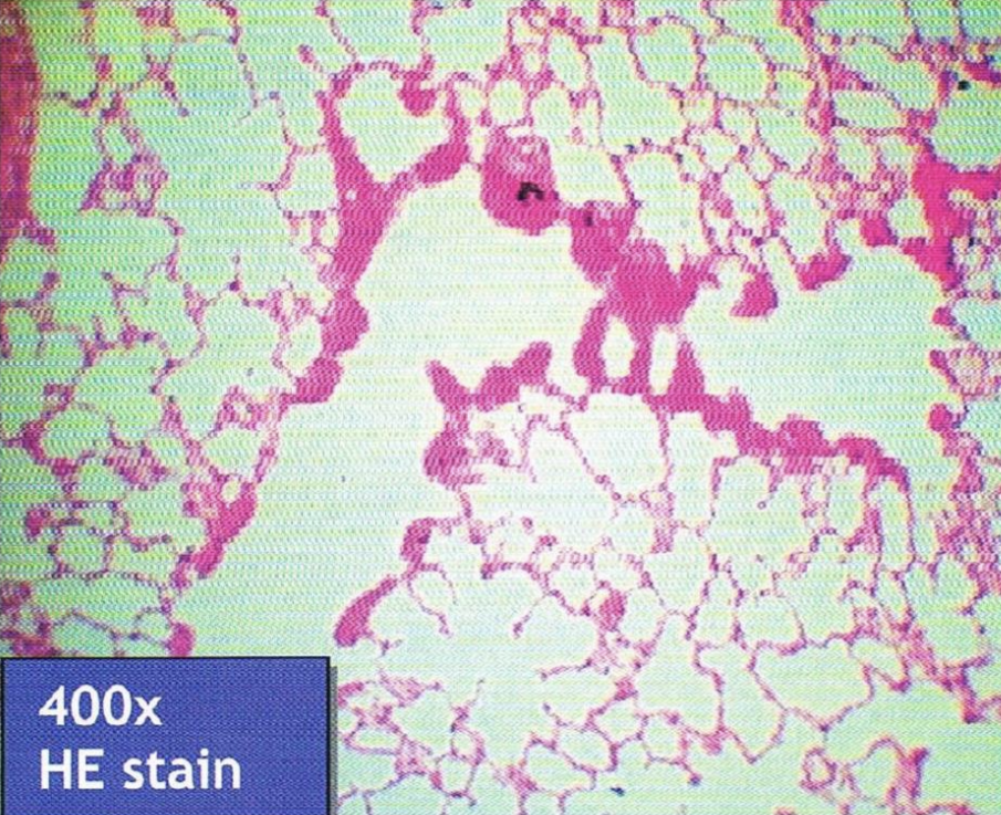
Identify the structure
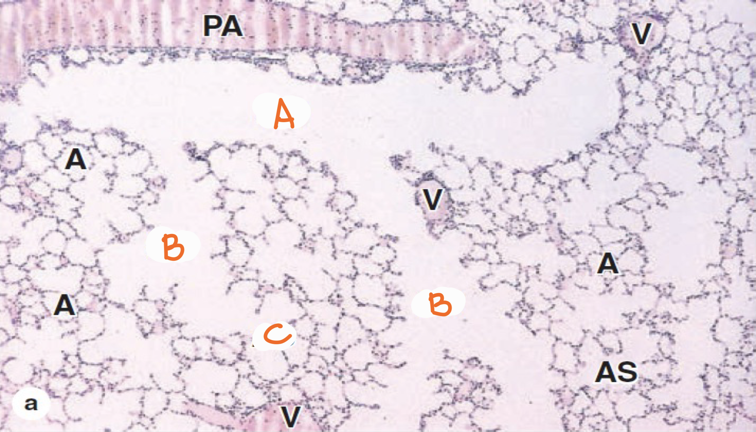
Lungs
A - respiratory bronchioles
B - alveolar ducts
C - alveolar sacs
Identify the structure
A - type 1 pneumocyte; lines 95% of alveolar surface
B - type 2 pneumocyte; secretes pulmonary surfactant
Identify the structure and their respective functions
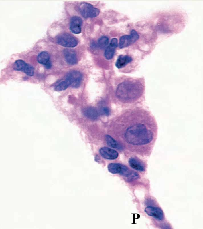
Type II pneumocystis
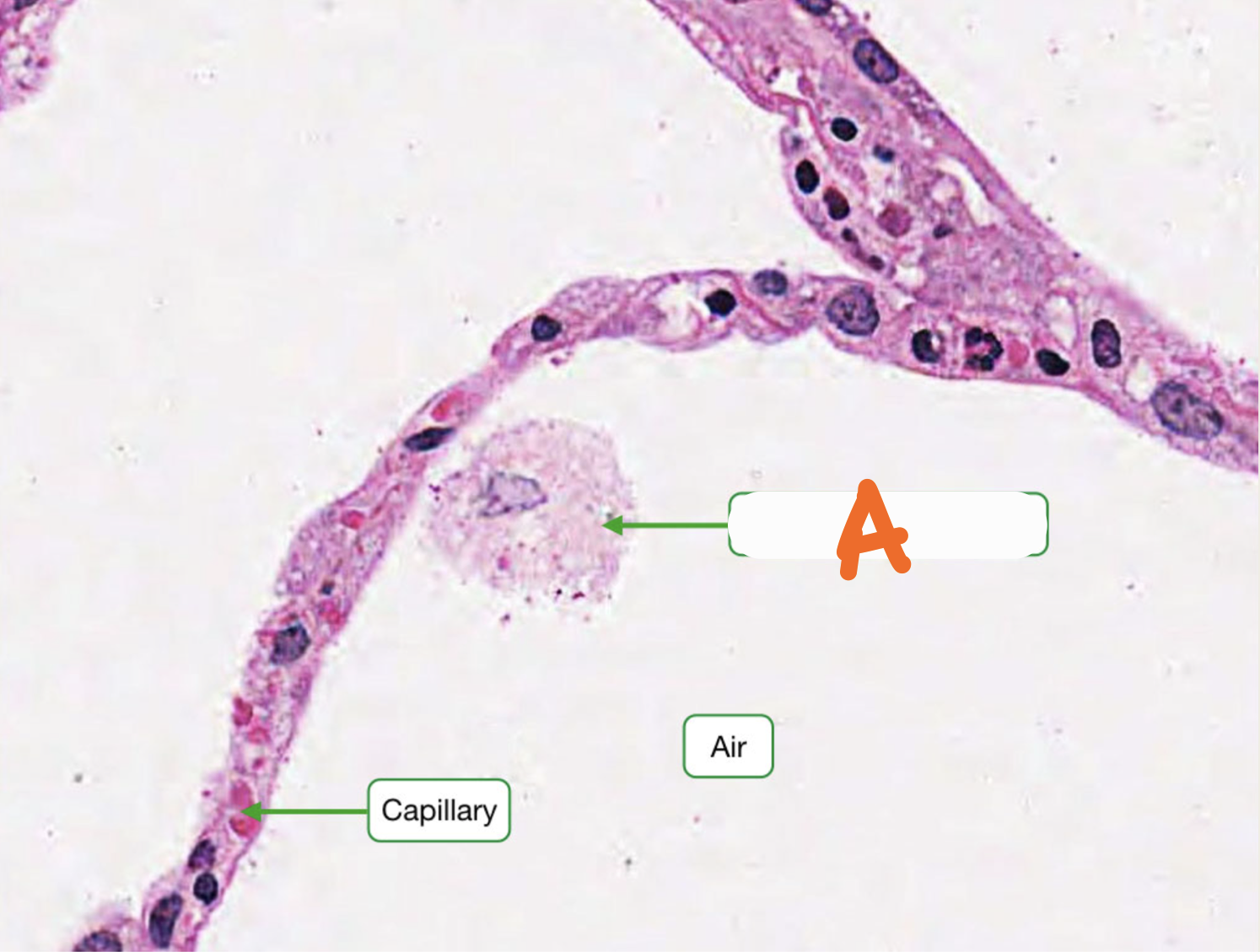
Identify the structure
A - alveolar macrophage
Identify the structure
parotid gland
submandibular gland
sublingual gland
Three types of salivary glands
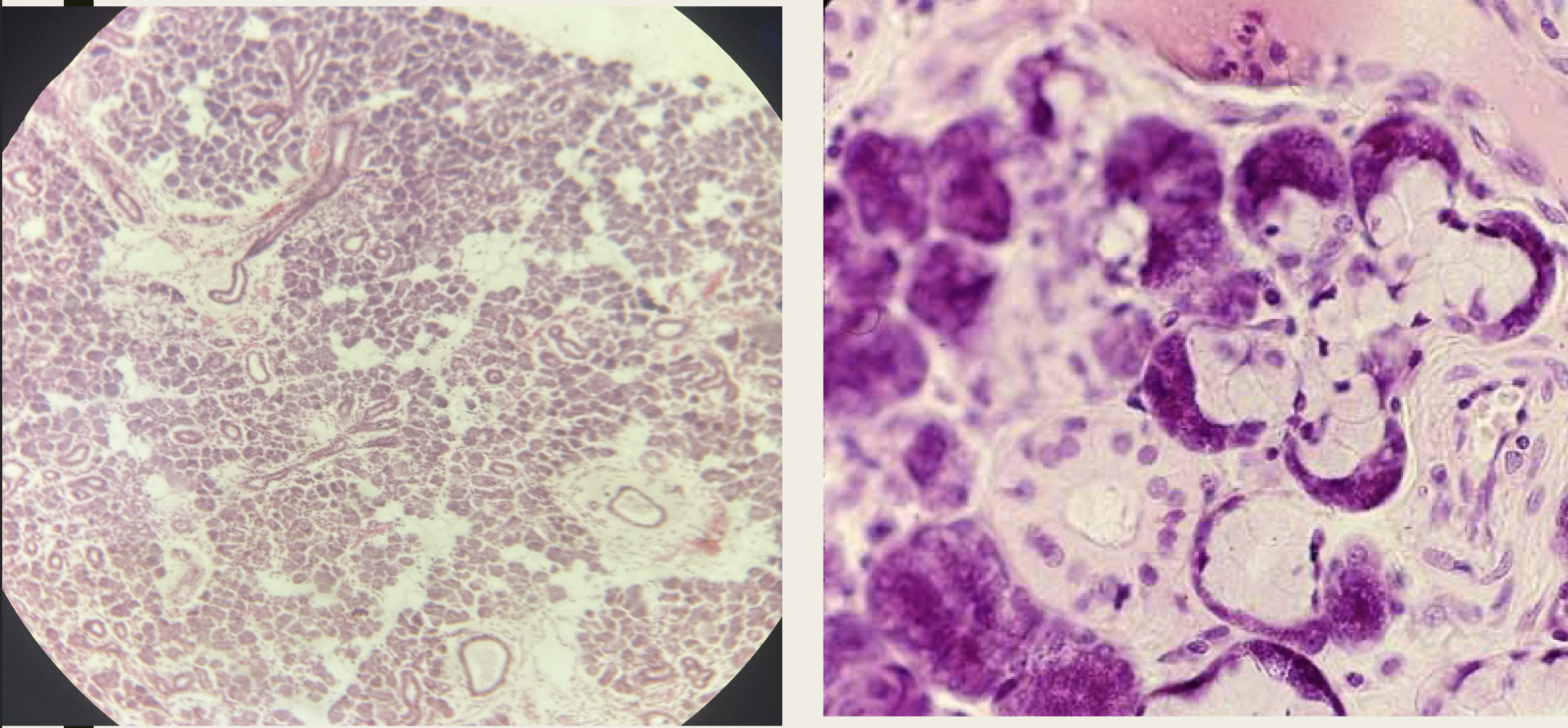
Parotid gland
A: purely serous acinus (branched acinar)
B: adipocytes
Identify the structure (A is type of gland and secretion)
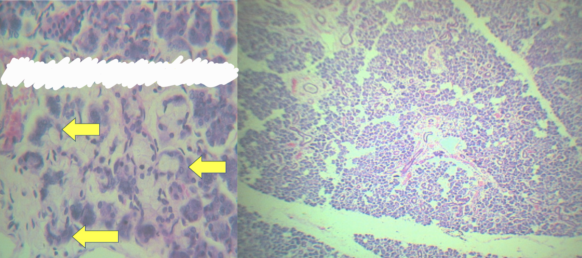
Parotid gland - interlobular duct
Identify the structure
Parotid gland - intercalated duct
Identify the structure
Parotid gland - striated duct
Identify the structure
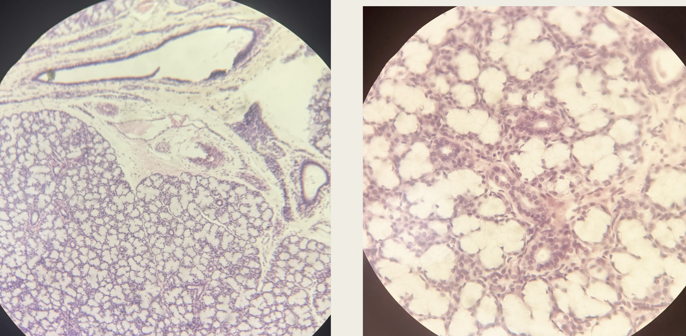
Submandibular/submaxillary gland
branched tubulo acinar gland
mixed predominantly serous
Identify the structure, gland, and secretion)
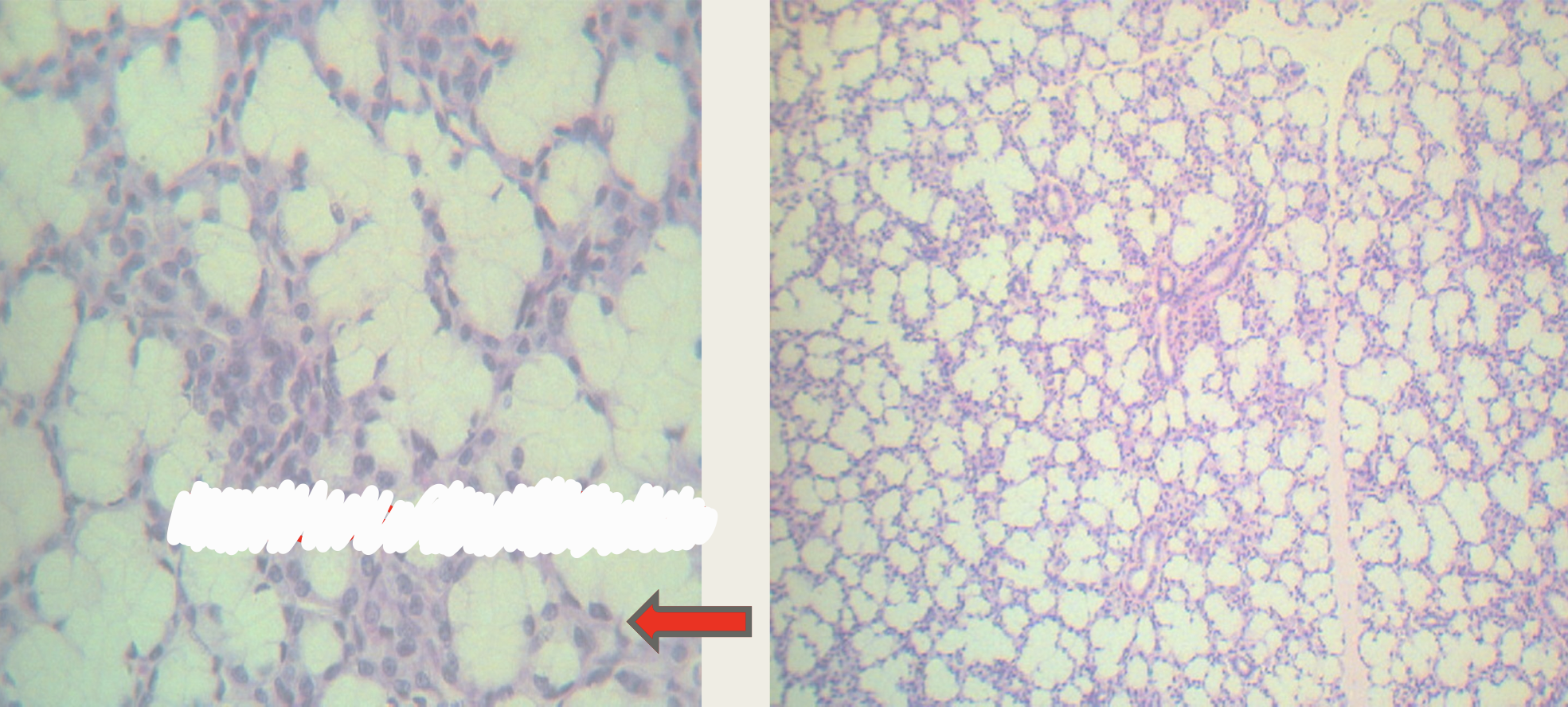
Submaxillary/submandibular gland & serous demilunes (demilunes of Gianuzzi)
Identify the structure
Sublingual gland
branched tubuloacinar gland
mixed predominantly mucous
Identify the structure, gland, and secretion
Sublingual gland and serous demilunes (demilunes of Gianuzzi)
Identify the structure
Sublingual gland
A - mucus acinus
B - serous acinus
C - serous demilunes (demilunes of gianuzzi)
Identify the structure
Liver
A - portal area
B - central vein
Identify the structures
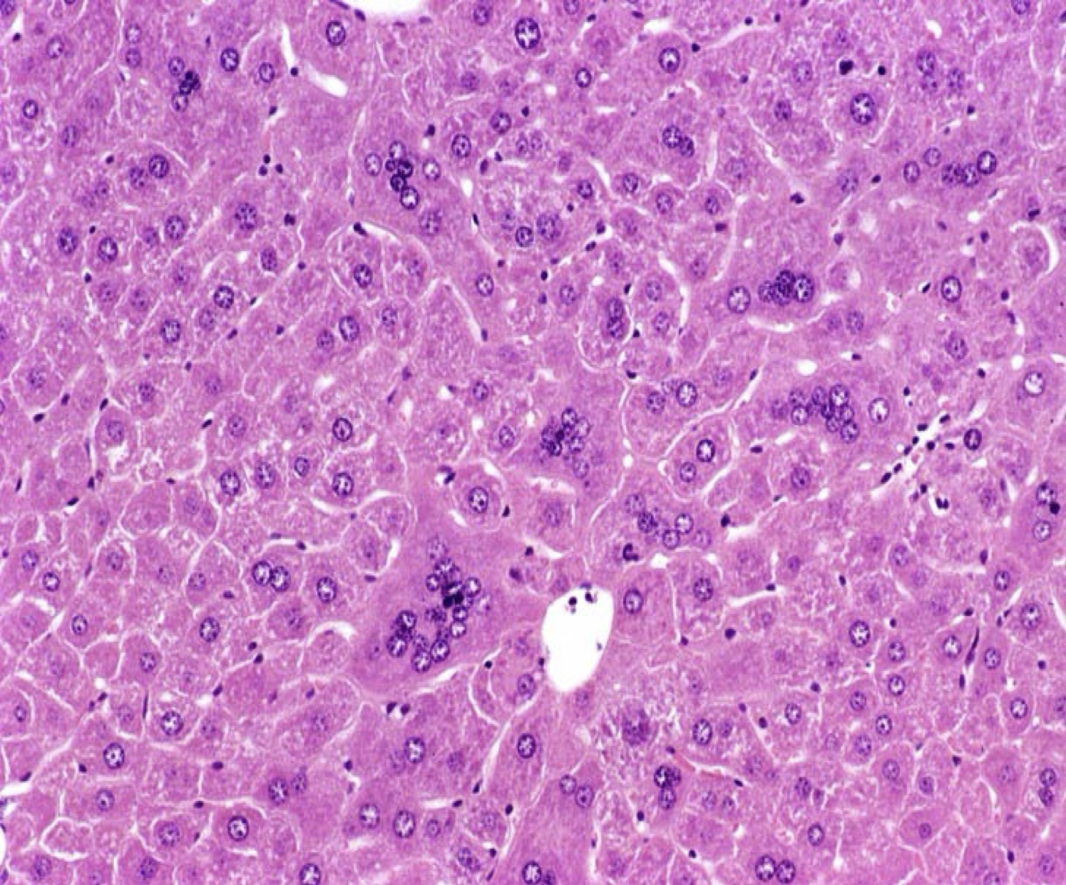
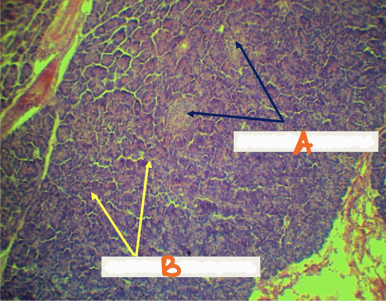
Organ - liver
Shape of cell - polygonal
Number of nucleus - mono and bi nucleated
Location of nucleus - central
Arrangement - rows, cords, columns
Identify the organ, parenchyma, shape of cell, number of nucleus, location of nucleus, and arrangement
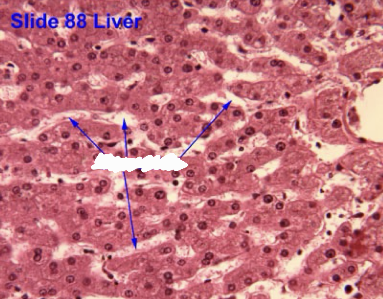
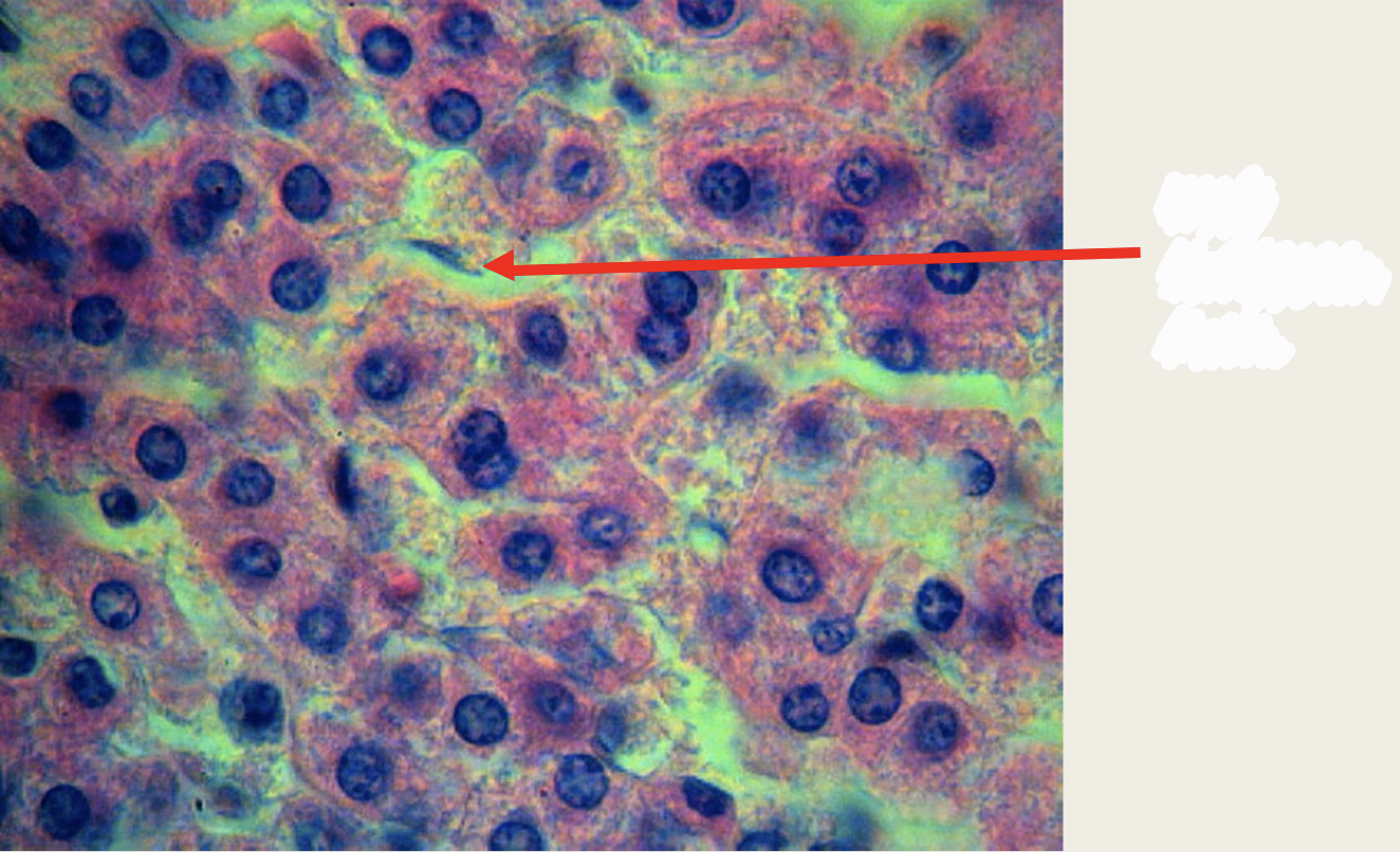
Liver - sinusoids
Identify the structure
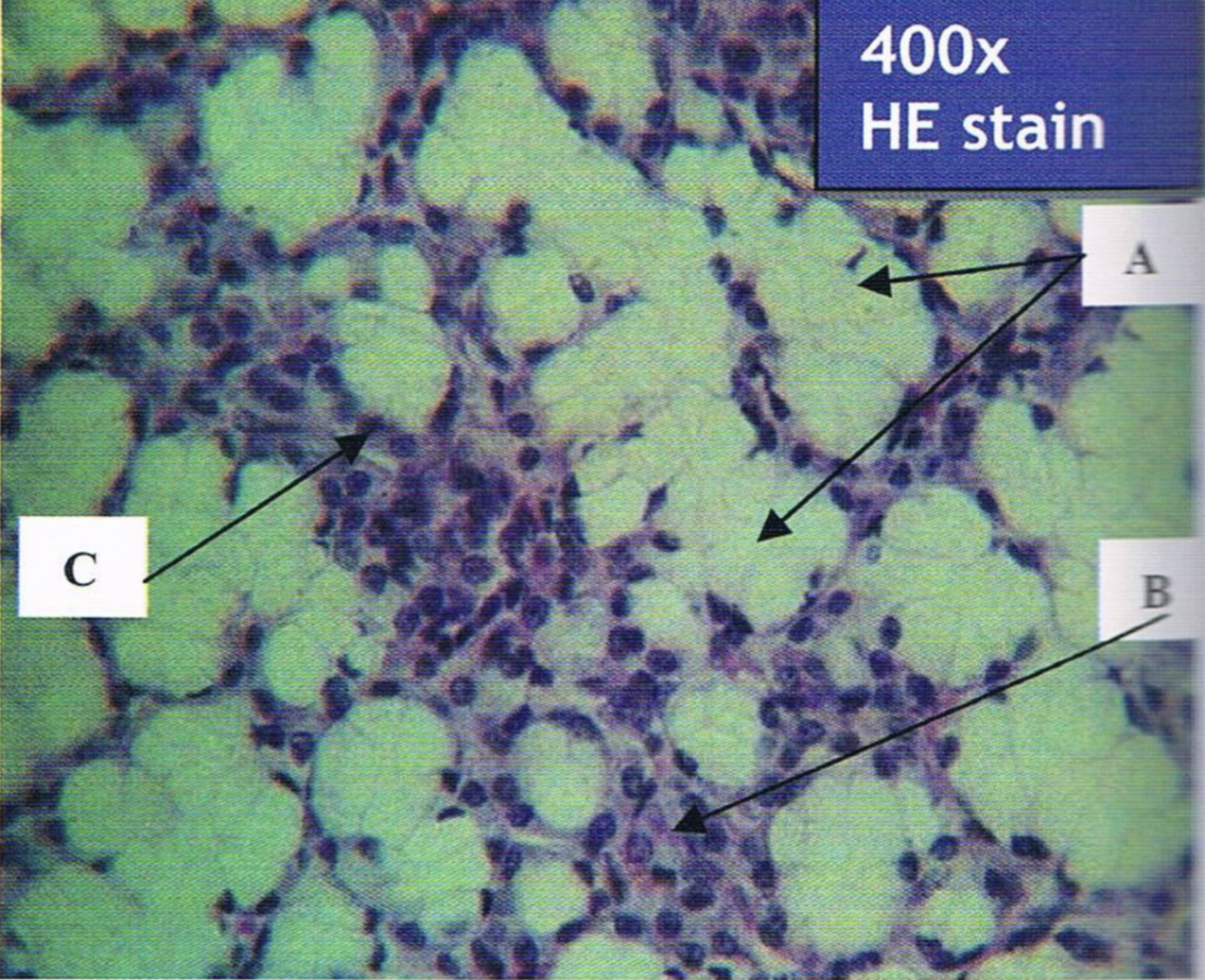
Liver - von kupffer cells
Identify the structures
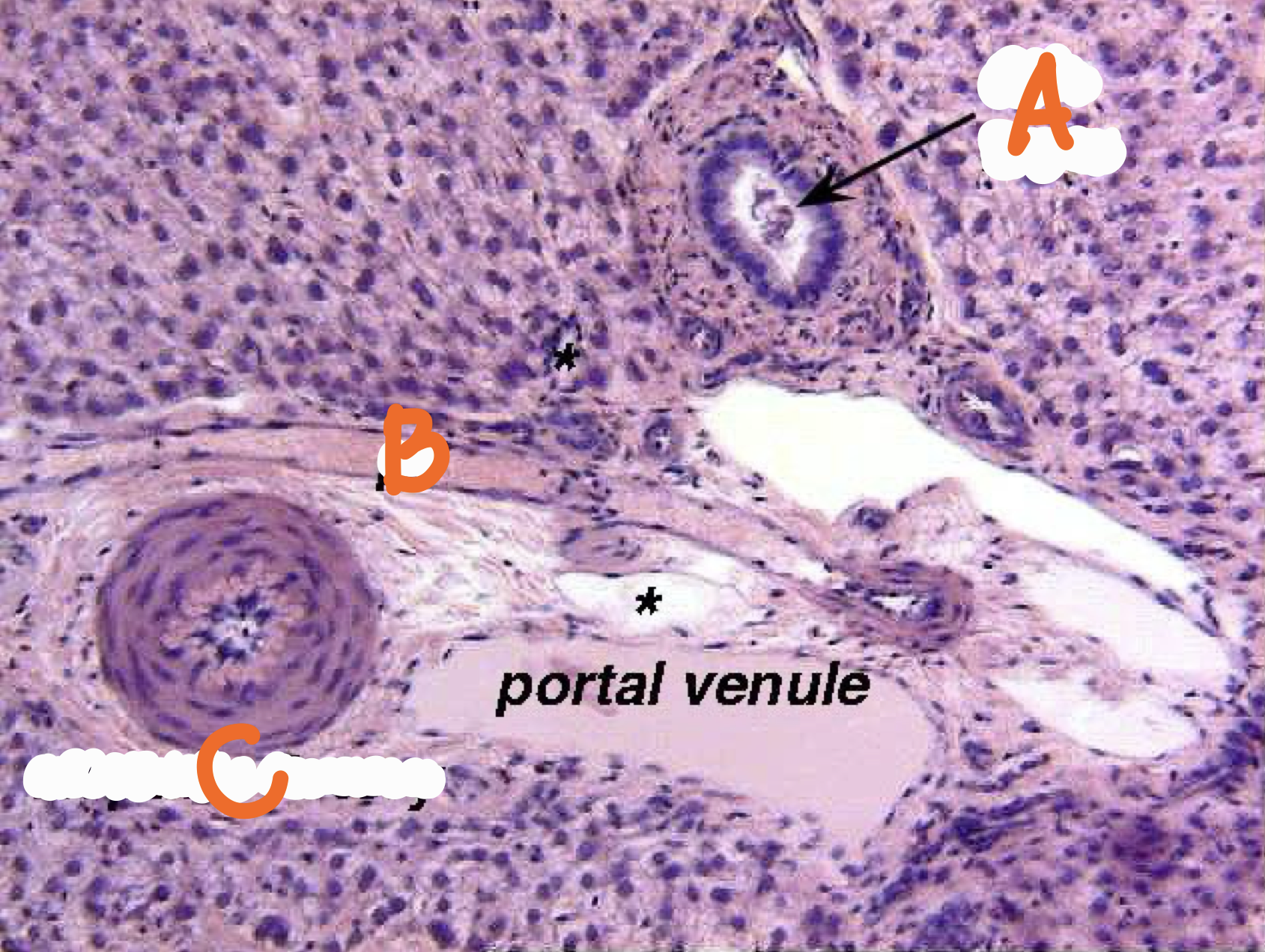
Portal triad
A - bile duct
B - hepatic artery
C - portal vein
Identify the structure
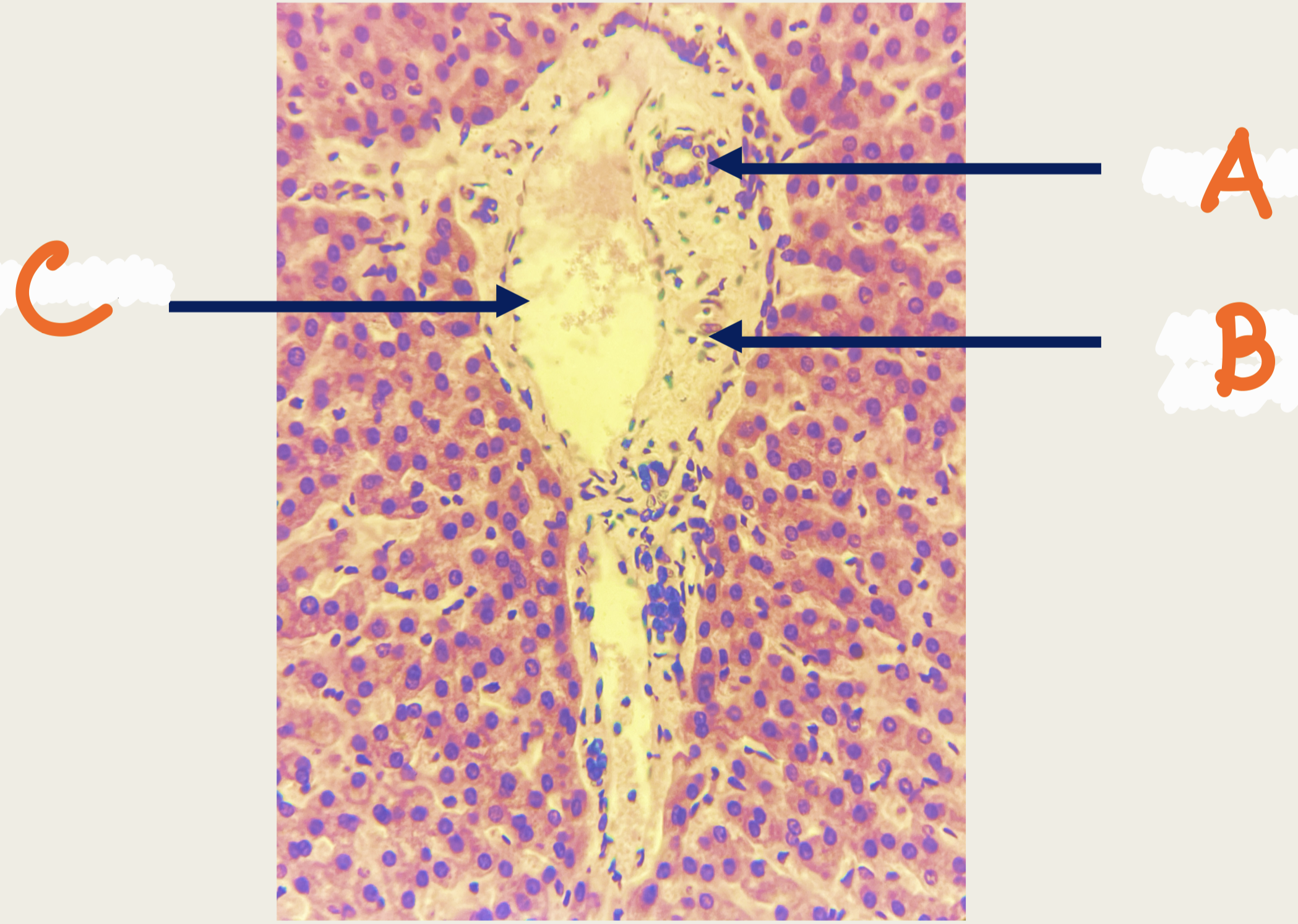
Portal triad
A - portal vein
B - bile duct
C - hepatic artery
Identify the structure
Portal triad
A - bile duct
B - portal vein
C - hepatic artery
Identify the structure
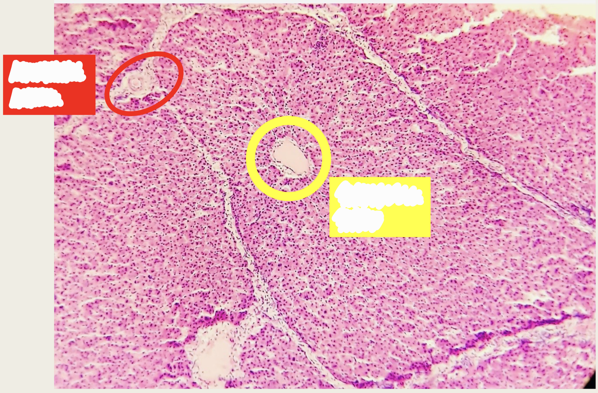
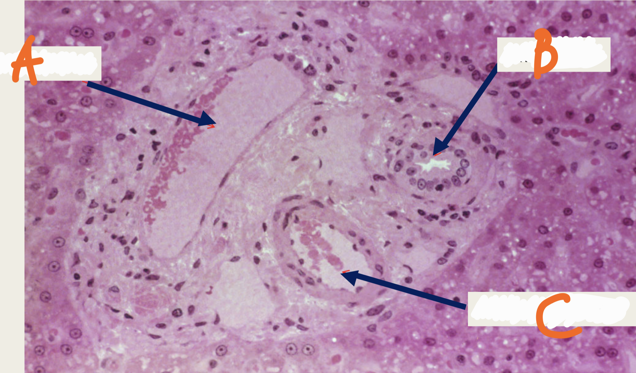
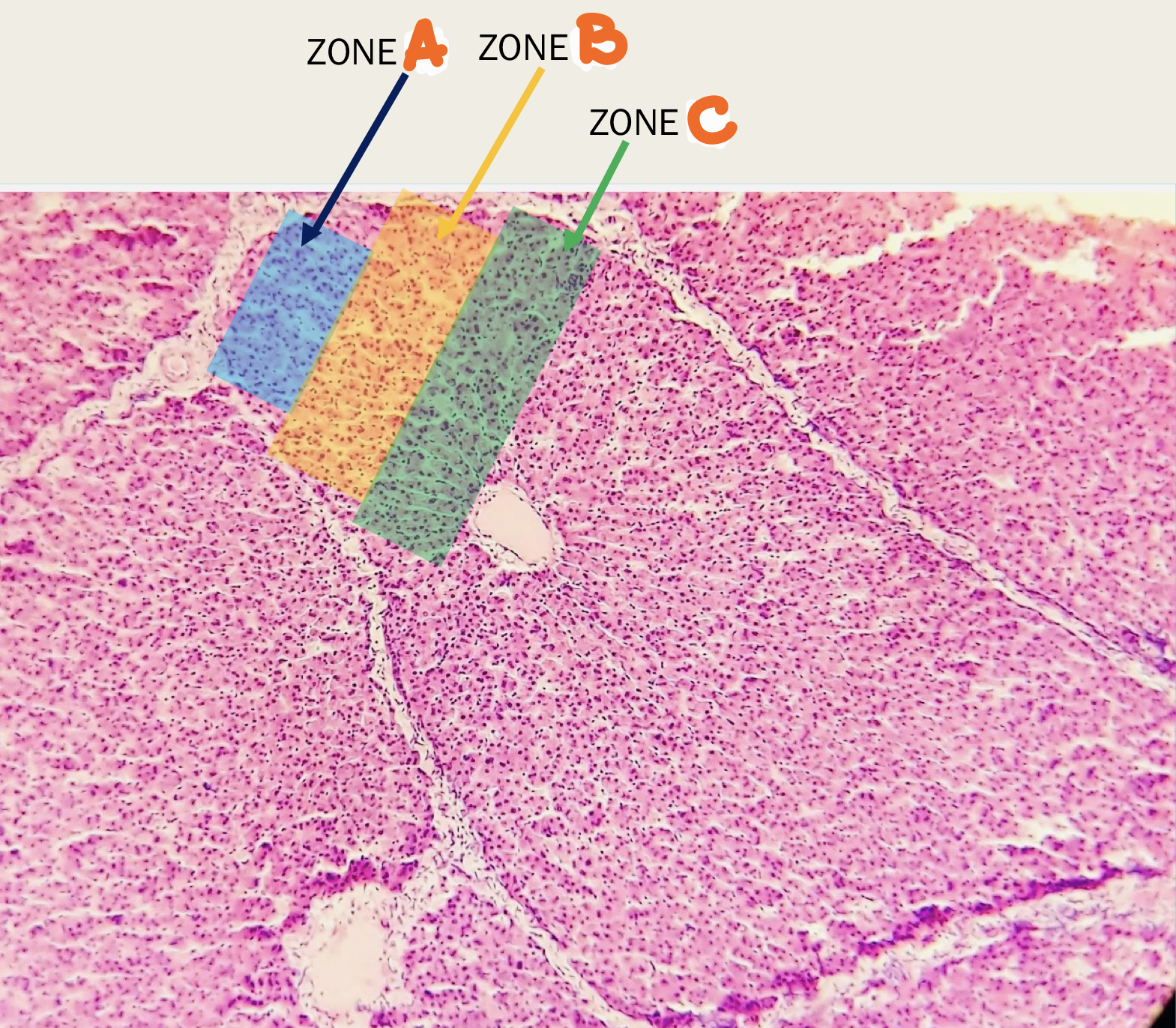
A - zone I (most oxygenated)
B - zone II (intermediate)
C - zone III (least oxygenated)
Identify the structure
Gallbladder
A - mucosa
B - tunica muscularis
C - tunica serosa
D - mesothelium
E - tunica adventitia
F - liver
Identify the structure
Gallbladder Mucosa
A - Simple columnar
B - Lamina propria
C - Rokitanski-aschoff (ra sinus)
Identify the structure
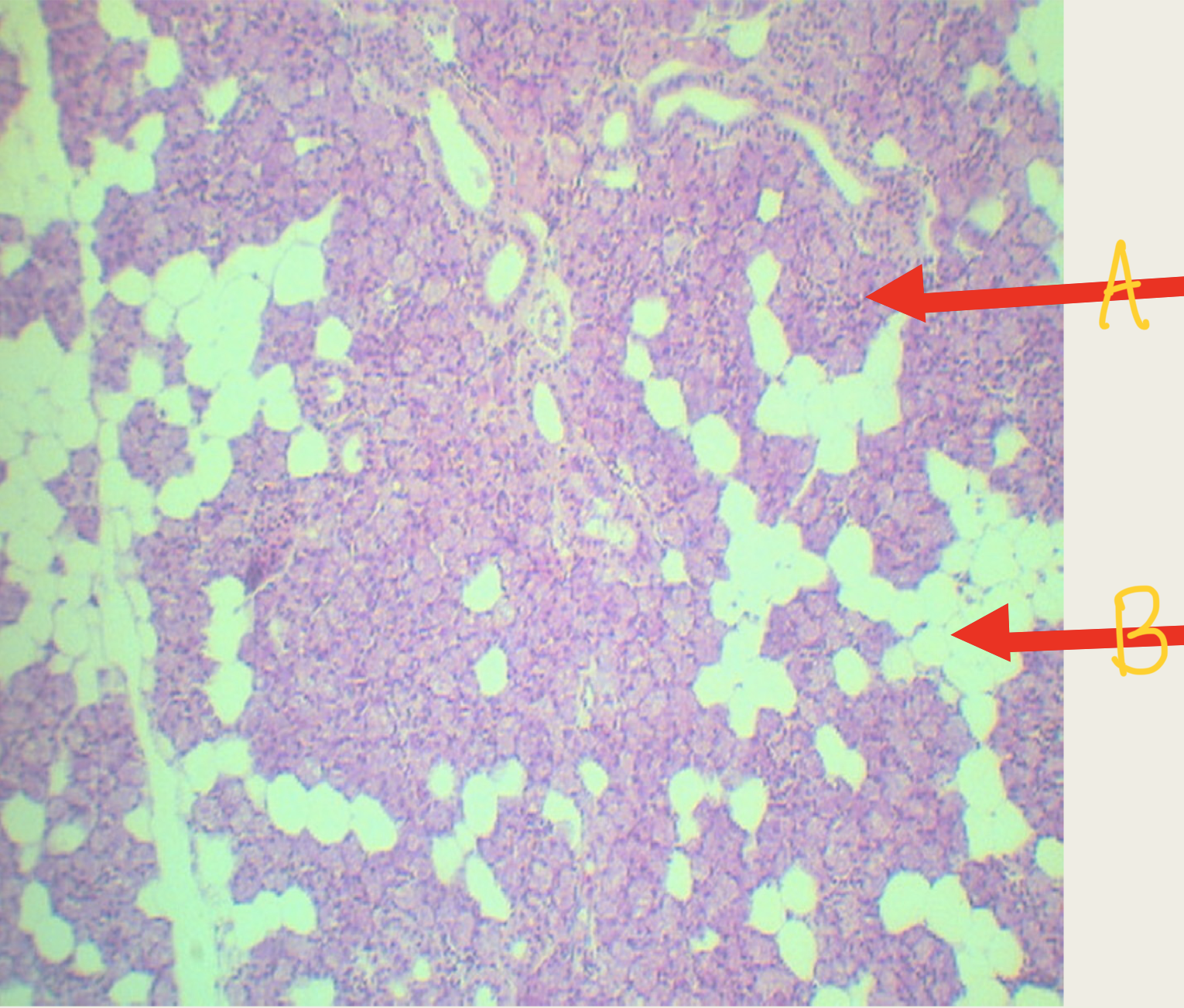
Pancreas
A - islets of langerhans
B - pancreatic acinar cells
Identify the structure
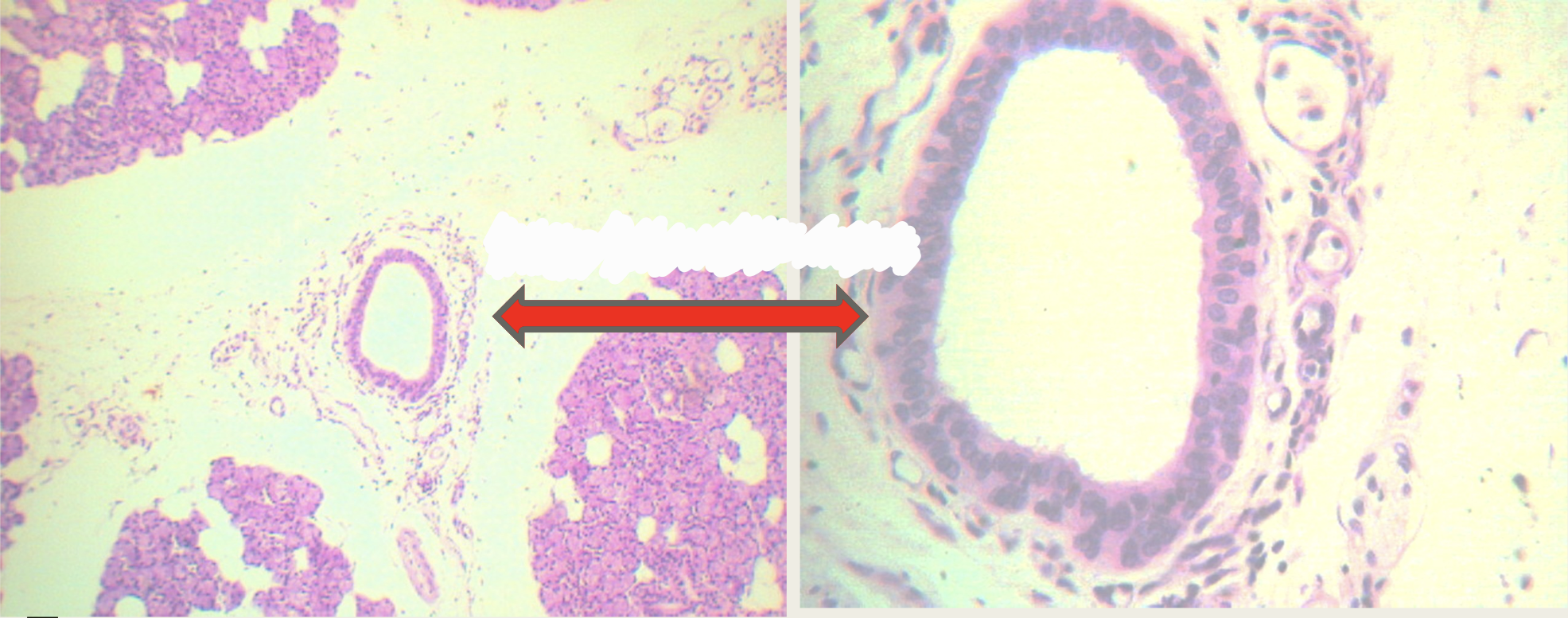
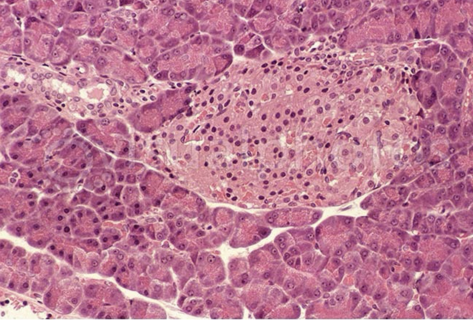
pancreas - islets of langerhans
identify the structure
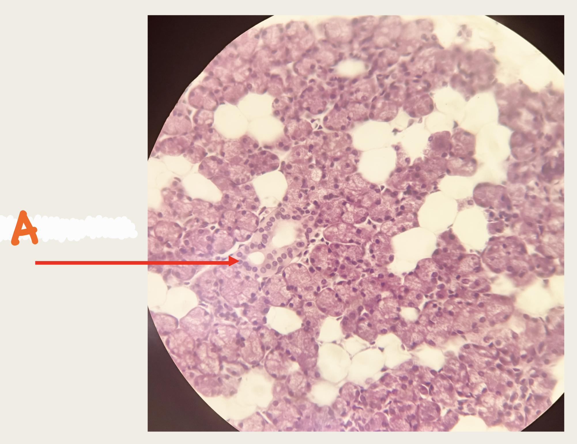
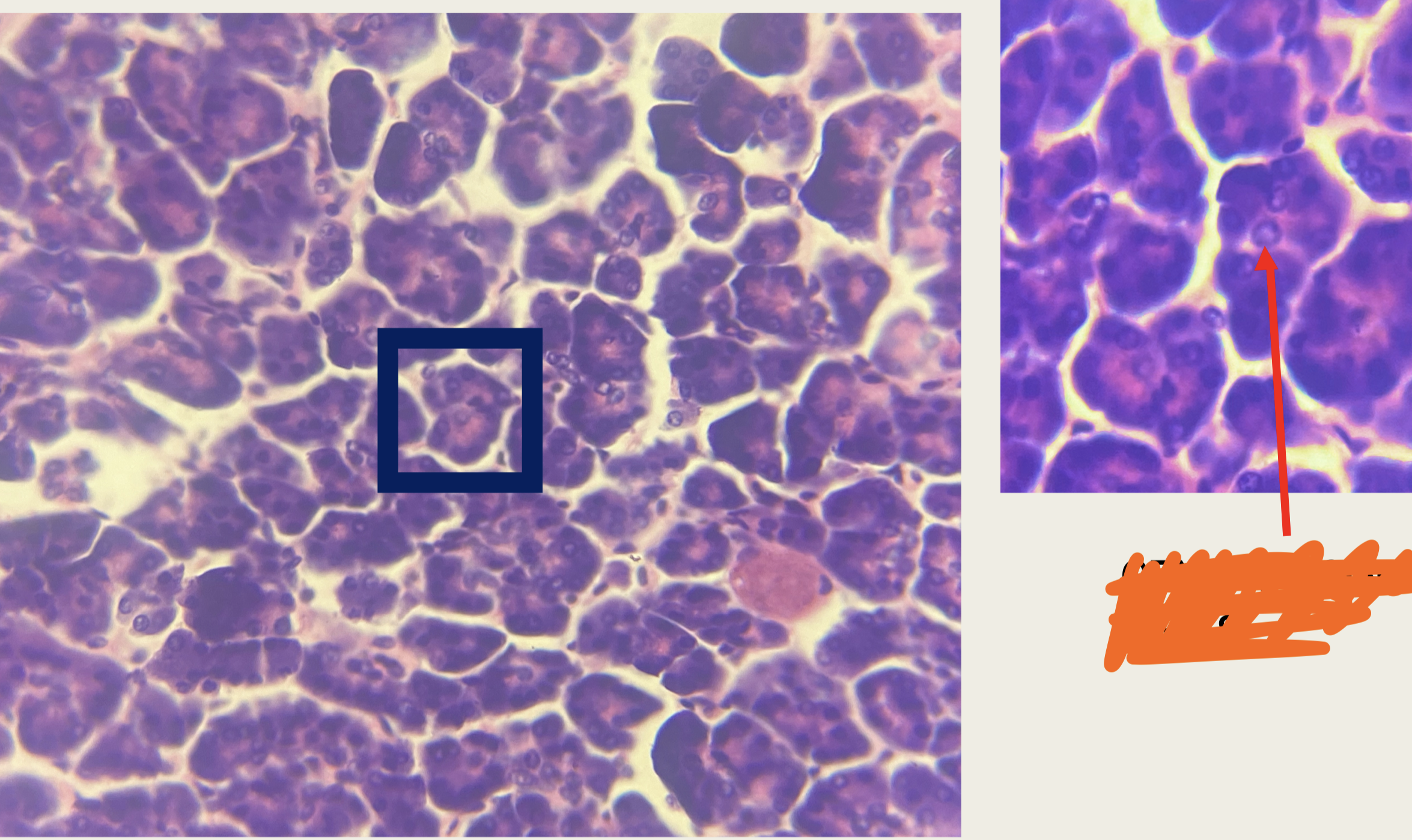
Pancreas
Square - pancreatic acinar cells
Pointed - centro acinar cells
Identify the structure
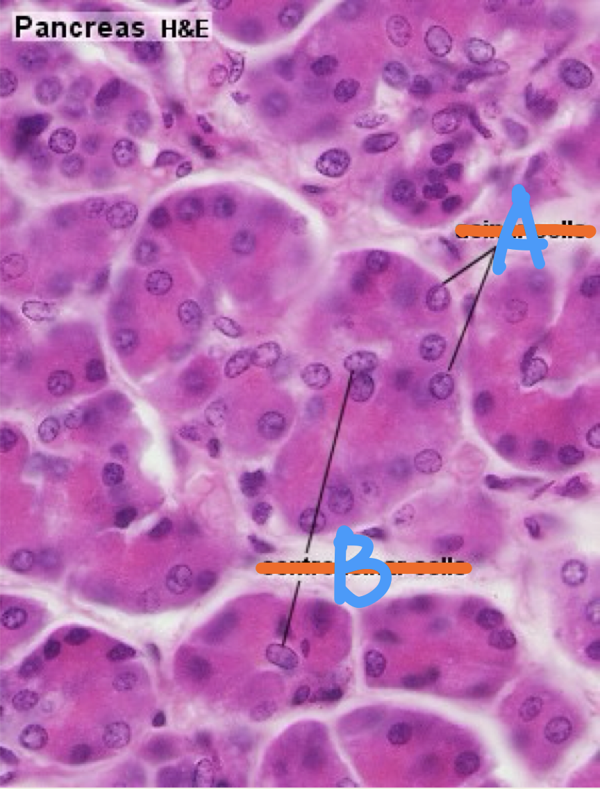
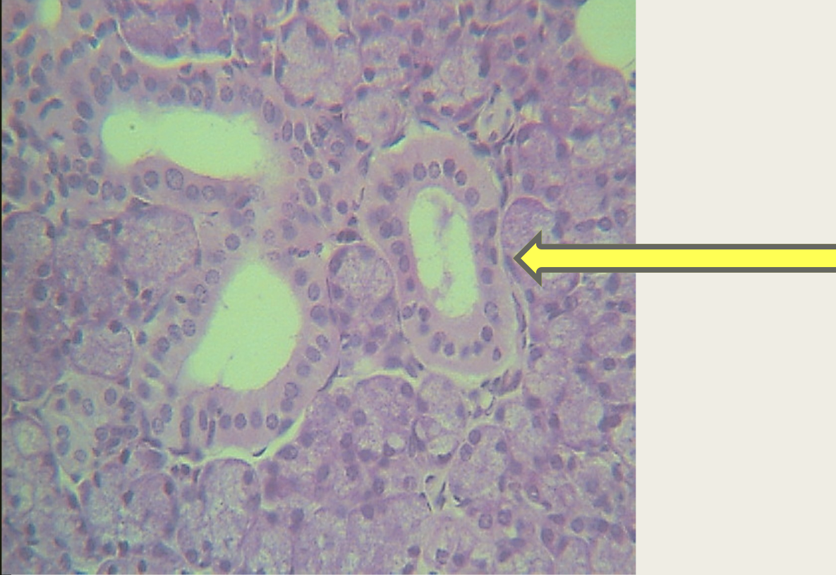
Pancreas
A - pancreatic acinar cells
B - centro acinar cells
Identify the structure