BIO2217 Basics
 Call Kai
 Learn
Learn Practice Test
 Spaced Repetition
 Match
Match Flashcards
 Knowt Play
1/159

Earn XP
Description and Tags
9/10-9/24 lectures
Name | Mastery | Learn | Test | Matching | Spaced | Call with Kai | Chat |
|---|
No analytics yet
Send a link to your students to track their progress
160 Terms
Anatomy
study of the physical structure of organisms (humans) and their parts
Physiology
study of the functions and processes of organisms (humans) and their parts and how they work together

Anatomic direction
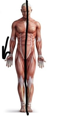
Anterior (ventral)

Anatomic direction
Posterior (dorsal)

Anatomical direction
Superior (relative)

Anatomical direction
Inferior (relative)

Anatomical direction
Medial (toward the midline)

Anatomical direction
Lateral (away from the midline)

Anatomical direction
Proximal (closer to the trunk)
Anatomical direction
Distal (further from the trunk)

Anatomical direction
Superficial (more external)

Anatomical direction
Deep (more internal)
Standard atomical position requires…
Upright position
Feet flat
Palms rotated anteriorly


Anatomic plane
Sagittal plane (slices vertically, dividing body into left and right)

Anatomic plane
Coronal plane (slices vertically, dividing body into back and front)

Anatomic plane
Transverse plane (slicing horizontally, dividing body into top and bottom)
Midsagittal
Along the midline
Parasagittal
Not along midline
Axial
Head, neck, trunk
Appendicular
Limbs

Cranial cavity

Vertebral cavity

Thoracic cavity

Diaphragm

Abdominal Cavity

Pelvic cavity
Levels of body organization (step by step format)
Chemical level (atom/molecule) > Cellular level (cells) > Tissue level (Epithelial tissue) > Organ level (Small intestine) > Organ system level (Digestive system), Organismal level (Human)
Anatomical varitation
Humans have a wide range of internal morphology and external appearance… anatomy is typically the same but physiology often differs because it relies on factors that often vary between individuals
Physiological variation
Sex, diet, age, activity level, environment, weight, and genetic backgorund
Gradient
Difference in value between one point and another… in A&P context often from lower to higher values/areas (passive)
Matter and energy spontaneously flow down _
Gradients
Homeostasis
State of steady internal conditions
Dynamic equilibrium
Physiological conditions fluctuate around an average value
Set point
Physiological average
Normal range
Range of values around set point that are considered typical and healthy
Homeostatic imbalances can cause…
Illness or death
Humans’ reliance on homeostasis has led to bodies having the ability to detect excessive change, referred to as…
Positive and negative feedback loops
Negative feedback loops
Opposes initial change and reduces stimulus (example: body temperature)
Positive feedback loop
Reinforces initial change and increases stimulus… requires external stimulus to end loop (example: blood clotting)
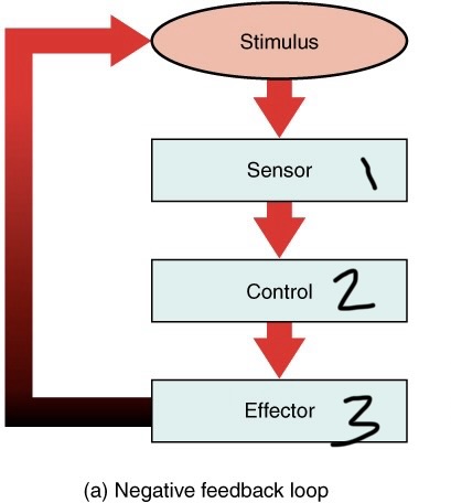
1.) Sensor: monitors condition, senses change
2.) Control: processes info. from receptor, communicates with effector if needed
3.) Effector: Cell/organ that carries out homeostatic mechanism

This is an example of…
Negative feedback loop

This is an example of…
Negative feedback loop

This is an example of…
Positive feedback loop
Potential energy
stored energy (example: chemical bonds)
Kinetic energy
Energy of motion (muscle contraction)
Potential and kinetic energy are…
Interconvertible (meaning they are easily converted into each other)
Chemical energy
energy stored in molecular bonds
Electrical energy
Generated by movement of charged particles
Mechanical energy
Transferred from one object to another to power movement
One type of energy can be…
converted into the other… Conversions often produce thermal energy
ATP stands for…
Adenosine Triphosphate
Adenosine Triphosphate (ATP) is the…
Primary energy source for cells… the hydrolysis phosphate bond releases energy converting the adenosine triphosphate (ATP) into adenosine diphosphate (ADP)
Adenosine diphosphate (ADP) becomes adenosine triphosphate by…
absorbing energy from food… phosphorylating adenosine diphosphate (ADP) becomes adenosine triphosphate (ATP) by absorbing energy
Bond
Electrical attraction between atoms that holds them together
Molecule
2 or more atoms bonded together
Compound
2 or more elements bonded together
Ionic bonds
transfer of electrons from one atom to another… forms ions… stay near each other due to unequal charge that attracts the other
Ions
Charged particles with unequal numbers of protons and electrons
Ions are attracted to each other…
When they have opposite charges due to transferring electrons to become more stable… strength depends on number of electrons exchanged
Electrolytes
Atoms of opposing charges hold together by ionic bond
Electrolytes do what in water?
Dissociate into ions… these ions conduct electricity which is important for nerve/muscle cell function (ex: NA+, K+, OH-)
Covalent bonds
Bond created that shares electrons between atoms… strength of bond depends on the number of electrons shared
Are ionic or covalent bonds stronger?
Covalent
Electronegativity
How strong protons in the nucleus attract electrons
Non-polar bond
A covalent bond with equivalent electronegativity (meaning that both atoms have the same charge in their nuclei, so electrons are shared equally between them)
Polar bond
A covalent bond with unequal electronegativity… this means that the electrons are not shared evenly between the two atoms because they have different charges in their nuclei…this creates a molecule with slightly negative and slightly positive sides
Polar molecules
Molecules with slightly positive and slightly negative regions
How do polar molecules interact with non-polar and other polar molecules?
Polar molecules repel non-polar molecules and attract other polar molecules
Plasma membrane
Flexible outer surface of a cell
Cytoplasm
Intracellular fluid containing organelles
Nucleus
DNA containing organelle… control center of cell… typically largest organelle in the cell

Label numbers 1-3
1.) Nucleus
2.) Cytoplasm
3.) Intercellular fluid
Plasma membrane
Barrier separating intracellular fluid (ICF) and extracellular fluid (ECF)… selectively permeable… made up of phospholipid bilayer
Phospholipid bilayer
Phosphate group + 2 fatty acid chains… head was hydrophilic and tails were hydrophobic… make up phospholipid bilayer to keep intracellular and extracellular fluid separate
Passive transport
Substances crossing phospholipid bilayer without the use of energy… substances diffuse until equilibrium is reached… examples: simple diffusion, facilitated diffusion, osmosis
Active transport
Substances need energy to pass through phospholipid bilayer… substances moving against concentration gradient require energy… there is primary or secondary active transport…
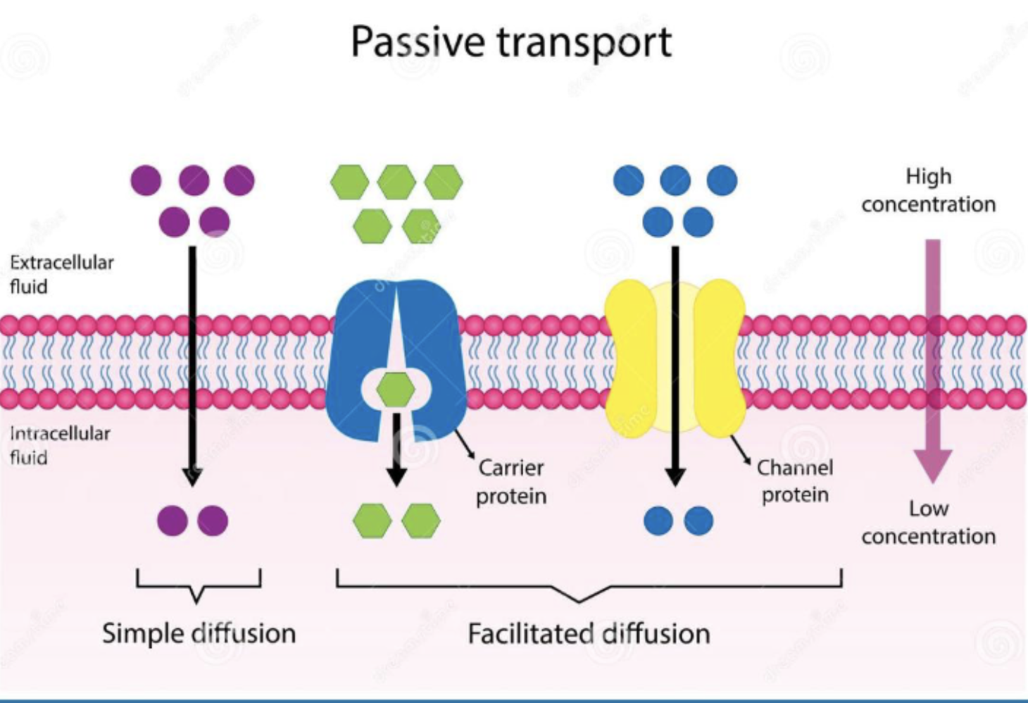
Simple diffusion
Non-polar, lipid-soluble (hydrophobic) substances diffuse directly through the phospholipid bilayer (ex: oxygen, carbon dioxide, steroid hormones)

Facilitated diffusion
requires involvement of protein channels or carrier proteins in plasma membrane (ex: glucose, amino acids, ions)
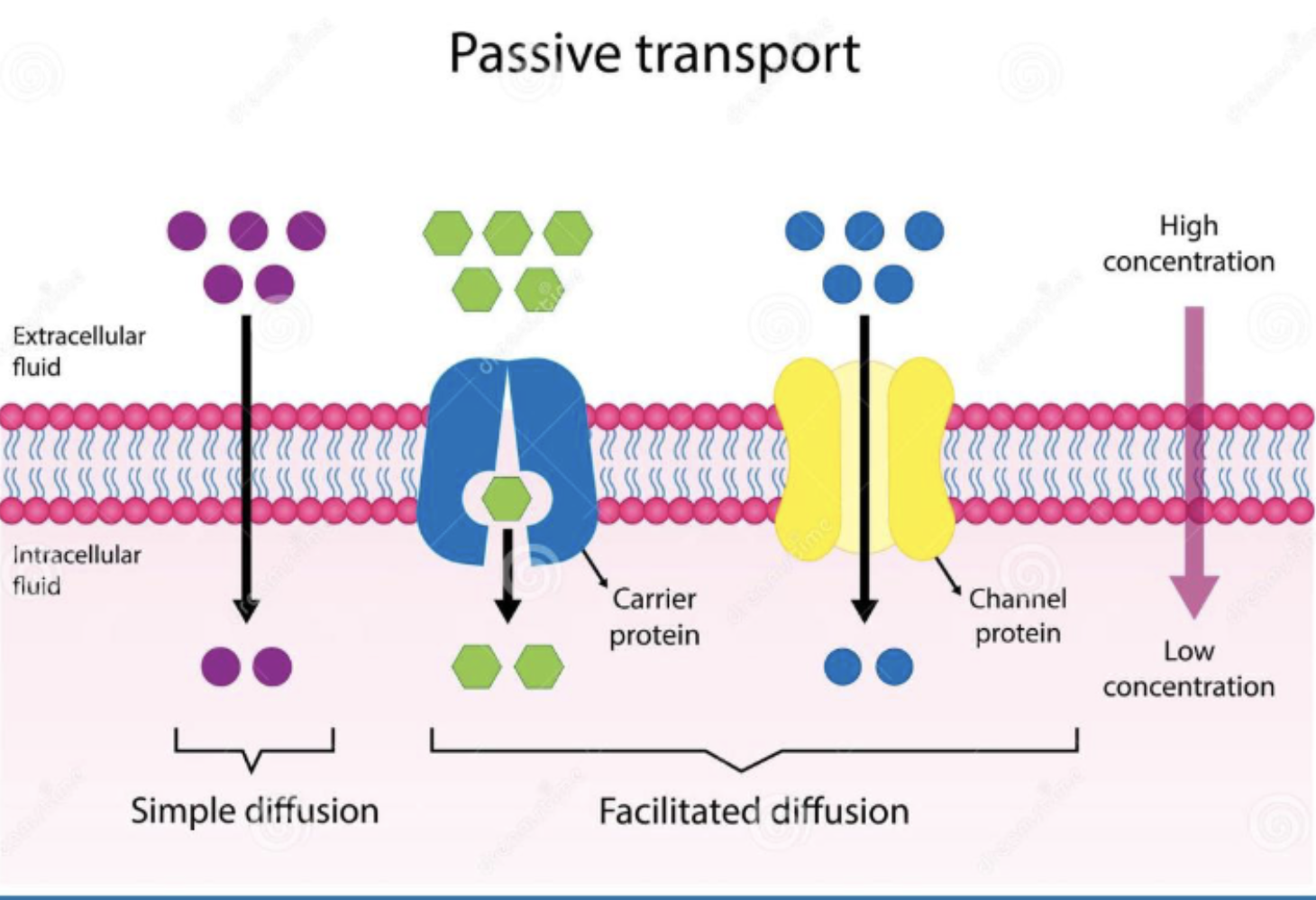
Protein channels
inside the plasma membrane… helps specific solutes get through… typically for an individual solute… always passive transport…

Carrier proteins
in plasma membrane… considered passive facilitated diffusion…

Carrier proteins:
1.) Uniporter:
2.) Symporter:
3.) Antiporter:
1.) carries a single solute
2.) carries 2 solute in the same direction
3.) Exchanges 2 solutes (one in each direction)

Osmosis
movement of water across the plasma membrane… moves along osmotic gradients (more water to less water/ less solute to more solute)… primarily through aquaporins… this occurs until solute concentration equalize… results in volume changes to both sides

Active transport
Uses energy to move a molecule against its concentration gradient… maintains/enhances the concentration gradients… important for nerve and muscle function
Phagocytosis
Active transport (needs ATP) for substances into the cell… cell surrounds substance to be engulfed… receptors bind to microorganisms/solid particles… engulfed substance digested internally… important mechanism for immune system
Tissue
Discrete population of related cells + extracellular matrix… different types: epithelial, connective, muscular, nervous
Epithelial tissue
Sheets of cells covering body surfaces, lining cavities and organs… Attaches to underlying connective tissue (basement membrane)… avascular, which means that it is nourished by diffusion from underlying connective tissue… since it relies on closeness to connective tissue, it limits the thickness
Glands
Specialized epithelial tissue
Connective tissue
Connect and anchor structures of the body… composed of cells (adipocytes, chondrocytes, etc), protein fibers (collagen for tensile strength, elastic for stretch and recoil, and reticular for support), and ground substance (ECF, proteins, carbohydrates) which cushions and supports tissue and maintains cells
Extracellular matix
Made up of the protein fibers and ground substance of connective tissue
Different types of connective tissue
Connective tissue proper (loose and dense), fluid connective tissue (blood and lymph), and supporting connective tissue (cartilage and bone)

Muscle tissue
Contractile cells, produce movement, highly vascular (lots of blood flow/nourishment)… different types: skeletal, cardiac, smooth
Nervous tissue
~10% neurons (which conduct electrical signals), ~90% neuroglia (which support neuronal functions)… ex: brain, spinal cord, nerves
Tissue regeneration
Replacement of damaged tissue with tissue of the same type (retains original function)… different tissues have different regeneration capacities… high capacity=epithelial tissue, bone, connective tissue… low capacity=cardiac muscle, nervous tissue… some injuries may fibrose instead (replacement of original tissue with connective tissue, where it becomes scar tissue instead and loses its original function)… collagen fibers support tissue repair, so larger injuries are more likely to scar
Fibrose
The process of damaged tissues becoming scar tissue… more likely for larger injuries for multiple reasons: the extracellular matrix is destroyed in larger injuries (making organized functional tissue regeneration difficult or impossible), in larger injuries the body prioritizes rapid repair (to prevent infection, fluid loss, and structural failure) so fibroblasts quickly proliferate and deposit dense disorganized collagen fibers to bridge the defect faster than functional tissue can proliferate… even tissues with high regeneration capacity (ex:epithelial tissue) cannot bridge massive gaps before collagenous connective tissue fills the space… since collagen connective tissue restores strength but lacks specialized functions, the replaces tissue becomes a permanent scar… scar tissue blocks contraction, nerve transmission, substance transport, and adhere to neighboring strucutres
Influences on tissue regenerative capacity… 5 answers
1.) extent of injury
2.) mitotic capactiy of cell population
3.) nutrition (ex: intake of protein, vitamin c, etc)
4.) blood supply (delivery of immune support and oxygen/nutrient supply to cells)
5.) age of the individual
Skin
Also referred to as cutaneous membrane… epithelial tissue + connective tissue… largest organ in the body
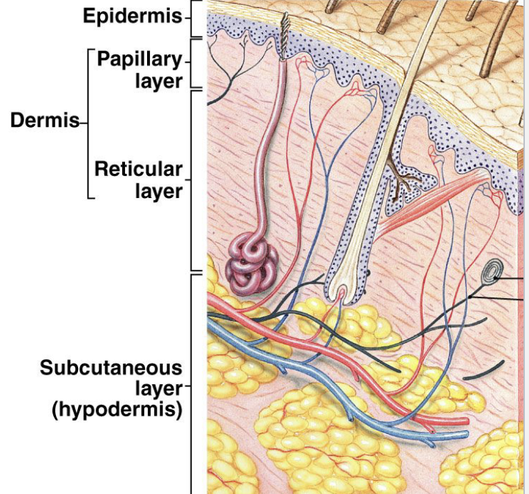
Epidermis
Keratinized stratified squamous epithelium… avascular (lacking blood cells or poor blood supply)… regenerates every 35-35 days
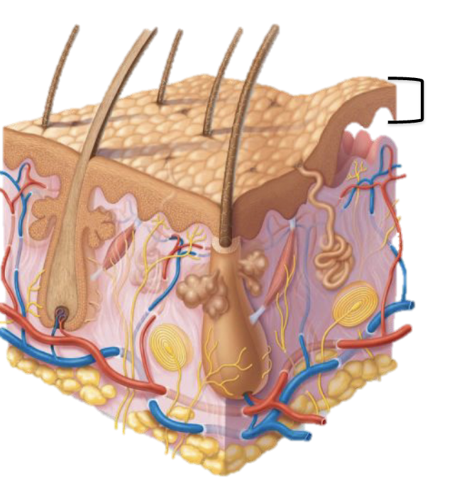
Cells that make up the epidermis
Keratinocytes (produce keratin (which provides skin with durability) and produce vitamin D precursor), melanocytes (produce melanin), Dendritic cells (immune protection), and tactile cells (sensory reception)
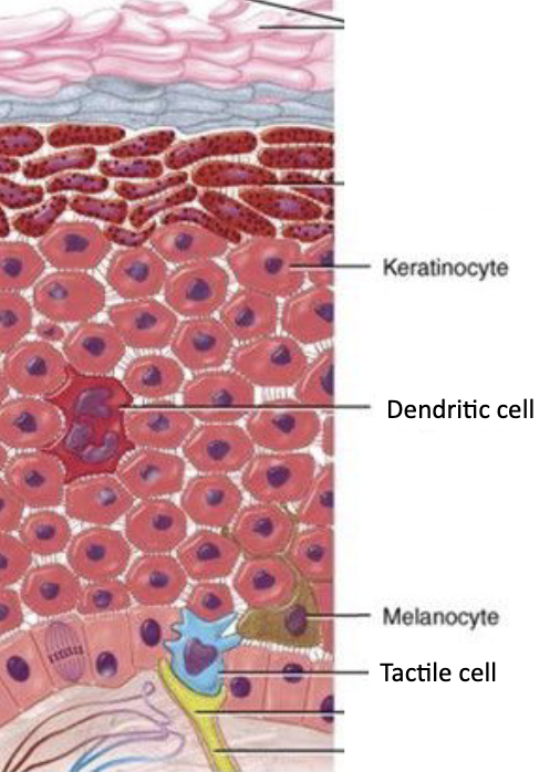
Keratinocytes
Cell of epidermis that produces keratin (which provides skin with durability) and produce vitamin D precursor

Melanocytes
Cell of epidermis that produces melanin
