(4) Microscopic Anatomy of the Nervous System
1/104
There's no tags or description
Looks like no tags are added yet.
Name | Mastery | Learn | Test | Matching | Spaced |
|---|
No study sessions yet.
105 Terms
sensory
functional type of neurons that carry impulses from sensory receptors into the central nervous
system; AFFERENT
**may be somatic or visceral
motor
functional type of neurons that carry impulses from central nervous system to effector organs; EFFERENT
**may be somatic or visceral
interneurons
functional type of neurons that form communication and integration networks between
sensory and motor neurons (i.e., they communicate between other neurons)
**these make up 99.9% of neurons in the body!
multipolar
anatomical type of neurons that have one axon and two or more dendrites (usually lots - forms a dendritic tree)
**includes all motor and interneurons
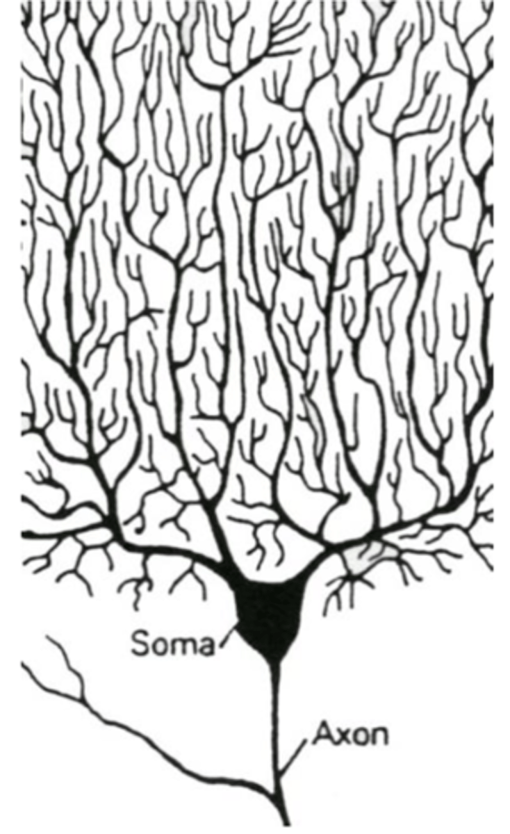
multipolar
What anatomical type of neurons are motor neurons?
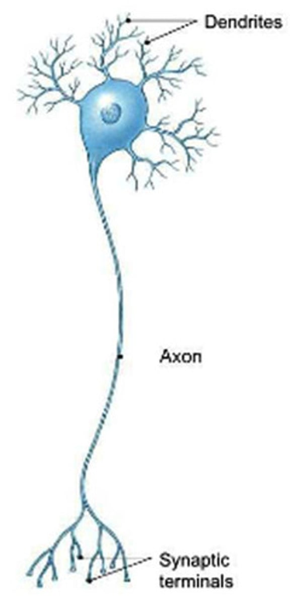
multipolar
What anatomical type of neurons are interneurons?
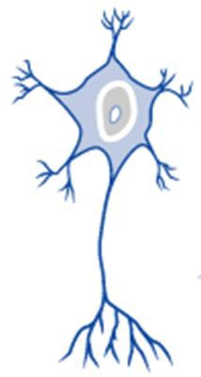
pyramidal cells
multipolar cells found exclusively in the gray matter of the cerebral cortex that have a triangular cell body and a single, long apical dendrite (oriented toward pial surface) among many smaller basal dendrites
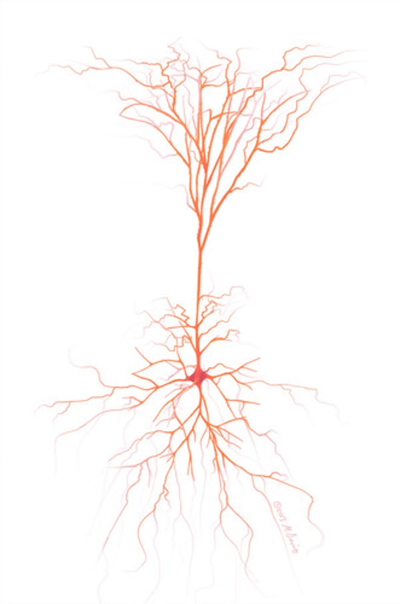
(gray matter of) cerebral cortex
Where are pyramidal cells found?
Purkinje cells
large multipolar neurons located exclusively in the gray matter of the cerebellar cortex that has a flask-shaped cell body with a large dendritic tree facing the pial surface
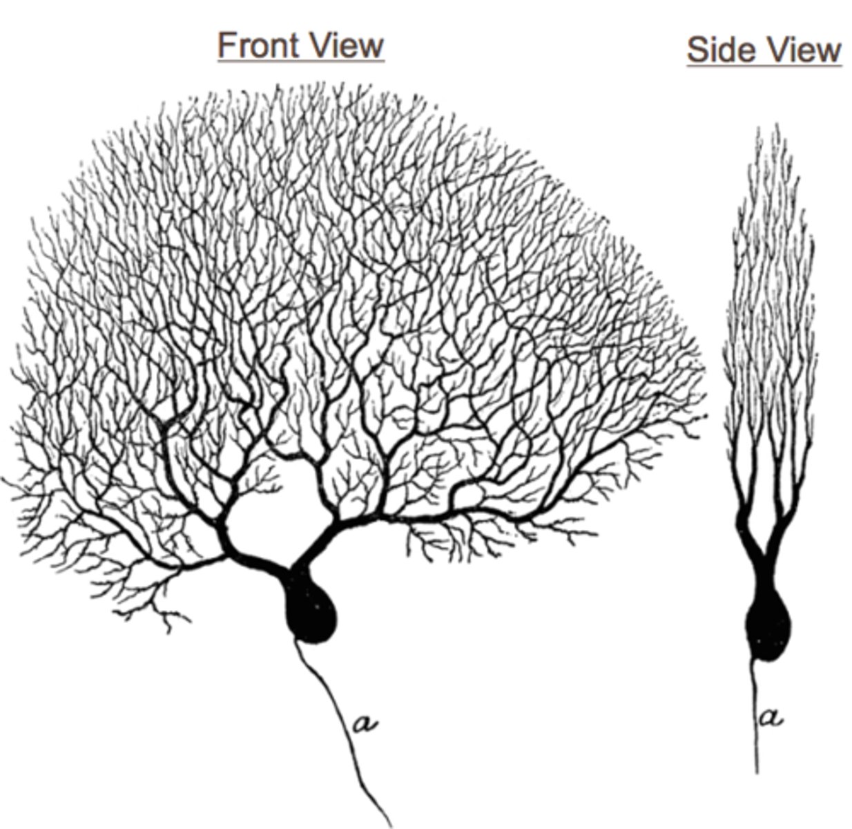
(gray matter of) cerebellar cortex
Where are Purkinje cells found?
stellate cells
smaller, star-shaped multipolar cells that are found in the cerebral and cerebellar cortices that have relatively short axons
cerebral and cerebellar cortex
Where are stellate cells found?
granule cells
small stellate-type multipolar cells that resemble grains of sand, found in the cerebellar cortex
cerebellar cortex
Where are granule cells found?
bipolar
anatomical type of neurons that have one axon and one dendrite
**includes types of sensory neurons associated with special senses and are found in the retina, vestibulocochlear apparatus (in the inner ear), and olfactory epithelium
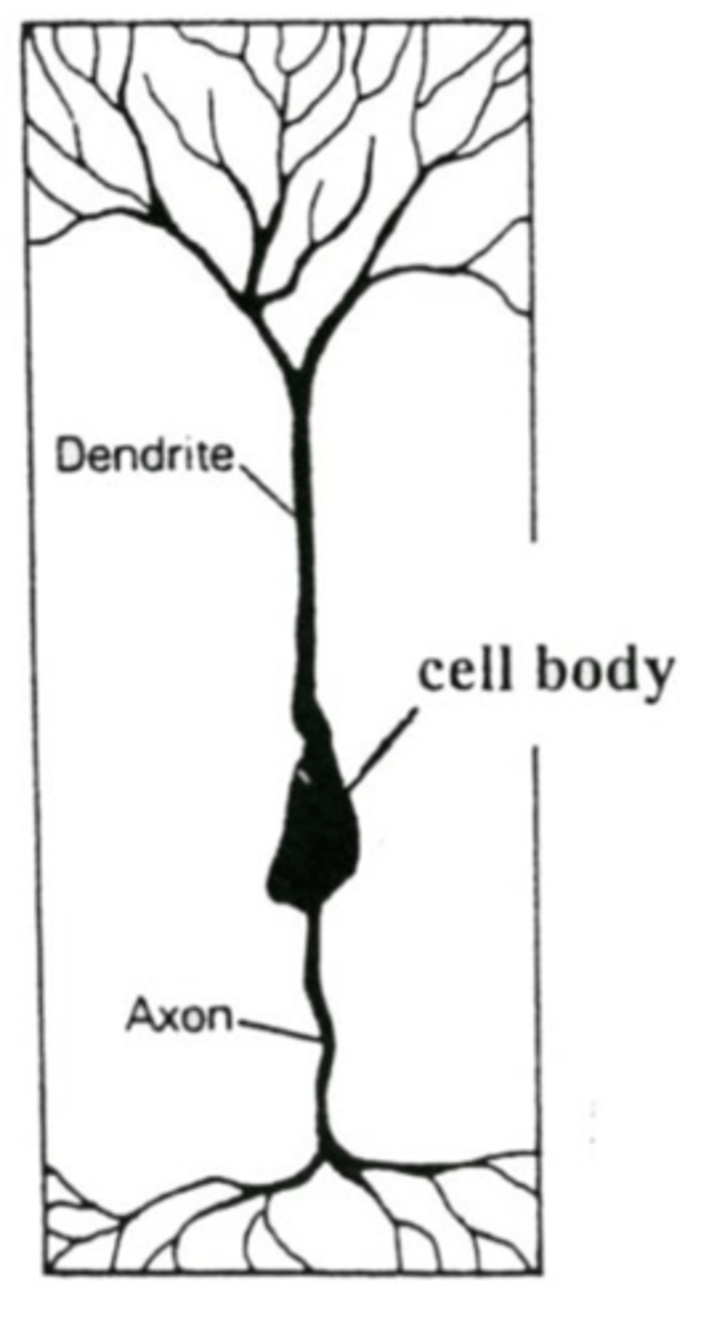
bipolar
What anatomical type of neurons are sensory neurons associated with special senses?
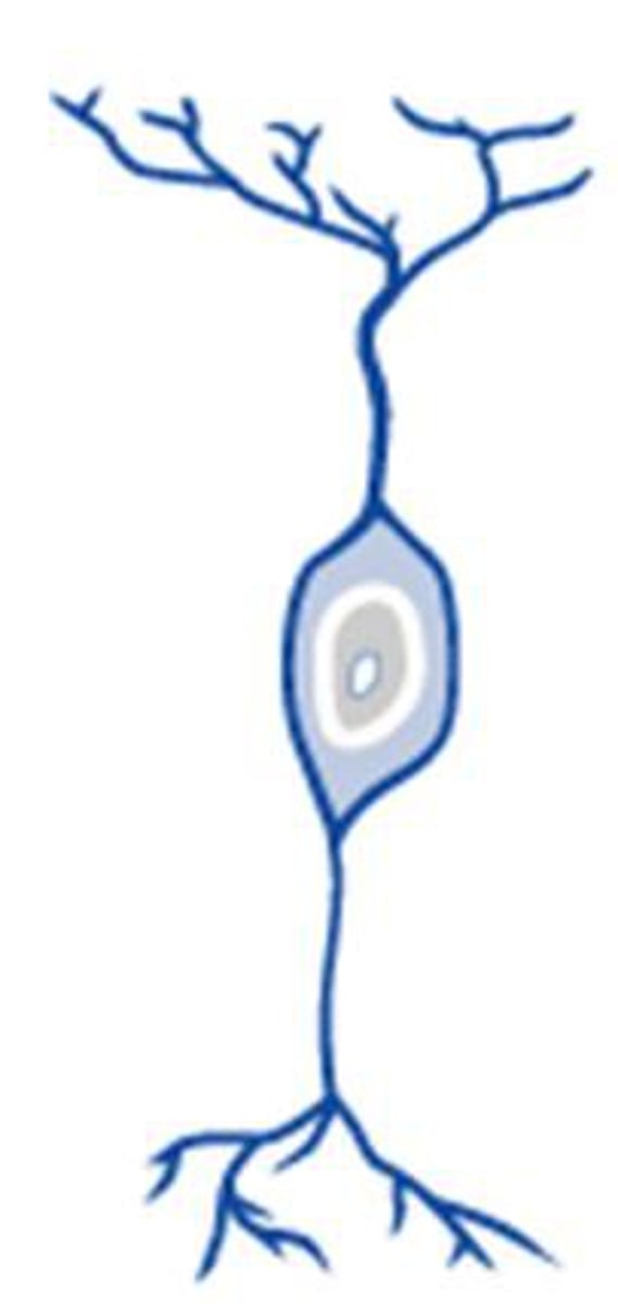
retina, vestibulocochlear area, olfactory epithelium
Where are bipolar neurons found? (3)
(pseudo)unipolar
anatomical type of neurons that appear as if only one process attaches to the soma, though actually, one axon and one dendrite that fuse together to form a single process that attaches to the perikaryon, and they split into two a short distance away
**located in sensory ganglia (e.g., spinal ganglia, trigeminal ganglion, etc.)
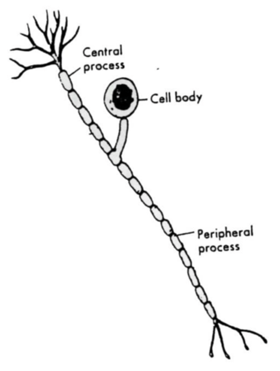
(pseudo)unipolar
What anatomical type of neurons are sensory neurons with cell bodies that are located in sensory ganglia? (e.g., spinal ganglia, trigeminal ganglion)
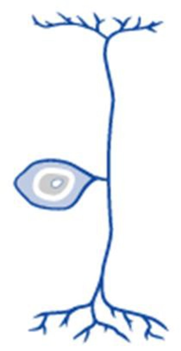
multipolar
(since they make up motor & interneurons)
What is the most common type of neuron: multipolar, unipolar, or pseudounipolar?
soma, perikaryon
two terms for the cell body of a neuron, the expanded portion of a cell that contains the cell nucleus and most of the usual organelles associated with cells
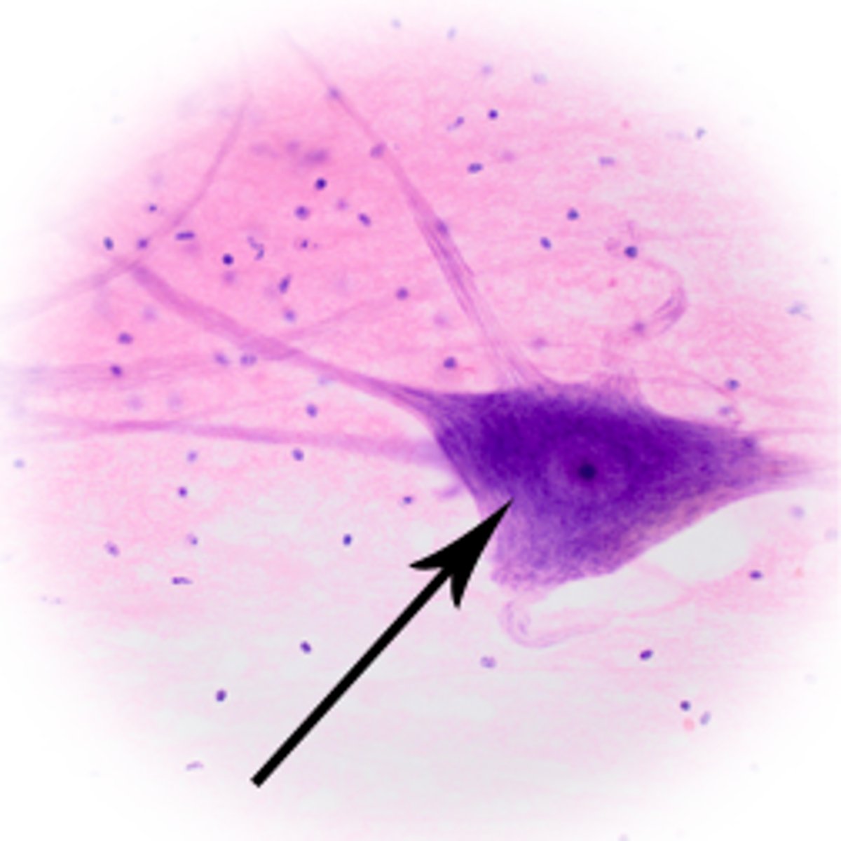
nucleolus
Neuron cell bodies typically have what prominent organelle?
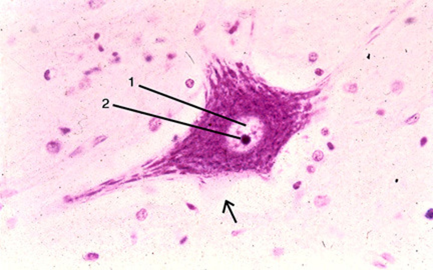
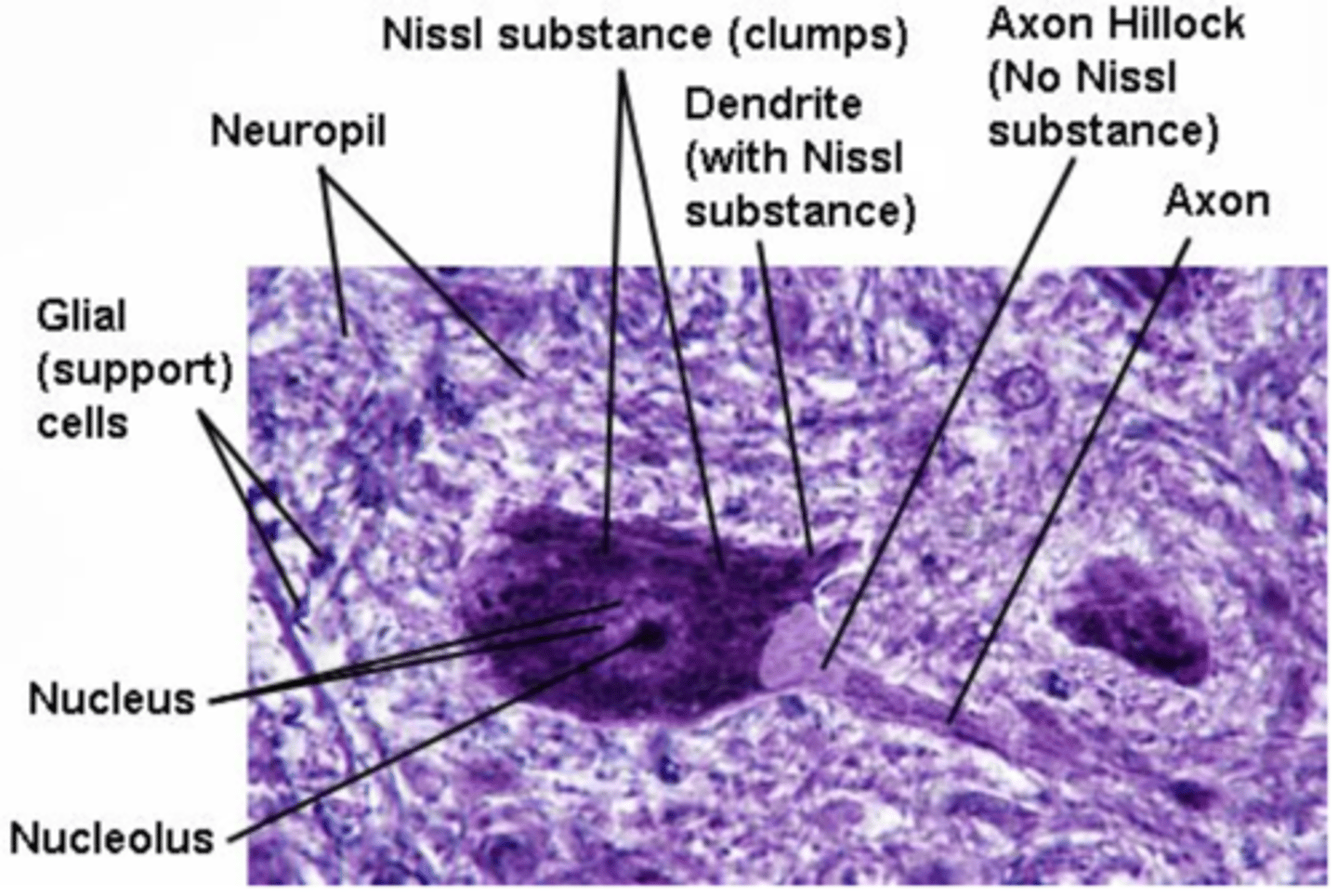
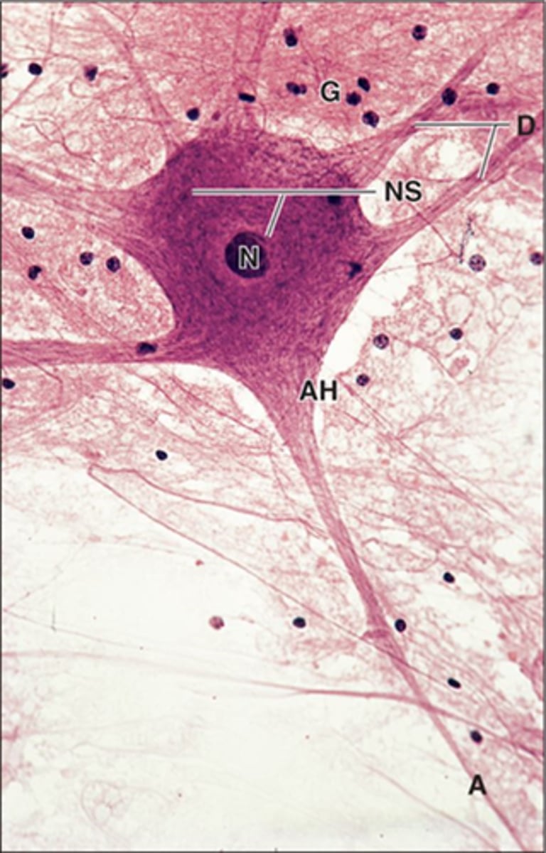
Nissl substance (bodies)
basophilic clumps in the cytoplasm of neurons that represent many free polyribosomes and rough ER
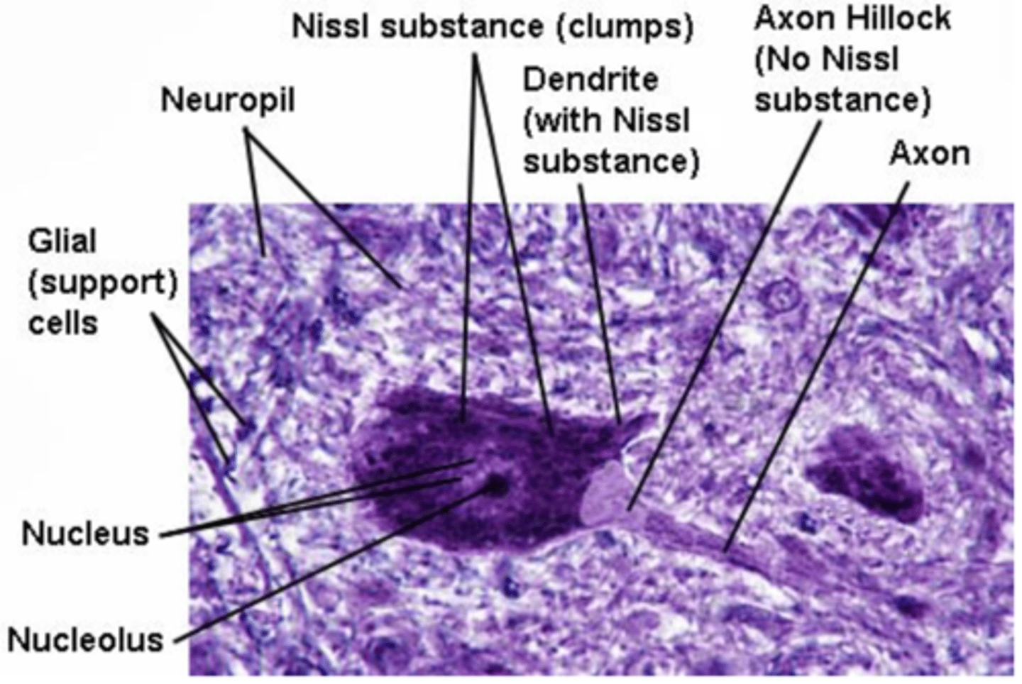
ribosomes, rough ER
What organelles does Nissl substance represent?
nucleus, axon hillock
Which parts of the neuron cell body don't have a lot of Nissl substance and therefore stain paler?
false
(so they are long lived and have a high metabolic activity to replace enzymes and other substances)
True or false: Adult neurons are capable of division.
dendrites
cytoplasmic extensions that carry information TOWARD the cell body of a neuron
arborization
formation of new dendritic trees and branches that greatly increases the receptive region of the neuron
dendritic spines (or gemmules)
small knob-like projections that can be found on some dendrites that have similar contents as the soma, except no nucleus, nucleolus, and Golgi
**so they DO usually have Nissl substance
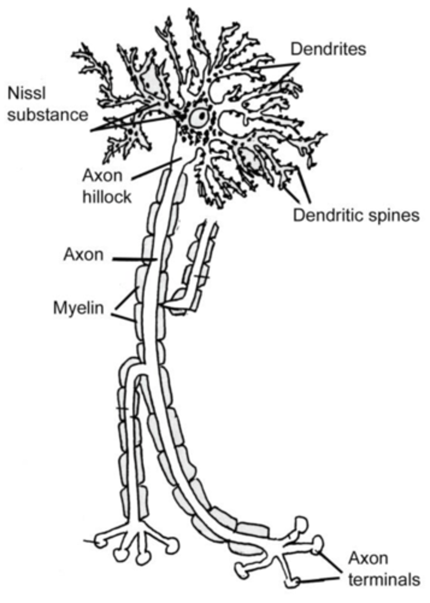
axon
cytoplasmic extension that carries information AWAY from the cell body of a neuron
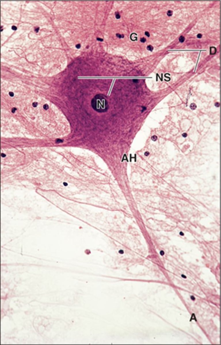
collateral branches
refers to branching of an axon
axon hillock
specialized site where the axon attaches to the cell body
**characteristically devoid of Nissl substance, but has neurofilaments, microtubules, membrane-bound vesicles, and a few mitochondria
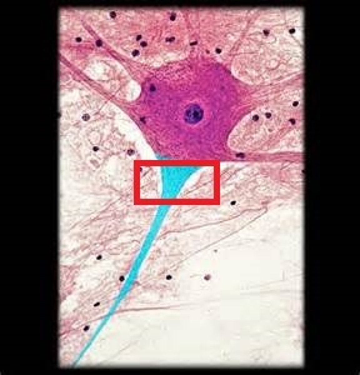
initial segment
region of the axon between the apex of the axon hillock and the beginning of the myelin sheath
**this is where an action potential is generated!
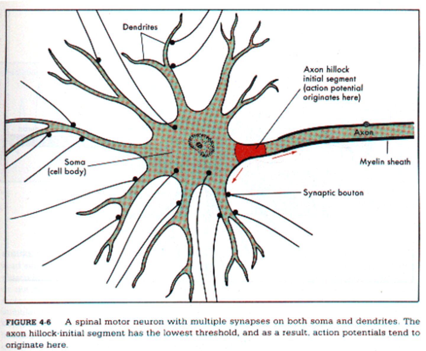
axon terminal (or bouton)
dilated region at the distal end of the axon that helps form a synapse with the next structure
projection
type of axon that connects the cerebral cortex with subcortical structures
association
type of axon that connects structures within the cerebral cortex (and usually on the same side)
synapse
Action potentials may be carried by several neurons arranged in a chain-like fashion known as a pathway. Individual neurons do NOT touch each other but transmit information from one to another by way of a special arrangement known as a what?
commissural
type of axon that connects similar structures on opposite sides of the CNS
chemical
one of the two physiologic types of synapses in which the conduction of the information in a nerve impulse from one structure to the next is achieved by release of chemical substances (known as neurotransmitters)
electrical
one of the two physiologic types of synapses that are basically gap junctions, since ions actually diffuse between the presynaptic and postsynaptic structures
**includes the gap junctions between smooth muscle cells and those between cardiac muscles cells, known as intercalated discs
chemical
Are way more human synapses chemical or electrical?
synaptic vesicles
membrane bound structures that contain neurotransmitters
**this is how to recognize the presynaptic component under a microscope
synaptic vesicles, thin presynaptic density
What are two observable characteristics of the PREsynaptic side of a synapse?
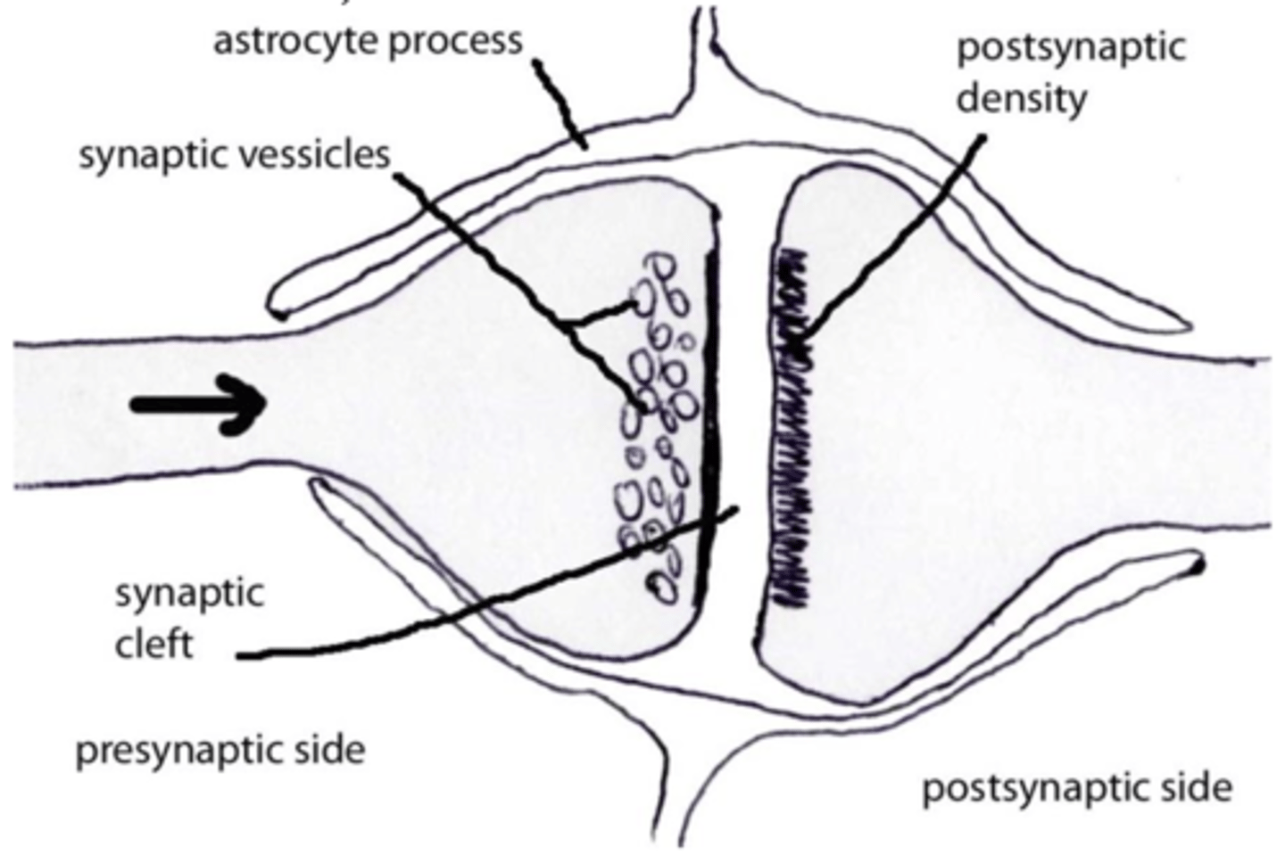
neurotransmitter receptors, thick postsynaptic density
What are two observable characteristics of the POSTsynaptic side of a synapse?
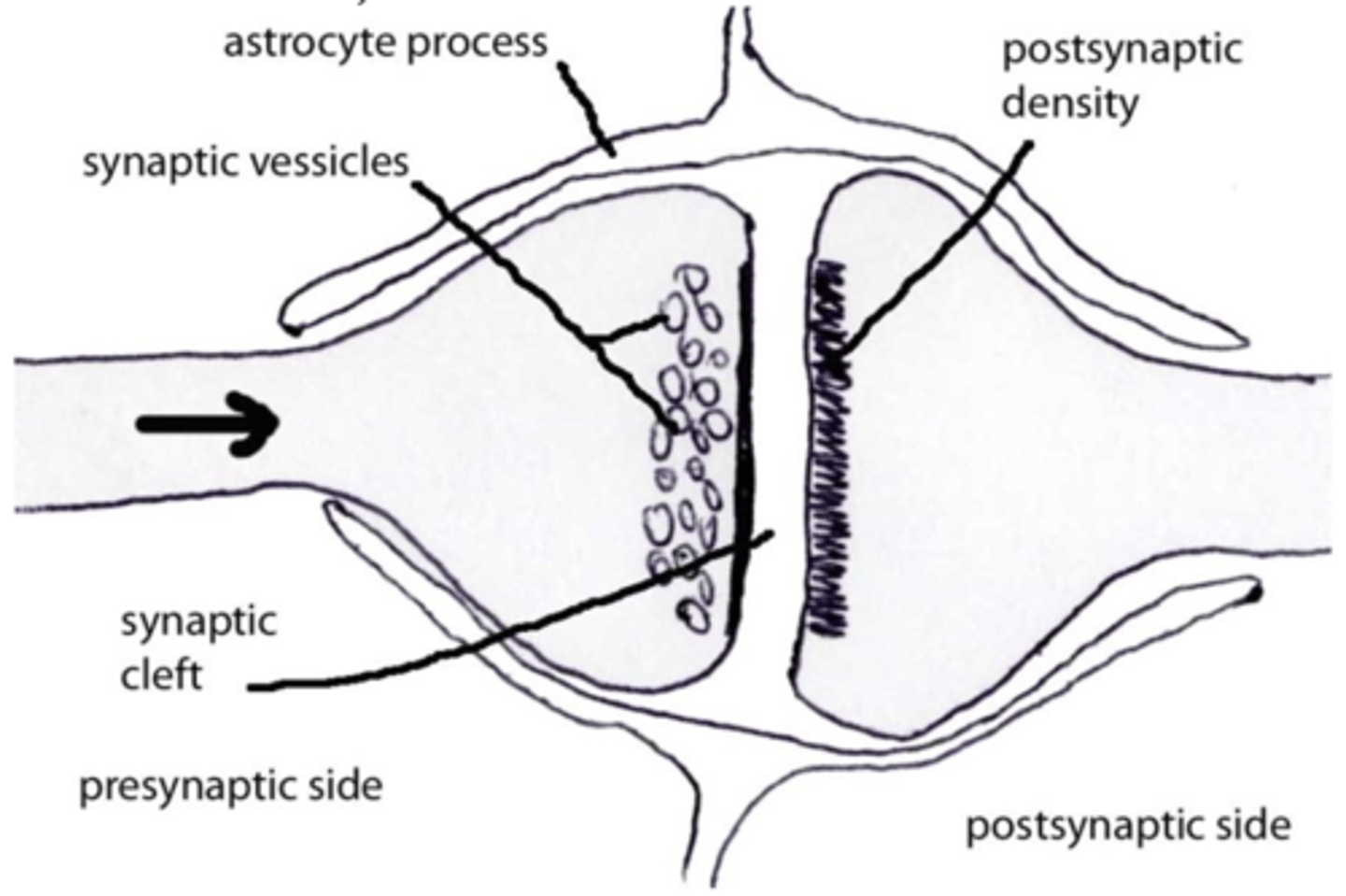
astrocyte processes
support cell processes (in the CNS) surrounding an entire synapse which help control the metabolic environment of the synapse
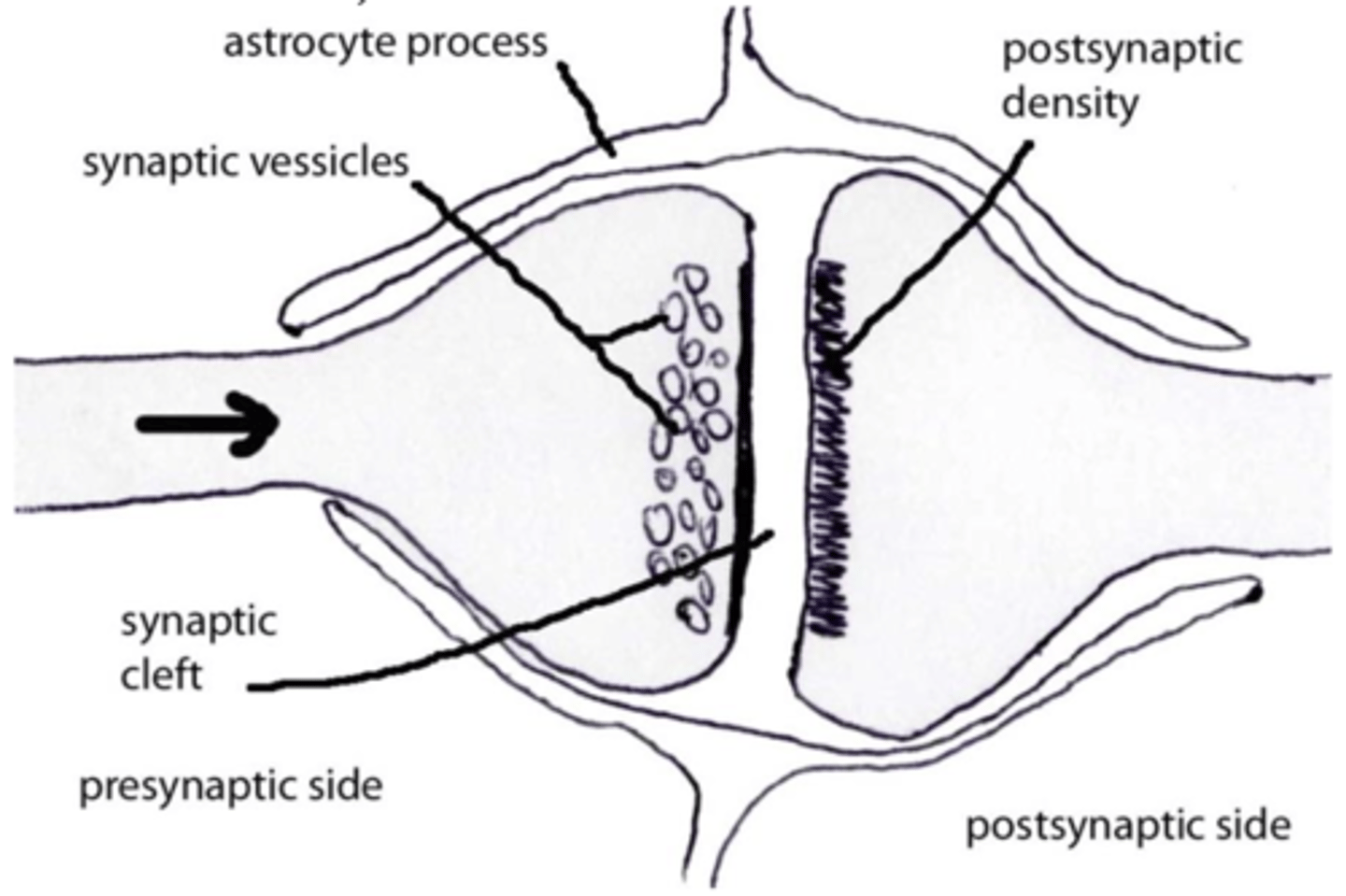
asymmetric
excitatory synapses in which the postsynaptic density is thicker than the presynaptic density
symmetric
inhibitory synapses in which the pre and postsynaptic densities are of similar thickness
slow
refers to axonal transport that carries components up to 5 mm/day that uses microtubules and neurofilaments
**usually carries cytoskeletal proteins and enzymes
fast
refers to axonal transport that carries components up to 400 mm/day that uses microtubules only (no neurofilaments)
**transports mitochondria, lysosomes, and vesicles
fast
Which type of neuronal transport uses microtubules only?
slow
Which type of neuronal transport uses microtubules and neurofilaments?
slow
Which type of neuronal transport carries cytoskeletal proteins and enzymes?
fast
Which type of neuronal transport carries mitochondria, lysosomes, and vesicles?
anterograde
direction of axonal transport that goes from soma to periphery
retrograde
direction of axonal transport that goes from periphery to perikaryon
both
Which mechanism of axonal transport can go in an anterograde direction: fast, slow, or both?
fast
Which mechanism of axonal transport can go in an retrograde direction: fast, slow, or both?
neuroglia
neuron supporting cells located in the CNS: includes oligodendrocytes, astrocytes, microglia, & ependymal cells
these physically protect neurons, electrochemically insulate them, and modulate the metabolic exchange of material in/out of the neurons
CNS
Are oligodendrocytes supporting cells found in the CNS or PNS?
CNS
Are astrocytes supporting cells found in the CNS or PNS?
CNS
Are microglia supporting cells found in the CNS or PNS?
CNS
Are ependymal cells supporting cells found in the CNS or PNS?
PNS
Are Schwann cells supporting cells found in the CNS or PNS?
PNS
Are satellite cells supporting cells found in the CNS or PNS?
Schwann cells
What are the most numerous supporting cells in the PNS?
Schwann cells
PNS supporting cells that are associated with the axons and:
- provide structural support
- mediate axon nutritional needs
- create myelin for many axons
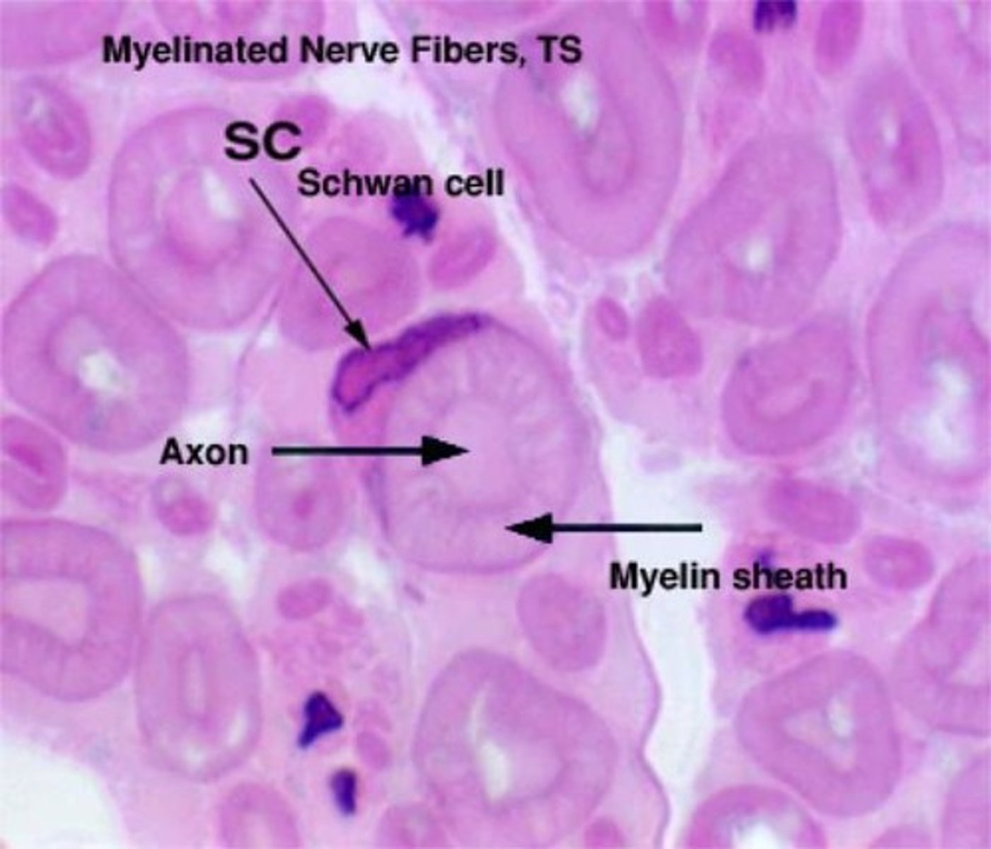
myelin sheath
lipid-rich layer directly surrounding an axon that acts as an electrochemical insulator
Schwann cells
What cells create the myelin sheath in the PNS?
neurilemma
additional thin external myelin sheath that is formed by Schwann cells ("squeezed" cytoplasm) and found just peripheral/external to the myelin sheath
represents Schwann cell cytoplasm and includes the nucleus and other organelles of the Schwann cell
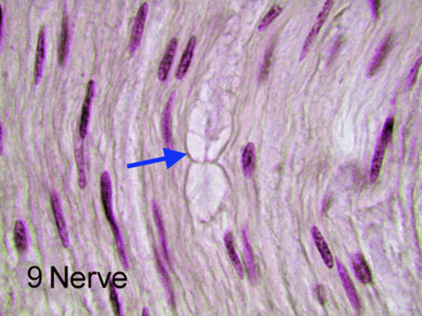
clefts of Schimdt-Lanterman
small islands of cytoplasm that can be found in certain regions of the myelin sheath; metabolically important for Schwann cells
node of Ranvier
junction between two adjacent Schwann cells and represents a small portion of the axon that does not have myelin
**interruption of the myelin sheath allows for saltatory conduction
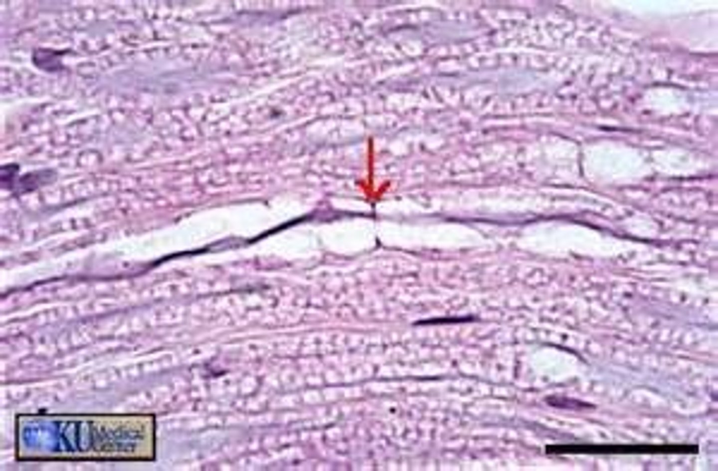
internode of Ranvier
region of myelin between two successive nodes of Ranvier; each one corresponds to one Schwann cell
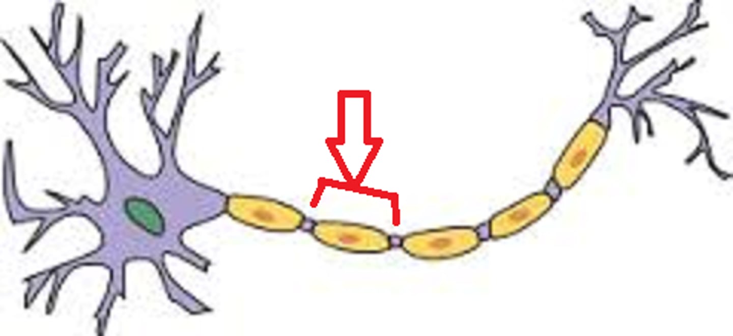
internode of Ranvier
What do we call one Schwaan cell covering the axon in myelin?
unmyelinated
refers to axons that do NOT conduct nerve impulses in a saltatory fashion (i.e. don't jump from node to node, continuous instead)
true
True or false: In the PNS, unmyelinated axons are still associated with Schwann cells.
grooves
In the PNS, unmyelinated axons are still associated with Schwann cells. Instead of multiple layers of Schwann cell membrane surrounding the axon (as in myelinated axons), the elongated Schwann cells develop what structures parallel to the long axis of the axons in which the axons reside?
**Each Schwann cell may have one ____ or over a dozen ____ and each ____ may contain one axon or several axons.
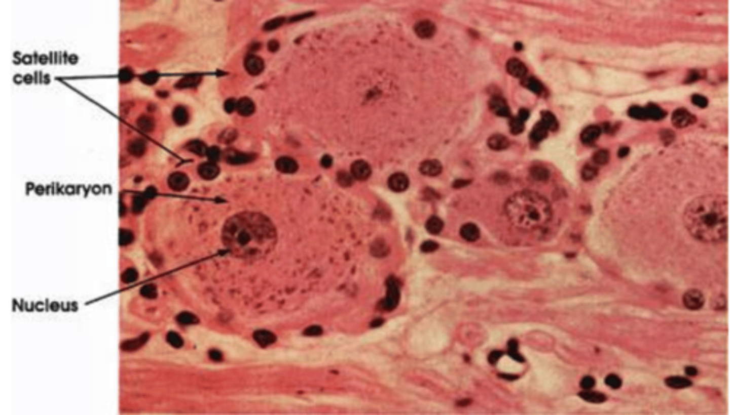
satellite cells (also called sustentacular cells)
PNS supporting cells that are associated with the cell bodies and are found in ganglia; they create the microenvironment for these neuron perikarya
single layer of cuboidal or squamous cells surrounding the soma
How do satellite cells usually appear?
oligodendrocytes (oligodendroglia, oligodendrogliocytes)
CNS supporting cells that produce and maintain the myelin sheaths associated with CNS axons
**Note: nodes of Ranvier are also present in the CNS because of these
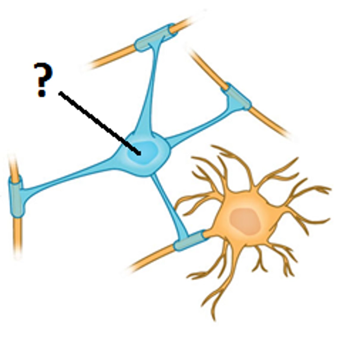
true
True or false: Each oligodendrocyte can myelinate more than one axon.
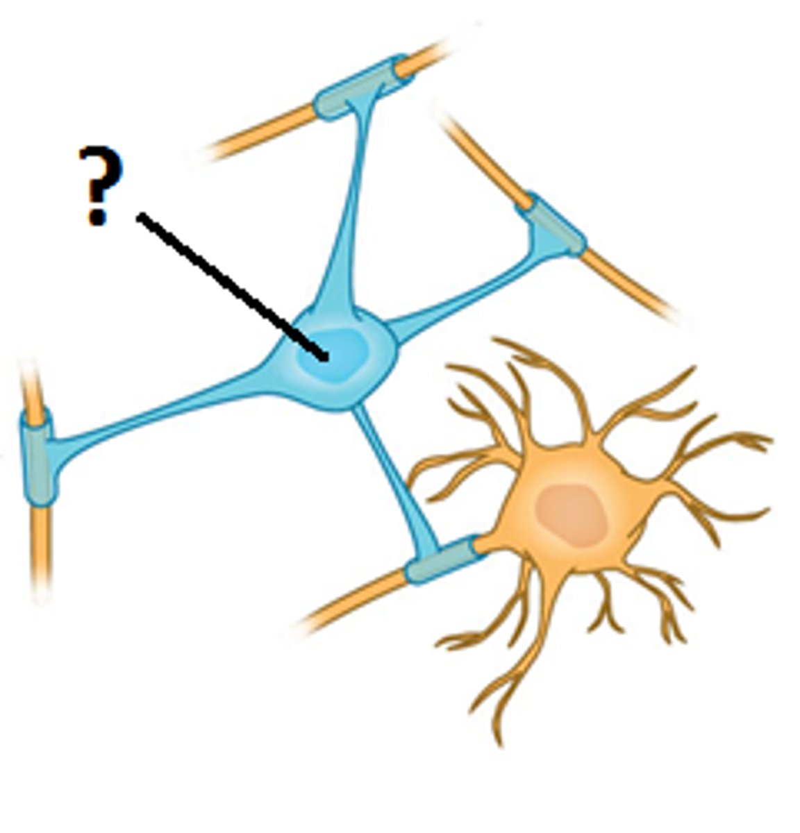
false
(astrocytes are associated with unmyelinated axons instead)
True or false: Similar to how in the PNS, unmyelinated axons are still associated with Schwann cells, in the CNS, unmyelinated axons are still associated with oligodendrocyte processes.
Schwann cells with both
In the PNS, what is associated with myelinated axons? Unmyelinated axons?
oligodendrocytes with myelinated, astrocytes with unmyelinated
In the CNS, what is associated with myelinated axons? Unmyelinated axons?
astrocytes
CNS supporting cells that are the largest glial cells and have processes that expand to form end feet, which surround all neuron cell bodies, unmyelinated processes, and synapses in the CNS to help control the metabolic environment of these structures, as well as blood vessels
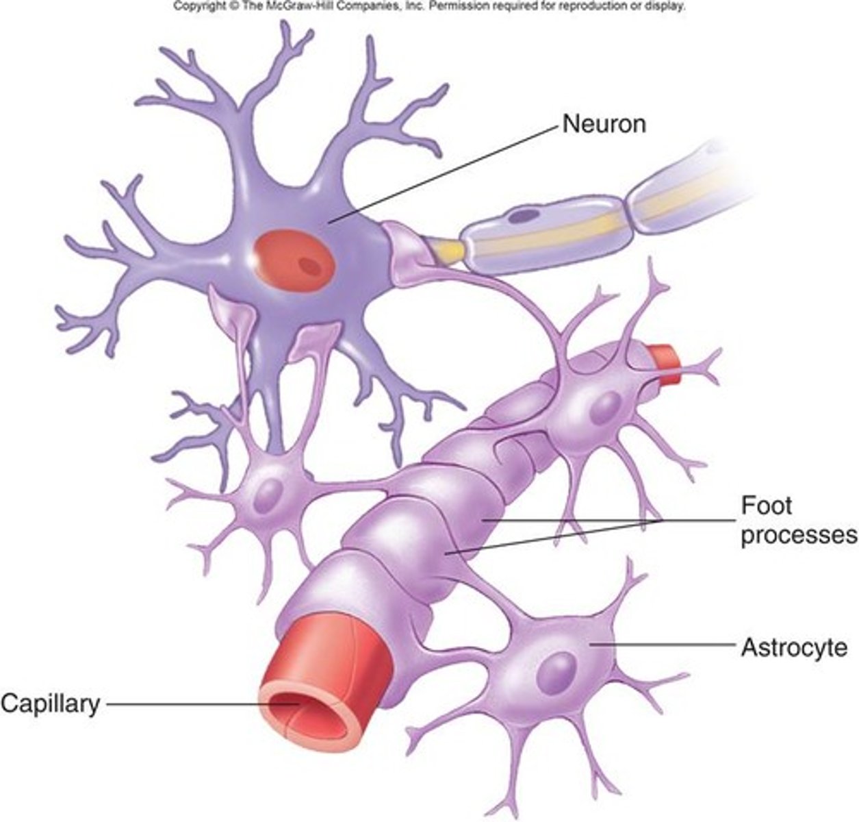
neuron cell bodies, unmyelinated axons, synapses, blood vessels
What all do astrocytes surround in the CNS? (4)
glial fibrillar acidic protein
special type of intermediate filament found in astrocytes in the CNS
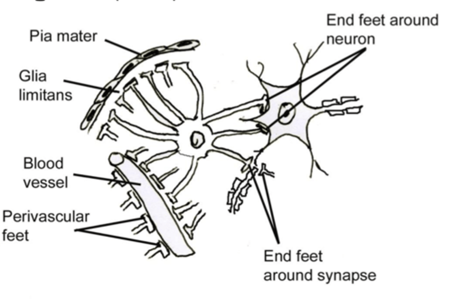
end feet
ends of astrocyte processes that expand to surround all neuron cell bodies, unmyelinated processes, and synapses in the CNS
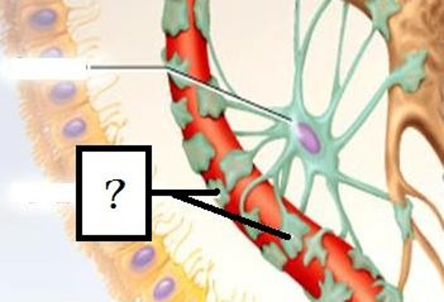
perivascular feet
astrocytic end feet that surround blood vessels in the CNS to help maintain the tight junctions between the epithelial cells that form the blood-brain barrier
glia limitans
Astrocytes adjacent to the pia mater (surrounding the surface of the CNS) form subpial feet that are associated with the basement membrane of the pia mater cells. These help form a relatively impermeable layer surrounding the central nervous system known as the what?
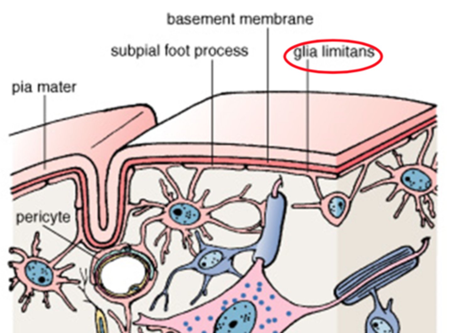
blood brain barrier, form the glia limitans
What are two functions of astrocytes in the brain?
gray matter
Where are protoplasmic astrocytes found?
**more numerous, shorter, and more branching processes
white matter
Where are fibrous astrocytes found?
**less numerous, longer, and straighter processes
microglia
CNS supporting cells that are actually small macrophages; therefore, these are the defense cells of the CNS
**originate from the embryonic yolk sac, NOT monocyte lineage
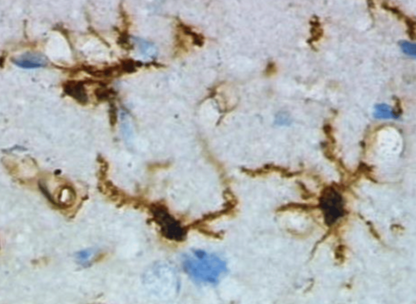
ependymal cells
CNS supporting cells that are the specialized simple cuboidal/columnar epithelium that lines the fluid-filled cavities of the central nervous system (i.e., the ventricles of the brain and the central canal of the spinal cord)
**basal domains of these cells are associated with astrocytic processes

choroid plexus
modified ependymal cells with their surrounding capillaries that produce CSF
(peripheral) nerve
bundle of neural fibers in the peripheral nervous system
nerve fiber
refers to an axon with its supporting cells
in the PNS: Schwann cells
in the CNS: oligodendrocytes or astrocytes
endoneurium
loose CT layer that surrounds each nerve fiber
perineurium
dense irregular CT layer that surrounds nerve fascicles (bundles of nerve fibers)
**metabolically active diffusion barrier that helps to form the blood-nerve barrier
epineurium
dense irregular CT layer that surrounds an entire nerve and fills in the space between nerve fascicles