Concepts of care for patients with skin conditions Exam 2
 Call Kai
 Learn
Learn Practice Test
 Spaced Repetition
 Match
Match Flashcards
 Knowt Play
1/52

There's no tags or description
Looks like no tags are added yet.
Name | Mastery | Learn | Test | Matching | Spaced | Call with Kai | Chat |
|---|
No analytics yet
Send a link to your students to track their progress
53 Terms
Pressure injuries
Loss of tissue integrity cause when skin and underlying soft tissue are compressed between a bony prominence and external surface
Results in reduced perfusion
Pressure inuries
Risk factors
Excessive interaction with moisture
Undernourishment
Aging skin
Mental impairment
Impaired nerves
Pressure injuries
Higher risk areas
Back of head and ears
Shoulder
Elbow
Lower back and buttocks
Inner knees
Heel
Shearing force
stationary and tissues below the skin moves
Friction
Surface rubs the skin and tears the epithelial tissue
Surface
Make sure your patients have the right support
Skin inspection
Early inspection means early detection
Show patients and carers what to look for
Nutrition/Hydration
Help patients have the right diet and plenty of fluids
Braden Scale
Predicting pressure ulcers
<18 needs to a turn patient (we turn them every 2 hrs)
Contributing factors to pressure ulcers
Immobility
Incontinence
Decreased sensory perception
Malnutrition
Cognitive problems
Contributing factors to pressure ulcers
Weight loss
Skin folds
Decreased level of consciousness
Impaired nutrition
Physical assessment for pressure ulcers
Inspect : esp bony prominences, BMI, skin, hair and nails
Mobility: loss of any
Wound assessment
Location
Size
Color
Extent of tissue involvement
Wound base
Exudate
Surrounding tissue
Epithelial & granulation
New tissue/scar tissue
Eschar
Necrotic tissue
Slough tissue
Will need to treat infection to see wound
Extra precautions of surrounding tissue
Check for foreign body to see if external factor is causing wound
Assessing patients with dark pigmented skin
Look for changes in skin color such as areas darker than surrounding skin
Use natural light to access color
Asses skin temp and edema
Ask about pain
Early sign of pressure related complications
Bacterial infection of skin and soft tissue
Cellulitis
Bacterial infection of skin and soft tissue
Undermining
Separation of skin layers around the wound margin
Stage 1 Pressure Injury
Intact skin with localized area of non-blanchable erythema
Changes in sensation, temp, or firmness
Color changes are not purple or maroon just pink
For darker skin loo faro darker areas and ask about pain
Stage 1 Pressure Injury
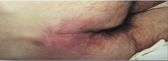
Stage 2 Pressure Injury

Stage 2 Pressure Injury
Partial-thickness loss of skin and exposed dermis
Wound bed is viable, pink or red and moist
May look like intact or ruptured serum-filled blister
Stage 3 Pressure Inury

Stage 3 Pressure Injury
Full thickness skin loss with adipose (fat) visible in the ulcer
Granulation tissue and rolled wound edges are often present
Underlining, slough and sometimes eschar tissue present
Stage 4 Pressure Injury

Stage 4 Pressure Injury
Full thickness skin loss with exposed or palpable fascia, muscle, tendon, ligament, cartilage or bone
Slough or eschar may be present
Undermining, rolled edges, tunneling often occur
High risk for infection
Osteomyelitis
High risk for this infection in stage 4 pressure injury
Unstageable injuries
Extent of damage cannot be confirmed because it is covered by eschar and slough
Unstageable injury

Suspected deep tissue injury
Purple or maroon localized area of discolored intact skin or blood-filled blister
Suspected deep tissue injury

Psychosocial assessment of pressure injury
Body image
SDOH
Laboratory assessment
Blood tests
Potential cultures
Diagnostic tests for pressure injuries
Arterial blood flow : if you don’t know the cause
Ex: not a nerve or turning issue
Duplex ultrasound : venous issue
Non surgical management: Dressings
Help remove debris off surface
Help protect healthy tissue
Make barrier from infection
Nonsurgical management: Debridement
Removing dead tissue
Debridement: Mechanical
Intrap and detach dead tissue
Wet or dry dressing put it on take it off and add new
Debridement: Topical
Type of debridement: Chemical
Use enzyme preparation (loosens UTO necrotic tissue)
Debridement: Surgical
Prior to starting dressing debridement and wound interventions
Surgical removal of dead tissue
Antibiotics
Drug therapy for wounds and pressure ulcers
Only to be used if patient has infection because can cause resistance
Nutrition therapy for wounds/pressure ulcers
Emphasize protein
Fats also necessary
Supplements/Alternatives for those who can’t eat
Adjuvant therapies
To be used only IN ADDITION to other interventions
Electrical stimulation
Low voltage to increase granulation
Hyperbaric oxygen
Enhances WBC to reduce swelling and kill bacteria
100 % oxygen tube
For life threatening wounds
Ex: burns
Topical growth factors
Something that’s naturally occurring and put artificially to enhance granulation
Ultrasound assured wound therapy
To cleanse and debread necrotic tissue
Negative pressure wound
Intermittent pressure helping to remove fluid bs promotes healing
Prevention infection
Monitor for signs and symptoms of infection
Report changes to primary healthcare provider
Maintain safe environment
CLEAN AND DRY
Home care management for wounds/pressure ulcers
Outpatient wound healthcare clinic
Home health care referral
Wedge that reduces pressure
Self management education for wounds/pressure ulcers
Well balanced diet
Increase fluids and protein
Self care and cleaning
Absorbent pads and barrier creams
Debridement: Natural
Let body self digest tissue