Exam 1 - Respiratory Block
 Call Kai
 Learn
Learn Practice Test
 Spaced Repetition
 Match
Match Flashcards
 Knowt Play
1/129

There's no tags or description
Looks like no tags are added yet.
Name | Mastery | Learn | Test | Matching | Spaced | Call with Kai | Chat |
|---|
No analytics yet
Send a link to your students to track their progress
130 Terms
what are some different types of URIs
Rhinitis, sinusitis, pharyngitis, tonsilitis, laryngitis, peritonsillar abscess
What is the most common cause for illness, reason for seeking health care and absences from school and work. May be minor, acute, chronic, severe, or life threatening. Can be treated in community settings: doctor offices, urgent care clinics, long-term care facilities, or self-care at home. Early detection of signs and symptoms and appropriate interventions can avoid unnecessary complications. Patient teaching focus on prevention and health promotion
Upper respiratory infections (URIs)
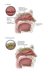
inflammation of the nose and sinuses
rhinitis and rhinosinusitis
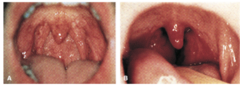
inflammation of the pharynx
pharyngitis
what do the assessment of patients with URIs include?
Health history
Signs and symptoms: headache, cough, hoarseness, fever, stuffiness, generalized discomfort, and fatigue
Allergies
Inspection of nose, neck, throat, and palpation of lymph nodes
what are some potential complications and collaborative problems with URIs
Airway obstruction (due to swelling)
Hemorrhage
Meningitis or brain abscess (nuchal rigidity) (due to chronic rhinitis or sinusitis)
Pneumonia
Sepsis
Medicamentosa (rebound congestion) (from nose sprays)
Acute otitis media (inner ear infections)
Trismus (TMJ spasm)
Dysphagia
Aphonia (loss of voice)
Cellulitis (periorbital cellulitis)
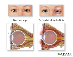
Infection of the eyelid and surrounding tissue, more common in children but can also occur in adults
periorbital cellulitis
what are some goals/planning for patients with URI
Airway management, reduce risk of aspiration
Pain management
Effective communication strategy
Normal hydration
what are some nursing interventions for patients with URIs
Elevate head (to decrease congestion)
Ice collar to reduce inflammation and bleeding
Hot packs to reduce congestion
Analgesics for pain
Topical anesthetics
Monitor for severe complications
Gargles for sore throat
Use alternative communication
Encourage liquids and use of room vaporizers or steam inhalation to keep secretions loose and moist for easier expectoration
Rest
what should you evaluate in patients with URIs
Maintenance of patent airway
Expresses relief of pain
Able to communicate needs
Evidence of positive hydration
Free of signs and symptoms
Absence of complications
what are some things to educate your patient about for URIs
Prevention of upper airway infections
Emphasize frequent hand washing
When to contact health care provider
Need to complete antibiotic treatment regimen
Annual influenza vaccine
Recognize signs of complications
what are some different examples of obstructions and traumas to the upper respiratory airway
Obstructive Sleep Apnea (OSA)
Epistaxis
Laryngeal Obstruction
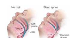
pharyngeal collapse during sleep, which leads to absence of breathing; can produce daytime drowsiness and elevated blood pressure
causes of this: loud snoring, obesity, large neck, being a male
obstructive sleep apnea (OSA)
what are some potential complications of obstructive sleep apnea (OSA)
Day time sleepiness
Hypoxemia/hypercapnia
Hypertension
Myocardial Infarction
Stroke
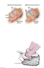
what are some ways to manage obstructive sleep apnea?
Medical Management
- Weight loss
- Mandibular advancement devices (MADS)
- CPAP/BIPAP
- Surgery
Patient education
Jaw thrust (common after surgery, first try to wake them up, if they dont wake up do a jaw thrust... get behind them, lay the head of the bed flat, put 4 fingers under the jaw and roughly thrust back)
Hemorrhage from the nose
Anterior septum, most common site
Serious problem, may result in airway compromise or significant blood loss
Risk factors, refer to Chart 18-5
epistaxis
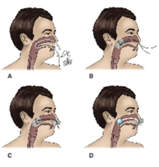
what are some medical managements for epistaxis
Identify cause and location (treat underlying cause)
Pinch soft portion of nose for 5 to 10 minutes, patient sits upright (DO NOT TILT HEAD BACK)
Phenylephrine spray, causes vasoconstriction so it will stop bleeding
Cauterize with silver nitrate or electrocautery
Gauze packing or balloon-inflated catheter inserted into nasal cavity for 3 to 4 days
Antibiotic therapy : only if there is an infection
what is the nursing management for epistaxis
Airway, breathing, circulation
Vital signs, possible cardiac monitoring and pulse oximetry
Reduce anxiety
Patient teaching:
- Avoid nasal trauma, nose picking, forceful blowing, spicy foods, tobacco, vigorous exercise
- Adequate humidification to prevent dryness
- Pinch nose to stop bleeding; if bleeding does not stop in 15 minutes, seek medical attention
what does the assessment of a patient undergoing laryngectomy look like?
Health history
Physical, psychosocial, and spiritual assessment (how they will cope because it will be life changing)
Nutrition, BMI, albumin, glucose, electrolytes (wait to eat until stoma is fully healed)
Literacy, hearing, and vision; may impact communication after surgery (make sure to establish different modes of communication for after the procedure)
Coping skills and available support systems for patient and family after surgery
what are some collaborative problems and potential complications for a patient undergoing a laryngectomy
Respiratory distress (due to occluded airway, mucus, edema)
Hemorrhage (surgical site is wet and moist so it is constantly open which can lead to irritation)
Infection (surgical site is wet and moist so it is constantly open which can lead to irritation)
Wound breakdown (surgical site is wet and moist so it is constantly open which can lead to irritation)
Aspiration (swallowing become difficult which can lead to this)
Tracheostomal stenosis (scar tissue formation, trouble removing mucus, respiratory distress)
what are some nursing interventions for a patient undergoing a laryngectomy?
Preoperative teaching
Reduce anxiety
Maintain patent airway, control secretions
Support alternative communication
Promote adequate nutrition and hydration
Promote positive body image, self-esteem
Monitor for potential complications
Self-care management; homecare (Chart 18-7, pg 522)
Motor vehicle accident, fall, work accidents, rib/sternal fractures can lead to lacerations
chest is struck by an object or fall.
blunt trauma

open wound through pleural space, severity depends on the location
object passes through the tissue, impalement
penetrating trauma
iatrogenic pneumothorax
Due to Medical Procedures
Spontaneous pneumothorax
Unknown Cause (VERY RANDOM)
Bleb
tension pneumothorax
Chest trauma
Emergency
air goes in but can not get out so it pushes on the lungs until it collapses
tracheal deviation!
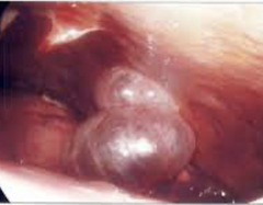
only known cause of a spontaneous pneumothorax, caused by ruptures of these air filled sacks
risk factors: smoking, tall and thin stature, male, genetics, prior occurrence
bleb
tension pneumothorax
MEDICAL EMERGENCY ... causes tracheal deviation
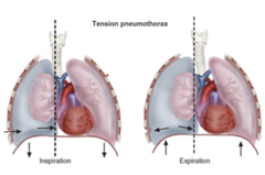
air in pleural cavity causing lung to collapse, always suspect this after blunt or penetrating trauma
Clinical manifestations
- Variable depending on severity
-- Mild tachycardia and dyspnea → severe respiratory distress (SOB, shallow breathing, low oxygen saturation, diminished lung sounds)
- Tension pneumothorax
-- tracheal deviation, neck vein distention, cyanosis, and profuse diaphoresis
Diagnosis - Chest Xray (if it is a tension pneumothorax do not wait to get an xray, you must treat it first)
Treatment - Chest Tube
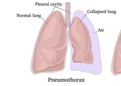
pneumothorax
pneumothorax
air in the pleural space
will be treated with chest tube
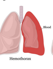
hemothorax
Blood in the pleural space
will be treated with chest tube
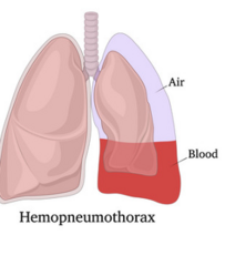
hemopneumothorax
air and blood in the pleural space
will be treated with chest tube
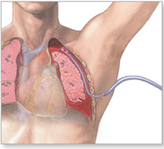
To remove air or fluid from pleural and/or mediastinal space
Reestablishes negative pressure
Lung re-expands
chest tube and pleural drainage
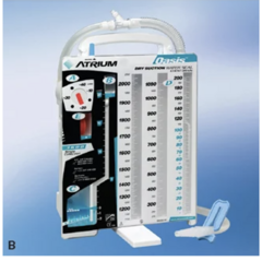
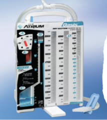
drain system
MAKE SURE C DOES NOT HAVE ANY BUBBLES IN THE COMPARTMENT.... THIS COULD MEAN AN AIR LEAK
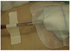
how should the nurse manage a patient with a chest tube?
Assess patient's clinical status
-- Vital signs, lung sounds (diminished), pain, subcutaneous empysema (air leaking under skin, feels like you are pressing on bubble wrap)
Dressing
-- Sterile, occlusive
-- Drainage site infection
Encourage deep breathing, range-of-motion exercises, incentive spirometry
how should the nurse manage the drainage system
Maintain drainage system
- Patency (NO LOOPS OR KINKS OR CLAMPS)
- Air leaks (tape all connections)
Bubbling
- Tidaling (ball moving up and down)
- Position below heart
- DO NOT milk or strip the tubing (basically dont squeeze the tube)
Assess drainage (mark with date, time, initials. if the patient has more than 200 CC in the first hour this could indicate the patient is bleeding more than normal.... after that if there is more than 100CC during the hour... this is concerning)
what are some issues with chest tubes
If break in system, place distal end in sterile water to maintain water seal
If chest tube comes out, place Vaseline gauze on incision and call provider
what are some complications with chest tubes
Re-expansion pulmonary edema
Vasovagal response
how do we remove chest tubes
When lungs re-expanded and drainage minimal
Pre-medicate prior to removal
Valsalva maneuver during removal
Apply occlusive dressing
Chest x-ray is done
Monitor for respiratory distress (bc the lung could possibly collapse again)
Can damage pleura, lungs, and internal organs
Clinical manifestations
- Pain
- Splinting
- Shallow respirations
Atelectasis and pneumonia
rib fractures
how do we treat rib fractures
NO strapping or binding chest
NSAIDs, opioids, nerve blocks
Patient teaching
Deep breathing and coughing
Incentive spirometry
Appropriate use of analgesics
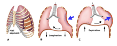
fracture of two or more adjacent ribs in two or more places that allows for free movement of the fractured segment
asymmetric movement when chest expands
flail chest
- increase HR
- pain
- splint
- crepitus
Diagnosis
- xray
Management
- Adequate airway and ventilation
- Oxygen therapy
- Analgesia
- Surgical fixation
flail chest clinical manifestations
what are the main symptoms of respiratory distress
Dyspnea
Cyanosis
Hemoptysis - coughing blood
Tracheal Shift
Diminished or absent breath sounds
Vital Signs
Frothy secretions
what are the main symptoms of cardiovascular compromise
Rapid, thready pulse (means heart is not pumping correctly)
Decreased BP with narrowed pulse pressure
Distended neck veins
Muffled heart sounds
Chest pain
Dysrhythmias
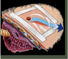
what are the emergency interventions (ABCs) due to chest trauma
Initial Administer O2 to keep SpO2 >90%
Establish IV access with 2 large-bore catheters and begin fluid resuscitation as appropriate
Remove clothing to assess injury
Cover sucking chest wound with nonporous dressing taped on 3 sides
what are the initial emergency interventions for chest trauma
Stabilize impaled objects (DO NOT REMOVE)
Assess for other significant injuries and treat appropriately
Place patient in a semi-Fowler's position or on injured side (it helps them breath easier)
After ruling out cervical spine injury
Administer analgesia
Prepare for emergency needle decompression
Potential Intubation due to respiratory distress
Chest Trauma: Emergency Management - Ongoing Monitoring
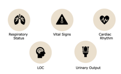
**urinary output because if the heart is not perfusing to the kidney.... the urinary output will drop
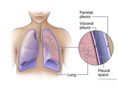
disorders that involve the pleural space
pleural conditions
inflammation of both layers of the pleura
pleuritic pain (on inhalation/exhalation and coughing), pleural friction rub
diagnostic tests: chest xray, sputum culture, thoracentesis
treat underlying cause, provide analgesia, teaching to splint the rib cage when coughing
pluerisy
accumulation of pus
clinical manifestations similar to pneumonia or acute respiratory infections
decreased breath sounds
chest ct/thoracentesis
antibiotics
nursing management
empyema
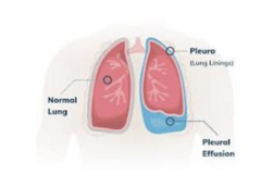
fluid collection in pleural space (an underlying cause could be cancer), you will hear diminished lung sounds
pleural effusion
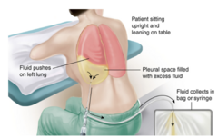
surgical puncture to remove fluid from the pleural space
relieves symptoms from the fluid but will not treat the underlying cause
thoracentesis
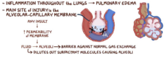
acute respiratory distress syndrome
Identification and treatment of underlying cause
Intubation, mechanical ventilation with PEEP to keep alveoli open
Treat hypovolemia to keep hemodynamically stable
Prone positioning is best for oxygenation, frequent repositioning to safeguard integumentary system
Nutritional support, enteral feedings preferred
Reduce anxiety, sedation, paralysis
Supportive care
medical managment of ARDS
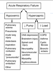
lungs are failing... if we can not ventilate, we are not getting oxygen
acute respiratory failure
Method of positive-pressure ventilation that can be given via facemasks that cover the nose and mouth, nasal masks, or other oral or nasal devices such as the nasal pillow (they have to be able to maintain their own airway)
Eliminates need for endotracheal intubation or tracheostomy
Continuous positive airway pressure (CPAP)
Bilevel positive airway pressure (BiPAP)
Noninvasive Positive-Pressure Ventilation
a mask that fits over the sleeper's nose and mouth, which is connected to a pump that pumps air into the person's airways, forcing them to remain open
continuous positive airway pressure (CPAP)
noninvasive spontaneous breath mode of mechanical ventilation that allows for the separate control of inspiratory and expiratory pressures; given via a mask
THIS CAN INITIATE A BREATH FOR THE PATIENT WHILE CPAP CAN NOT !!!!!!
bilevel positive airway pressure (BiPAP)
fluid in lungs
cardiogenic (heart failure) vs noncardiogenic (injury to the capillary beds from smoking, trauma, etc) - use diuretic to get rid of fluid
life threatening
pulmonary edema
PAP >30 mm Hg with heart failure
increased blood pressure which leads to right sided heart failure ... treat the underlying cause
pulmonary hypertension
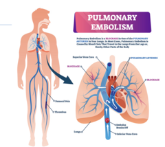
blockage in one of the pulmonary arteries in your lungs. in most cases it is caused by a blood clot that travels to the lungs from the legs and rarely other places in the body
symptoms will vary on the size of the clot
- SOB, chest pain, increase heart rate, anxiety
give heparin (less serious cases) or TPA (severe cases) to dissolve clot
can put a filter in IVC to catch future clots
pulmonary embolism
speak to respiratory status. not just RR and oxygenation, but tachycardia and acute hypertension can also be signs of respiratory distress because the body is compensating for low oxygen by increasing heart rate and pressure
vital signs
what can indicate respiratory distress
labored breathing, cyanosis, change in LOC, flared nose/ribs
what will you hear when auscultating breath sounds during respiratory distress
1. crackles: fluid
2. wheezing: narrowed airways
3. coarse: mucus buildup
4. diminished/absent: fluid/blood
Measures acid-base balance
respiratory will usually come to the bedside to draw from the wrist. after.... monitor site for bleeding, hematoma, notify provider of the results. this is done for anyone in respiratory distress because it tells us right away how severe the problem is and how immediately we need to act
Arterial Blood Gases
Thin tube with camera at the end is put down mouth or nose to view airways.
prior to, patient must be NPO and consented. post procedure, monitor for aspiration and pneumothorax. used more for a dx COPD or monitoring progression of lung diseases, not used in acute situations
Bronchoscopy
Measures lung volumes, flow rates, and gas exchange
non invasive, no anesthesia so no post procedure risks, before make sure that patient does not smoke or use inhaler for 6 hours
NEVER USE IN AN EMERGENCY SITUATION
Pulmonary Function Tests
what complications can occur due to URIs
periorbital cellulitis, pneumonia, occluded airways, meningitis
what nursing interventions/education can be implemented
no crowded places when sick, wash hands, VACCINES, antibiotics (if bacterial) - MAKE SURE TO FINISH THE ANTIBIOTICS
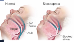
airway collapses when you sleep (gurgling noise and snoring)
risk factors: obesity, big necks, men, loud snoring
complications: hypertension, MI, stroke
treat with: CPAP, mouth piece, or surgery
after surgery with an oxygen drop: first try to wake them up... if they do not wake up, do a jaw thrust
obstructive sleep apnea
put palms on their cheeks
fingers under the mandible
thumbs used to open mouth
thrust head back
jaw thrust
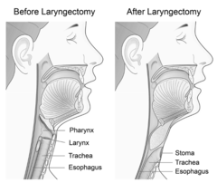
surgical removal of the larynx
complications: airway obstruction, aspiration, infection, scar tissue stenosis, mucus obstruction
laryngectomy
common complication where a connection forms between the pharynx can the skin or stoma. fluids can leak through this fistula and enter the airway, leading to aspiration
pharyngocutaneous fistula (PCF)
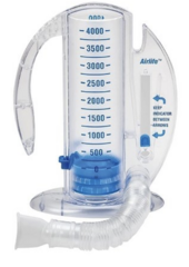
what is this? how is it used? what is it used for?
it is a spirometer. the patient is going to suck in, taking 10 breaths SLOWLY, once an hour (set a goal and follow the ball on the side to make sure they are sucking at a slow enough pace). it is used to encourage slow deep breathing to PREVENT ATELECTASIS
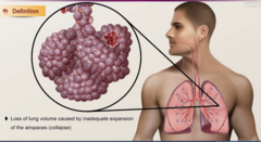
collapse of alveoli. these are little air sacs at the end of the lung where gas exchange takes place. it can be acute or chronic, but more commonly acute. there are lots of risk factors for this but most important: IMMOBILIZATION, SHALLOW BREATHING, and ANESTHESIA. not moving around as much and shallowing breathing means those alveoli arent being fully inflated, therefore more likely to collapse.
atelectasis
do these for an assessment and diagnosis for __________
increased work of breathing, retractions in between the ribs, decreased SPO2 on pulse ox, abnormal ABGs, decreased breath sounds or coarse/crackle sounds
patients can be asymptomatic, but if it progresses they can experience respiratory distress symptoms. if there is mucus build up they are at risk for infection. as it gets worse they could have increased HR and RR. they may also have chest pain that worsens with inhalation. they can also have central cyanosis that can be seen if it is really severe. most patients have anxiety and want to sit up to help lungs expand more easily.
use chest xray to diagnose this (it will clearly show a collapse in one or more lobes)
atelectasis
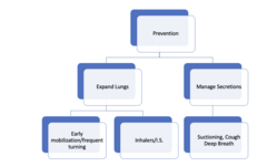
nursing interventions for atelectasis
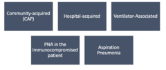
CAP is most common
hospital is while in hospital ... not before admission
ventilator is caused by bacteria getting into the lungs due to ET tube
immunocompromised get opportunistic and rare pneumonia (fungal)
pneumonia classifications
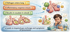
pneumonia pathophysiology
what are some risk factors for pneumonia
CHF, DM, CF, COPD, HIV/AIDS, Ca, alcoholism, increasing age
GET THIS VACCINE
reduces the incidence of pneumonia, hospitalizations for cardiac conditions, and deaths in the older adult population.
two types of vaccines
recommended for all adults 65 and older as well as 19 year olds and older with weakened immune systems
pneumococcal vaccination
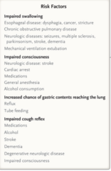
risk factors for aspiration pneumonia
mainly anyone with impaired swallowing, decreased LOC, or decreased cough reflex
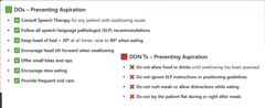
DOs and DONTs to prevent aspiration in at risk patients
Varies depending on type, causal organism, and presence of underlying disease
Respiratory symptoms: Productive cough, crackles/coarse breath sounds, orthopnea, central cyanosis, pleuritic chest pain, tachypnea, tachycardia, and respiratory distress
Generalized symptoms Other: fever, chills, headache, myalgia, rash, flushes cheeks, change in LOC or mental status, fatigue
Gradual onset - URI
clinical manifestations of penumonia
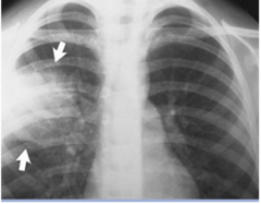
made by a history and physical, chest xray, blood cultures to confirm or rule out bacteremia, sputum examination and culture. bronchoscopy may be used in severe cases.
in this photo it indicates a more opaque left side which shows fluid or mucus build up indicative of pneumonia in the area.
for planning you want to ensure hydration and nutrition, make sure the patient understands the treatments and preventative measures, monitor for complications, and try to prevent complications... do discharge planning specifically to the patients needs
some nursing diagnoses: ineffective airway clearance, impaired gas exchange, activity intolerance
diagnosis of pneumonia
what are some ways we can treat pneumonia
1. antibiotics (only if it is bacterial and should be based on culture and sensitivity)
2. antipyretics (fever)
3. antitussives (cough)
4. decongestants (congestion)
5. antihistamines
what are some nursing interventions for pneumonia
1. hydration/nutrition/rest (give fluids to ensure adequate hydration as fevers are common and causing fluid loss. also fever and infection increases metabolic rate, causing fluid loss and weight loss. patients may also have decreased appetite or unable to eat - therefore nutrition is important. rest and activity as tolerated are important)
2. oxygen/CPT/IS (if gas exchange is impaired, IS is to expand lungs and prevent atelectasis, CPT to break up and drain mucus) - CPT is chest physiotherapy, this is when they pound on the chest to get the patient to cough, but now we have vibrating vests that help as well (cystic fibrosis patients especially)
3. patient education (finishing antibiotics, decreasing spread, and explaining all the medical therapies to the patient and family)
4. gerontologic considerations (more difficult to treat and higher mortality. diagnosis can be missed bc they may not have classic symptoms)
what are some complications of pneumonia
1. sepsis and septic shock (SEPSIS IS GOING TO BE THE NUMBER ONE COMPLICATION OF INFECTION. the main symptoms for sepsis are fever, chills, confusion, SOB, rapid heart rate, and pale sweaty skin.
2. respiratory failure (indicated by severe SOB, rapid or shallow breathing, cyanosis, confusion, extreme fatigue, rapid/irregular heartbeat, excessive sweating)
3. pleural effusion
4. delirium (ACUTE change in mental status and has identifiable cause - very common in elderly when they have infectious process going on. if your patient was AAOx4 and now they are not, suspect delirium and notify the provider)

Debbie is a 69-year-old female with a persistent productive cough with green sputum that started 4 days ago. Three days ago, she was started on prednisone 40 mg PO daily, Augmentin XR 2000 mg PO BID, and azithromycin 500 mg PO daily. She complains of intermittent chills, fever of 102 overnight, and difficulty breathing. She also admits to using her albuterol inhaler every 1-2 hours with no improvement. She called 911 and was brought to the ED where you are assuming care. She has a history of COPD, HTN, high cholesterol, and anxiety. She also has a history of smoking 1 pack per day of cigarettes.
Which findings from the present problem are most important and considered clinically significant?
What condition is the patient likely experiencing?
the highlighted information is clinically significant, while the difficulty breathing is the most important (A,B,Cs)
the patient most likely has pneumonia
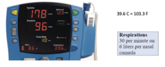
Which vital signs are recognized as most important and clinically significant?
Most important: SPO2, RR, temperature
Clinically significant: ALL OF THEM
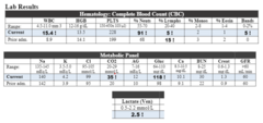
Which lab results are recognized as most important and clinically significant?
1. WBCs increased from prior admit and high - suggest an immune response - increased neutrophils suggest bacterial infection
2. CO2 - end stage COPD patients or patient in exacerbation retain CO2, could be indication of poor gas exchange
3. glucose - prednisone/stress on the body (not significant until it is in the 300s)
4. lactate - MOST CONCERNING, sepsis, could go into septic shock, lead to organ failure, and hemodynamic instability
Which of the following orders does the nurse anticipate the MD to order, Which one should be done first?:
A.Chest X-ray
B.NPO
C.IV antibiotics
D.Sputum Culture
E.Bed rest
F.Titrate supplemental oxygen to achieve oxygen saturation >90%
order A, C, D, and F
do first: F.Titrate supplemental oxygen to achieve oxygen saturation >90% - DO THIS FIRST BECAUSE OF OXYGEN!!!!!!
Vancomycin IVPB is prescribed, and the nurse administers it as ordered. 10 minutes into the infusion, the patient complains of shortness of breath and itching. What should the nurse do first?
A.Call the provider
B.Slow down the infusion, because it is a vesicant
C.Stop the infusion
D.Monitor oxygen saturation
E.Tell the patient this is a normal reaction
C.Stop the infusion
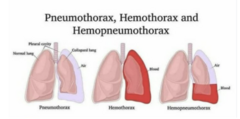
explain what a pneumothorax, hemothorax, and hemopneumothorax is and what the S/S are, how to diagnose it,what the treatment is, and how lungs sounds would sound.
pneumothorax: air in the pleural space, chest trauma causes this
hemothorax: blood in the pleural space, chest trauma causes this
hemopneumothorax: air and blood in the pleural space, chest trauma causes this
S/S: resp distress, tracheal deviation (tension pneumothorax), check by xray
diagnose by: XRAY
treat: chest tube
diminished breath sounds because there is air or blood blocking the lungs, so the sounds would be diminished
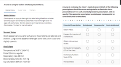
Fill in the bubbles
Obtain ABGs: anticipated
Prepare for insertion of chest tube: anticipated
Obtain intravenous access: anticipated
Computed tomography (CT) of the chest: nonessential
Pulmonary function tests (PFT): nonessential (NEVER USE IN EMERGENCY SITUATIONS)
Thoracentesis: contraindicated (it wont reinflate the lungs, it will only remove the fluid)

What is this?
What are the most important nursing goals? to address?
flail chest (asymmetrical chest movement)
1. oxygenation (mechanical ventilation
2. help with pain
3. fluids
4. get patient ready for surgery (NPO)
**make sure ribs do not puncture the surrounding organs so prep for surgery
Kevin Long, a 40-year-old patient, is admitted with the diagnosis of small cell carcinoma of the left lower lobe. The patient had a left lower lobectomy removing the cancerous mass. After surgery, the patient has a chest tube to a closed water-seal drainage system.
1.What are the nursing responsibilities when caring for a patient with a chest tube to a drainage system?
1. assess the patient first. check for color, LOC, airway patency, respiratory rate, depth, rhythm, chest movement (symmetry), lung sounds, lips, and nail beds. assess the chest tube insertion site for intactness, dressing occlusive, crepitus, listen for air leak at the tube site. assess connections. assess tidaling in the waterseal chamber. fluid flunctuates in the tubing with breathing. if pt is on wall suction, nurse will disconnect suction to assess. examine drainage color, consistency, amount, clots. more than 200 in the first hour and more than 100 in an hour after that is CONCERNING (call provider). mark drainage at least once a shift (time, date, initials). NO LOOPS or CLAMPS. keep below the chest. OBSERVE BUBBLES IN CHAMBER C... bubbles indicate air leaks. keep VASELINE GAUZE, 4x4s, PADDED HEMOSTAT, and STERILE WATER AT BEST SIDE
Kevin Long, a 40-year-old patient, is admitted with the diagnosis of small cell carcinoma of the left lower lobe. The patient had a left lower lobectomy removing the cancerous mass. After surgery, the patient has a chest tube to a closed water-seal drainage system.
2.The chest tube is accidentally disconnected from the drainage system, and the drainage system is cracked. What should the nurse do?
place tube immediately in a bottle of sterile water at the bedside. pt will need a NEW drainage system. the bottle of sterile water is a temporary measure. assess vital signs and breath sounds in all fields. assess for dyspnea, anxiety, skin temperature, moisture level, complaint of pain, and arrhythmias
Kevin Long, a 40-year-old patient, is admitted with the diagnosis of small cell carcinoma of the left lower lobe. The patient had a left lower lobectomy removing the cancerous mass. After surgery, the patient has a chest tube to a closed water-seal drainage system.
3. The patient has to be transferred, what do we do with the chest tube?
DO NOT CLAMP, DO NOT EMPTY, DO NOT MILK, simply unhook from suction and keep BELOW LEVEL OF HEART