MICI 3114
 Call Kai
 Learn
Learn Practice Test
 Spaced Repetition
 Match
Match Flashcards
 Knowt Play
1/12

There's no tags or description
Looks like no tags are added yet.
Name | Mastery | Learn | Test | Matching | Spaced | Call with Kai | Chat |
|---|
No analytics yet
Send a link to your students to track their progress
13 Terms
What are the differences between prokaryotes and eukaryotes?
Prokaryote= before nucleus, smaller, unicellular, archaea bacteria
Eukaryote= new nucleus, larger, specific organelles, uni or multicellular, Specialized cell types (Immune, organ cells, blood cells etc) Slime molds, Yeast and other fungi Plants and Animals
What are the features of Archaea?
Prokaryotic, unicellular, diverse ecological niches, similar protein translation and transcriptional paradigms to eukaryotes ( no pathogens described to date)
Incredible biotech and industrial uses ( heat stable enzymes)
all life forms require water to survive
increase pressure to prevent water from boiling above 100 degrees
water can exist in different forms depending on the pressure (archaea - some can also survive these extreme heats)
How are Archaea distinct prokaryotes?
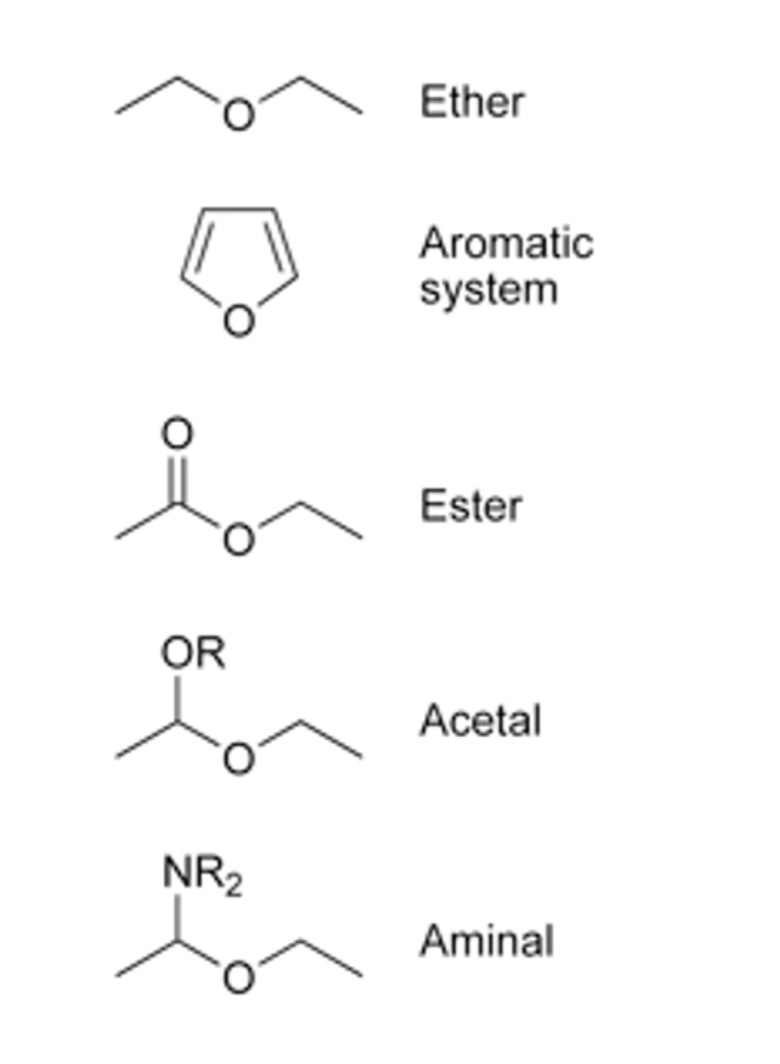
• Ether linked lipids (More stable?, For harsher environments?)
• A hybrid of bacterial and eukaryotic cellular functions
• Prokaryotic, although they have histones, transcription machinery like eukaryotes
• No introns and a single chromosome (like bacteria)(generally)
• Swim by using a distinct structure not like bacterial or eukaryl flagella
• One archaeon grows above 121 degrees Celcius! ( higher than an autoclave!)
• PCR and thermostable polymerases
• Proofreading activity in a single unit!
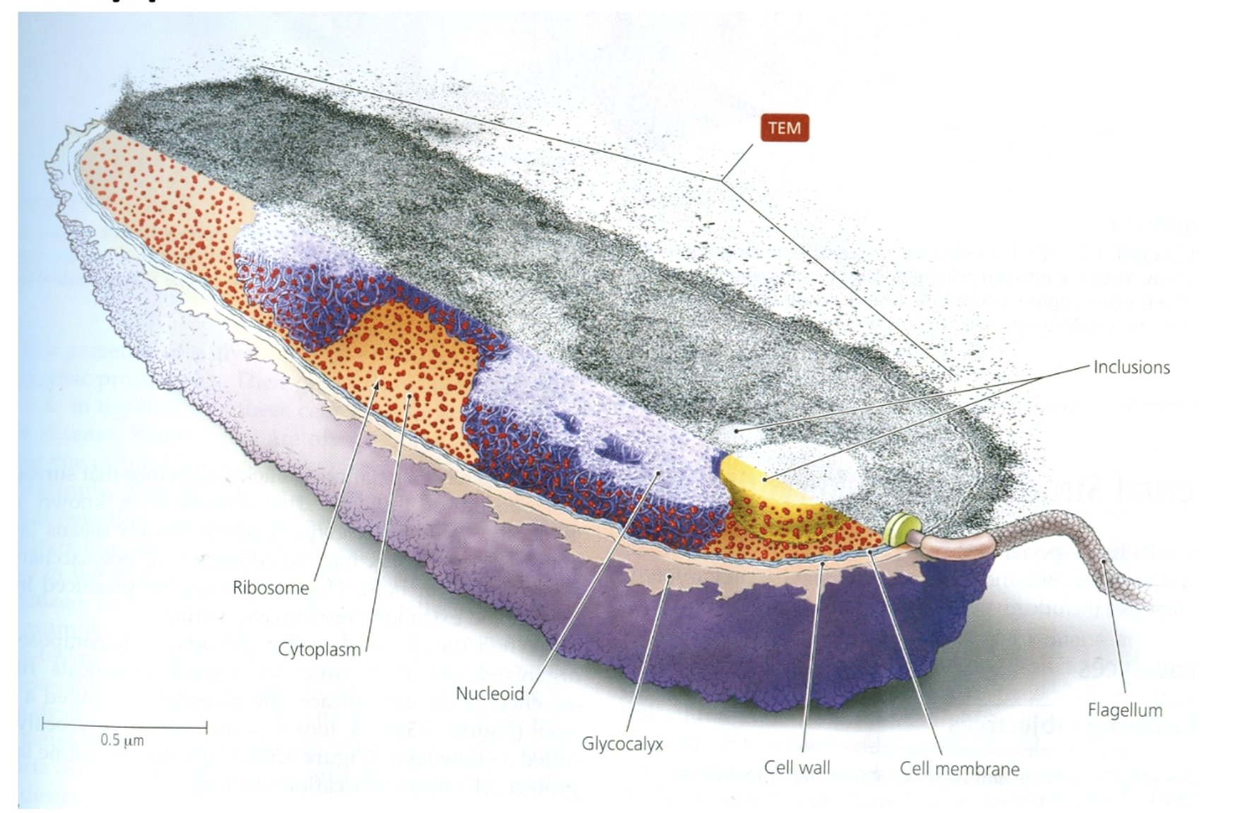
What are the physical components of a prototypic bacterial cell?
Ribosome
Cytoplasm
Nucleiod
Glycocalyx
Cell wall
Cell membrane
Flagellum
Inclusions
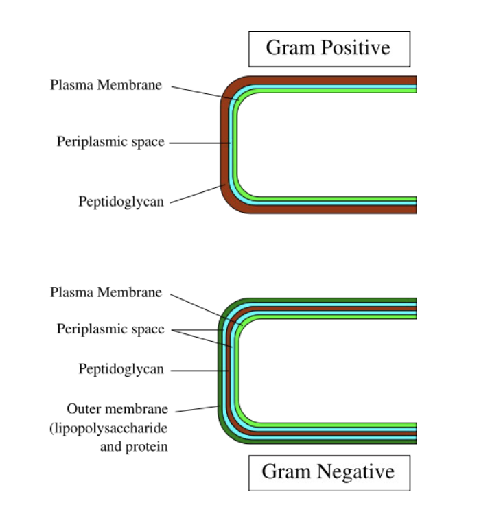
What are the features of the Gram Staining process?
• After the dutch scientist Christian Gram
• Crystal violet forms a complex with iodine that cannot ‘escape’ from thick cell wall
• Not perfect for identification purposes but generally useful for rudimentary (basic) analyses
Gram + has thick peptidoglycan
Gram - has thin peptidoglycan and has an outer membrane
flood cell with crystal violet then add iodine ⇒ complex that is too big to leak out the thick peptidoglycan in gram + and stain purple
gram - has saffron that stains on pink (opposite to the image)
What are the features of the peptidoglycan layer?
• Some bacteria have mDAP: Meso-diaminopimelate, a derivative of lysine in their tetrapeptide
• Some bacteria use a pentaglycine chain to cross link glycans
peptidoglycan= polymer of N-Acetyl Glucosamine (NAG) and N-Acetyl Muramic Acid (NAM) it is a crosslinked network (cage around bacterial cell) provides cell rigidity and cell shape and are crosslinked by the tetrapeptides (two of them) (most made like this but there are alternatives)
peptidoglycan peptide (peptides) , glycan (sugar) mesh work that provides structure for the exterior of the cell
some bacteria might have mDAP (specific a.a.)
pentaglycine chain

What are the features of the Gram positive cell wall?
• Thick peptidoglycan layer
-Cross-linked NAG and NAM polymers
• Lipotechoic acids (LTA) are a major component of Gram positive cell walls
• LTAs are recognized by components of the innate immune system in higher eukaryotes
-Toll like receptors

What are the features of the Gram Negative outer membrane?
• A SECOND lipid bilayer that contains polysaccharide
→Polysaccharide is linked via a lipid moiety to form lipopolysaccharide (LPS)
• Core polysaccharide
→Ketodeoxyoctonate (KDO), heptoses, glucose, galactose and N-acteylglucosamine
• Connected to the core is O-specific polysaccharide (hexose sugars)
• Lipid A (part of LPS) is embedded in the OM
→In an ester linkage with a disaccharide
→KDO is covalently linked to the disaccharide of Lipid A
KDO is exclusive to Gram negative bacteria (no OM in Gram positives!)
Lipid A – Endotoxin that can cause severe illness
outer membrane is a problem for antibiotics that want to target and kill the cell intracellularly many layers for the drug to get through
use porin molecules or channels (the tubes image bottom right)
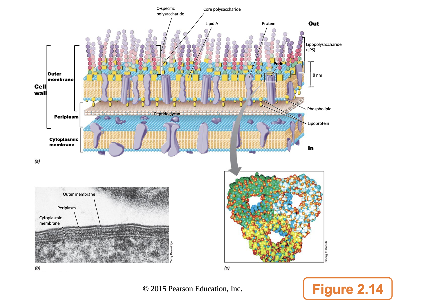
What is the general composition of LPS?
lipid A linked to KDO liked to the core polysaccharide and the o-specific polysaccharide
n= repeated length of the sugar (often signature of specific strand of bacterial cells)
lipid A closest to cytoplasmic membrane
O-specific antigens would bind to the o specific polysaccharide

What is a bacteria capsule?
• Dense, well defined polysaccharide or protein layer closely surrounding a cell
→Often serves a protective function
→Bind water, may serve to resist desiccation?
• Can promote adherence to host cell surfaces (promoting colonization)
→Or can aid in cell to cell contact to promote biofilm formation and microcolonies
• Encapsulated bacteria resist phagocytosis
• Capsules require resources (sugars)
→Bacteria may not ‘build’ capsule when nutrients are plentiful
sugars tend to be sticky
can help bacterial cells stick together to create biofilm and form layer
phagocytosis: macrophages engulf and eat foreign particles
encapsulated bacteria have additional layer making it difficult for the marcrophages to recognize as something to destroy
capsules are energy intensive and takes ressourcces to assemble it
What is the surface layers (S layers)?
• Protein ‘coat’ or layer that forms on the outer surface of bacterial cells
• The S-layer proteins (usually 1 or 2) are secreted out of the cell in copious amounts and are generally triggered by an environmental cue (ion, metal, etc) to form a highly ordered lattice
→ Crystallization
→ S-layer protein is often glycosylated
• Examples of S-layer functions:
→ protect the bacterial cell from exterior forces or attacks
→ adhere to surfaces, aid in biofilm formation
→ aggregate with other cells
proteins are often glycosylated→ added sugar makes them sticky and also identifying
image:
organisms form S layers- main point is the surface layer proteins are secreted across the cytoplasmic membrane and peptidoglycan once their they crystalize and perform their functions
can also form S layers

What are the general features of cell division? When bacterial cells divide, what happens to their cell walls?
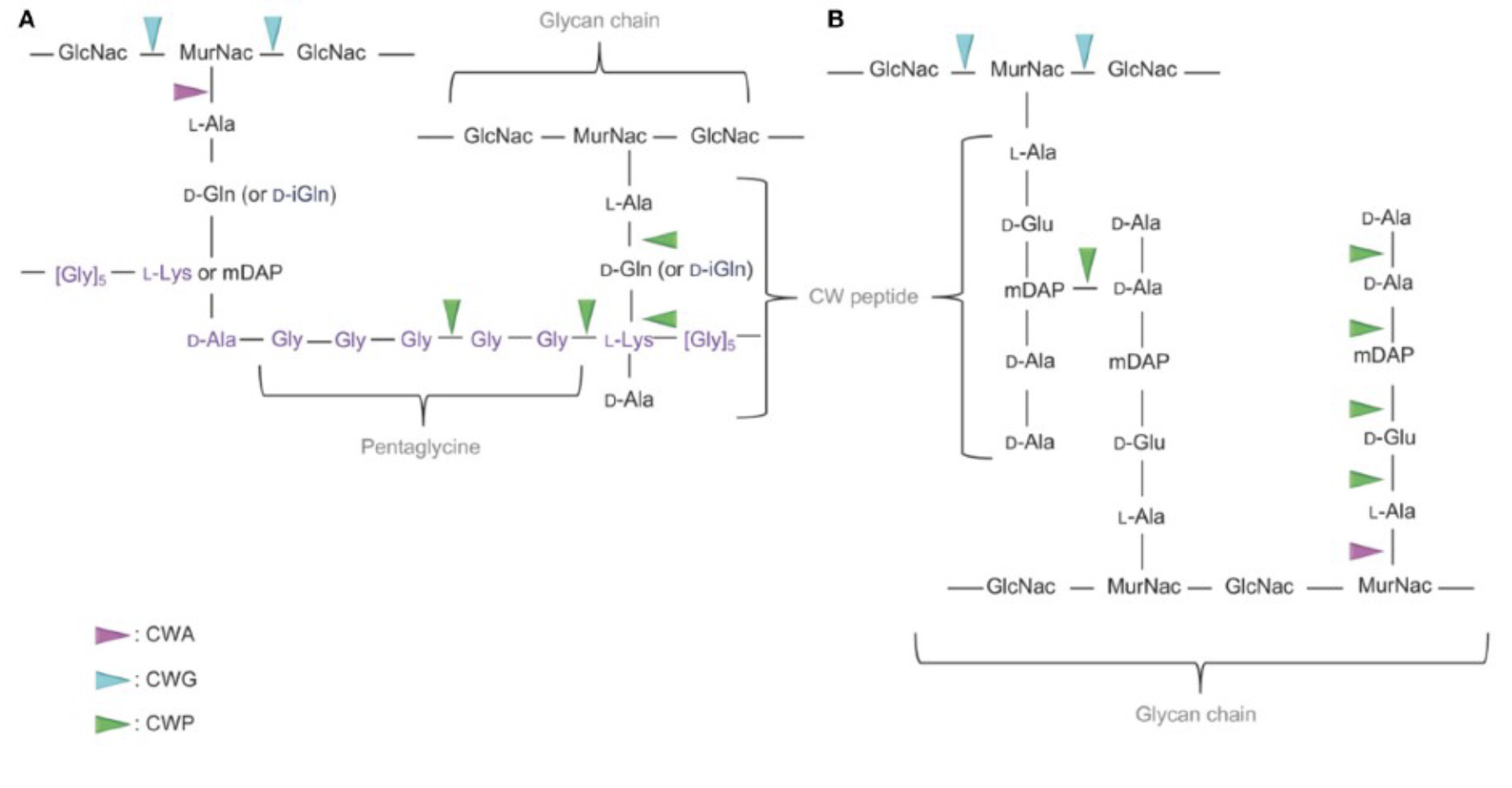
• Cell wall hydrolases (enzymes) must degrade the cell wall at a specific site (septum) to separate the daughter cells
• 3 classes of cell wall hydrolases:
• Cell wall amidase
• Cell wall glycosidase
• Cell wall peptidase
covalent bonds (hard to break w/out energy or enzymes)→ peptidoglycan
if cell is going to replicate needs to break the peptidoglycan via cell wall hydrolases (need water to work with enzyme to break the bonds) break the bonds at specific sites at the septum to create two even cells
amidase breaks amide bond
glycosidase breaks sugar or glucose bond
peptidase breaks peptide bond
breaking glycine bonds at NAM and NAG repeats
CWA amidase
CWG is glycosidase
CWP is peptidase
What are the features of cell wall recycling and repair?
• Bacteria have machinery to regenerate, repair, and even recycle peptidoglycan
• Remember that NAG and NAM are rich in carbon and nitrogen
• These are important commodities in nutrient limiting conditions
bacteria can suffer damages and the cell needs to repair peptidoglycan
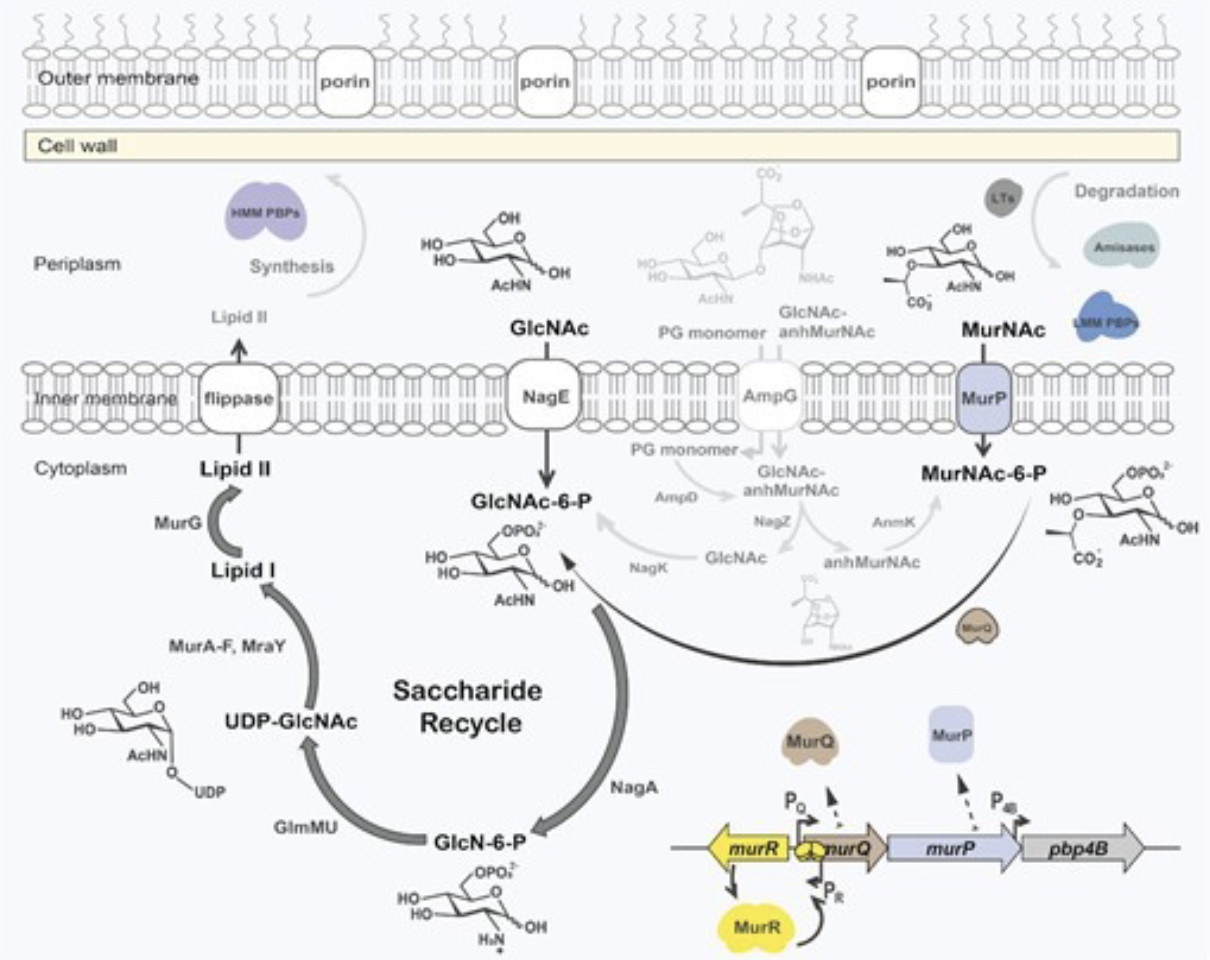
MurP and MurQ facilitate cell wall recycling in bacteria
MurP –MurNAc phosphotransferase
MurQ- MurNAc-6-P etherase
can repair under these conditions by taking the old building blocks and recycling them via using a series of enzymes MurP (MurNAc phosphotransefare that is found in the cytoplasm) and MurQ
cover this in a later lecture
bacteria can recycle peptidoglycan and use these enzymes to do it