Unit 2- Intro to cross sectional anatomy
 Call Kai
 Learn
Learn Practice Test
 Spaced Repetition
 Match
Match Flashcards
 Knowt Play
1/53

There's no tags or description
Looks like no tags are added yet.
Name | Mastery | Learn | Test | Matching | Spaced | Call with Kai | Chat |
|---|
No analytics yet
Send a link to your students to track their progress
54 Terms
cross sectional anatomy overview
physical relationship among structures
3D
evaluation of soft tissue structures
slices taken in a sequence
sagittal/median plane=
left/right (X)
axial or transverse plane=
inferior/superior (Z)
coronal or frontal plane=
posterior/anterior (Y)
thickness of a slice is dependent on…
technical settings (ex: thinner slices= more detail)
slice thicknesses vary from several__ to ___
millimeters to centimeters
slices overall are determined by
procedure protocols (institution controls protocol)
scout is also known as
scanogram
breathing can drastically affect
image quality
this modality is 3D and taken around a single axis of rotation
CT
when was CT developed & what was the first scan
1971; brain scan 2.5 hours
scans are taken on 1 plane & reconstructed to create…
sagittal & coronal planes
___represents the densities of each tissue
hounsfield unit
bone HU
10000 (bright white)
water HU
0 (grey)
air HU
-1000 (black)
what year was MRI discovered
1973
what utilizes a powerful magnetic field to align the magnetization of some atoms in the body
MRI
human body is what percent of hydrogen?
80%
how MRI works
a “COIL” is placed around the area of interest to obtain the info of that specific area of anatomy.
magnet aligns the single protons in the hydrogen nuclei within the field
radiowaves enter patient from another angle- the radiowaves cause the nuclei to flip positions and either align with the magnet or against it
at termination of radio signals, nuclei return to OG position by spinning like a top back into their varied position
energy that is released from that relaxation is used to generate images
T1 weighted images
fat appears BRIGHT
water appears DARK
best for ANATOMY
T2 weighted images
water appears BRIGHT (white)
fat appears DARK
best for PATHOLOGY
____ is a contrast used in MRI to improve clarity of the images
gadolinium
iodine vs gadolinium
gadolinium is a safer contrast due to less adverse reactions & eliminated through kidneys easier
example of gadolinium visibility
inflammation
tumors
blood vessels
some blood supply within organs
MRI advantages over CT
no radiation
superior contrast resolution
superior tissue characterization
visualization of angiography WITHOUT using contrast
ultrasound uses
high frequency sound waves w/ a transducer generated at 20,000 vibrations per second (hertz/second)
megahertz used in ultrasound
3-20 mHz
how does ultrasound work
reflected soundwave echoes are recorded & shown as an image in real time
transducer transmits inaudible soundwaves into the body as it echoes from the body’s fluid & tissues, transducer records the strength and character of the waves, then displayed
each kind of tissue has a different___ in ultrasound
attenuation coefficient
what is an attenuation coefficient?
how much of the sound wave will get absorbed
how much passes through
how much bounces back
piezoelectric crystals
converts electricity into soundwaves and then echoes into electricity
2 functions of the transducer
generates sound waves- speaker
detects reflected sound waves- like a microphone
purpose of the gel in ultrasound
creates a surface to help the probe transmit sound waves
curvilinear transducer overview
low frequency 5mHz
abdominal, obstetrics, pelvis
linear transducer overview
high frequency 15 mHz
great detail
superficial structures
thyroid, scrotal, musculoskeletal, soft tissue masses
intraoperative probe
very high frequency
directly on organ or vessel
liver transplants, vascular stuff
endocavity transvaginal transrecetal probe mHz
8
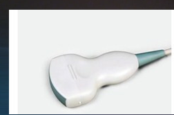
what probe is this?
curvilinear

what probe is this?
linear

what probe is this?
endocavity, transvaginal, transrectal

what probe is this?
intraoperative probe
what are the imaging planes in ultrasound?
long (sagittal)
transverse
**occasionally oblique images are obtained**
describes the tissue in relation to another tissue, or as compared to its normal state
echogenicity
each tissue type, such as a liver, spleen, or kidney, has a particular___ in its normal state
echogenicity
in an abnormal state, the echnogenicity of an organ can be….
altered (hyperechoic, hypoechoic, anechoic)
observations of echogenicity can help the radiologisy
to categorize the type of disease process involved
hyperechoic is
white/ bright structure
hypoechoic is
grayish black
anechoic
no color, entirely black
abnormality of kidney; accumulation of calculi within the renal pelvis and kidney calices
staghorn calculus (coral calculi)

what organ scan is this?
liver (can see hepatic tree)

what organ scan is this?
kidney (bean shape)
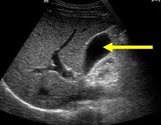
what organ scan is this?
gallbladder