30 Clinical Signs and Diagnostic Tests for Nasal Disease
 Call Kai
 Learn
Learn Practice Test
 Spaced Repetition
 Match
Match Flashcards
 Knowt Play
1/54

There's no tags or description
Looks like no tags are added yet.
Name | Mastery | Learn | Test | Matching | Spaced | Call with Kai | Chat |
|---|
No analytics yet
Send a link to your students to track their progress
55 Terms
Maxillo: respiratory epithelium
Ethmo: olfactory epithelium
What is the difference between the lining of the maxilloturbinates and the ethmoturbinates
Cribriform plate
What separates the ethmoturbinates from the brain?
1. nasal discharge
2. sneezing
3. stertor (snoring, snorting)
Name the most common clinical signs of nasal disorders:
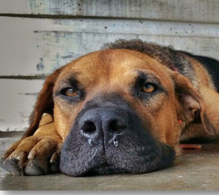
Serous (clear, watery): may be associated with viral disease
Describe this nasal discharge. It can be normal, but what MIGHT is be associated with?
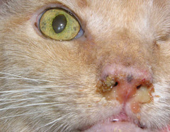
Mucopurulent (white, yellow or green + thick): associated with inflammation (viral, bacteria, fungal, foreign bodies, oral dz, lower airway dz)
Describe this nasal discharge. What is it associated with?
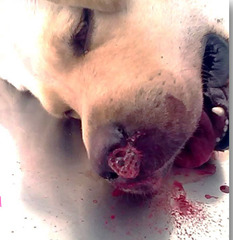
Hemorrhagic epistaxis: associated with trauma, locally aggressive disease, hypertension, coagulopathies
Describe this nasal discharge. What is it associated with?
Facial deformity + mucopurulent discharge
Describe what is happening in this cat
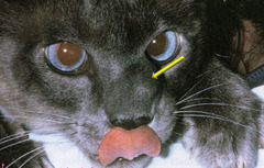
Depigmentation and ulceration of nasal planum = blastomycoses
Describe what is happening in this dog
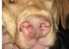
Depigmentation and ulceration of nasal planum = aspergillosis
Describe what is happening in this dog
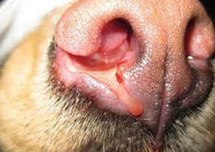
Phases I thru IV
How many phases are there for diagnostic approach of chronic nasal discharge
Phase I noninvasive
This phase of chronic nasal discharge diagnosis includes history, PE, fundic exam, fecal float, nasal swabs and viral testing
Phase II requiring general anesthesia
This phase of chronic nasal discharge diagnosis includes *nasal radiography, rhinoscopy,* dental rads, nasal biopsy with histopath, and CT
Phase III
This phase of chronic nasal discharge diagnosis includes CT, MRI, and frontal sinus exploration which you usually refer for
Phase IV
This phase of chronic nasal discharge diagnosis includes repeat phase II in several months using CT, MRI, exploratory rhinotomy with turbinectomy, usually a referral
4 views: lateral, open mouth VD, intraoral DV, skyline frontal sinuses
How many views should you get when doing nasal radiographs in phase II of chronic nasal discharge?
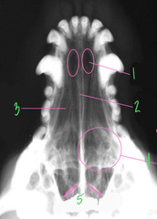
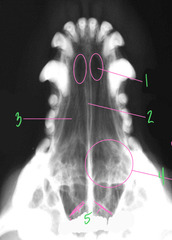
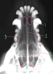
Palatine fissure
What is 1 in this normal radiograph?
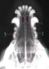
Nasal septum, vomer
What is 2 in this normal radiograph?
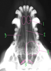
Maxilloturbinates aka nasal conchae
What is 3 in this normal radiograph?
Ethmoid turbinates
What is 4 in this normal radiograph?
Cribriform plate
What is 5 in this normal radiograph?
Abnormal: loss of trabecular detail
Is this normal or abnormal?
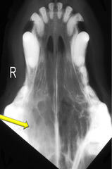
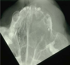
Abnormal: loss of trabecular detail and soft tissue swelling
Is this normal or abnormal?
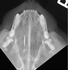
Abnormal: metallic object
Is this normal or abnormal?
Abnormal: loss of detail
Is this normal or abnormal?
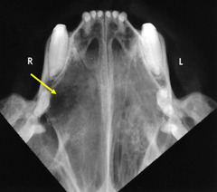
Normal
Is this normal or abnormal?
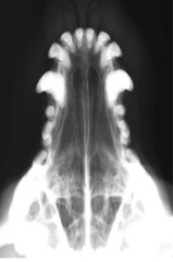
Abnormal
Is this normal or abnormal?
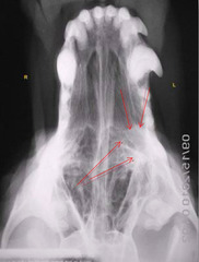
Abnormal: lysis of vomer bone (neoplasia)
Is this normal or abnormal?
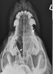
Neoplasia: commonly causes lysis of vomer bone
Aspergillus: commonly causes roughening of vomer bone
What is the difference between neoplasia and aspergillus and what they will do to the vomer bone (NORMALLY but not ALWAYS)?
Abnormal: extension of abnormality into frontal sinus
Is this normal or abnormal?
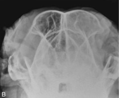
Eye
What is 1?
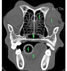
Ethmoid turbinates = normal
What is 2?
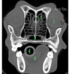
Normal nasopharyngeal meatus
What is 3?
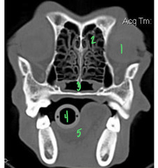
Et tube
What is 4?
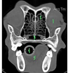
Tongue
What is 5?
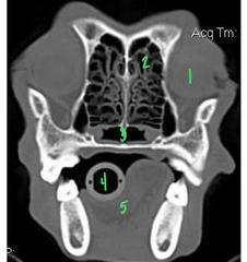
Normal
Is this normal or abnormal?
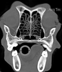
Abnormal
Is this normal or abnormal?
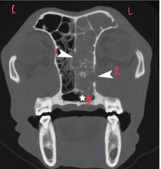
lysis of vomer bone
What is 1?
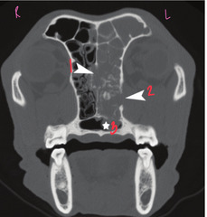
Lysis of nasal cavity into orbit = may present with exopthalamos or hard to retropulse
What is 2?
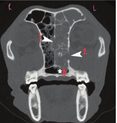
Tumor extended into nasopharyngeal meatus = common sign of malignancy
What is 3?
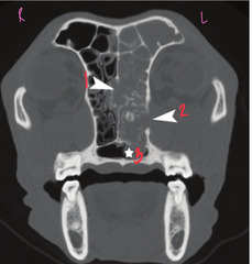
Bony lysis
What is a HALLMARK of neoplasia in the nasal cavity?
Abnormal = Lysis thru vomer bone and hard palate
Is this normal or abnormal?
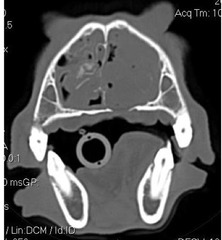
Rhinoscopy. Look at normal side first under general anesthesia
What is this, and how do you decide where to look first?
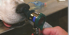
Rigid rhinoscopy = never blindly pass anything into the nose. measure medial canthus and do not go beyond that because the cribriform plate and brain are beyond this
What is this and what do you have to keep in mind if you do this?
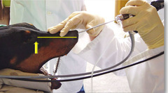
Rigid
Would you get biopsies and cultures with *rigid or flexible* rhinoscopy
Flexible rhinoscopy (pass into oral cavity and retroflex to see caudal aspect of nasopharynx)
What is this?
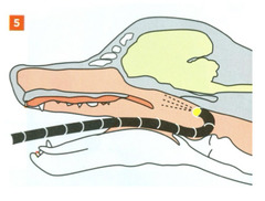
Nasopharyngeal disease
Sterterous breathing is a hallmark of disease where?
Cryptococcus neoformans
What is this?
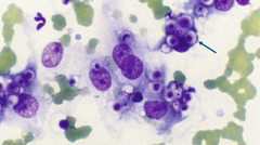
Cryptococcus in feline (though FNA may also be used)
Nasal swabs are least invasive but typically considered to be nondiagnostic due to nonspecific results. Which problem is the exception to this?
True
TRUE OR FALSE: animals must be under anesthesia for a nasal flush
Nasal flush
This diagnostic nasal test is minimally invasive and involves saline flush from internal nares rostrally towards external nares to produce a cytologic sample
Pinch biopsy (alligator biopsy)
This diagnostic nasal test is *invasive* and coagulation panels and buccal bleeding time (BMBT) *need to be collected beforehand*, where a minimum of *6 samples* should be collected from small forceps to collect tissue samples
Measuring to medial canthus and taking a pinch (alligator) biopsy
What is this?
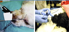
Pinch biopsy
What is the *preferred nonsurgical method of specimen collection* for nasal disease?
Turbinectomy
This is the MOST AGGRESSIVE AND INVASIVE diagnostic nasal test which is performed thru a rhinotomy incision
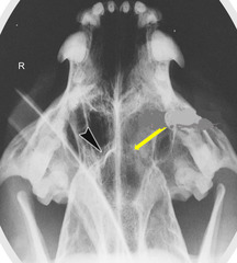
On right side of image: missing cribriform plate due to left nasal tumor
Describe what is happening here