Muscle Tissue: Structure and Function Overview
1/111
There's no tags or description
Looks like no tags are added yet.
Name | Mastery | Learn | Test | Matching | Spaced |
|---|
No study sessions yet.
112 Terms
What is the sodium-potassium ratio?
3 sodium ions are pumped out while 2 potassium ions are pumped in.
Functions of Muscular System(Easy Question on test!)
movement, generate heat, venous return(push vein blood back to the heart), smooth muscle(maintains good blood pressure; movements within visceral organs encourage transport), cardiac muscle(pumps blood throughout the body)
3 types of muscle tissue
skeletal, cardiac, smooth
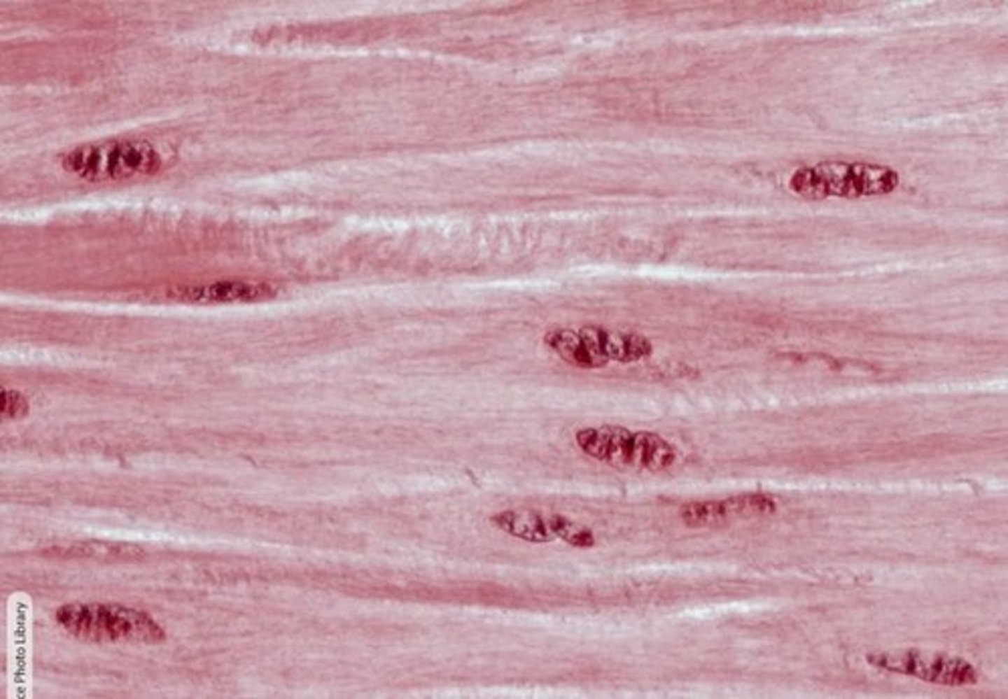
Smooth Muscle
Maintains blood pressure; aids in organ transport.
spindle shaped, no striations, involuntary, found in organs and blood vessels
Cardiac Muscle
- Pumps blood throughout the body.
- muscle found only in the heart
- striated because of overlapping cells.
- cells are branched in shape
- has intercalated discs between cells
- has gap junctions between the cells so the calcium flows quickly across the cells as the entire heart depolarizes as one in order to pump as one
- single nuclei per cell
- involuntary
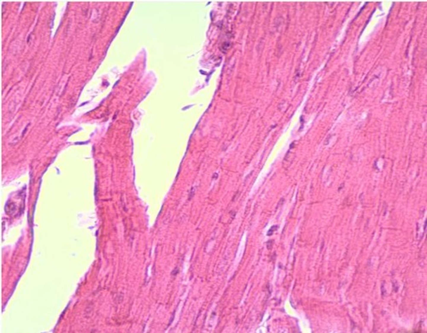
Skeletal Muscle
- super long muscle cells that are fibers
- striated(because of overlapping cells)
- voluntary
- multinucleated (because in the fetus, the skeletal muscle cells start fusing together into one long fiber cell which is why skeletal muscles are multinucleated)
Characteristics of all muscle tissue
excitability, contractility, elasticity, extensibility
Excitability
Ability to depolarize(going through a change in electrical charge) and generate action potentials(an electrical wave that moves down the membrane).
Contractility
all muscle contracts to create force by shortening when actin protein is pulled by myosin protein.
All muscle needs Ca2+ to contract.
All muscle needs ATP+ to relax.
What do all muscles need to contract?
Calcium
What do all muscles need to relax?
ATP
Elasticity
Ability to recoil to original length after stretching.
Extensibility
Ability to stretch beyond original length.
Striations
Pattern of overlapping actin and myosin in skeletal muscle.
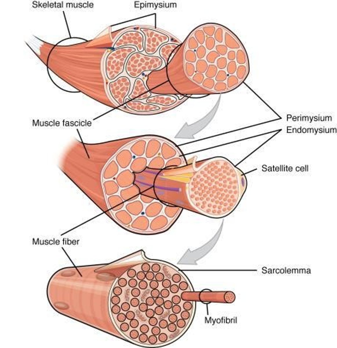
Epimysium
Connective tissue wrapping around an entire muscle. It holds fascicles together and separates the muscle from other body structures
Perimysium
Connective tissue wrapping around muscle fiber bundles called fascicles.
fascicle
bundle of muscle fibers
Endomysium
Connective tissue surrounding individual muscle fibers in order to protect each muscle fiber.
What are muscle fibers?
It's another name for muscle cells.
"myo" prefix meaning
muscle
"sarco" prefix meaning
flesh
Tendon
Connective tissue connecting muscle to bone.
It is made of collagen from the 3 mysia layers that meld together.
How do tendons connect muscles to bones?
The collagen of the tendon melds into the collagens of the periosteum which is how tendons connect muscle to bone.
Aponeurosis
Broad, flat tendon connecting muscle to bone.
Some muscles connect to a broad flat tendon instead of connecting directly to bone. Ex: Latissimus dorsi.
Fascia
Connective tissue sheets beneath the skin that help with anchoring and separating muscles.
Myoblasts
Embryonic cells that fuse to form muscle fibers. After the cells fuse, the nuclei remain which is why a muscle fiber is multi-nucleated.
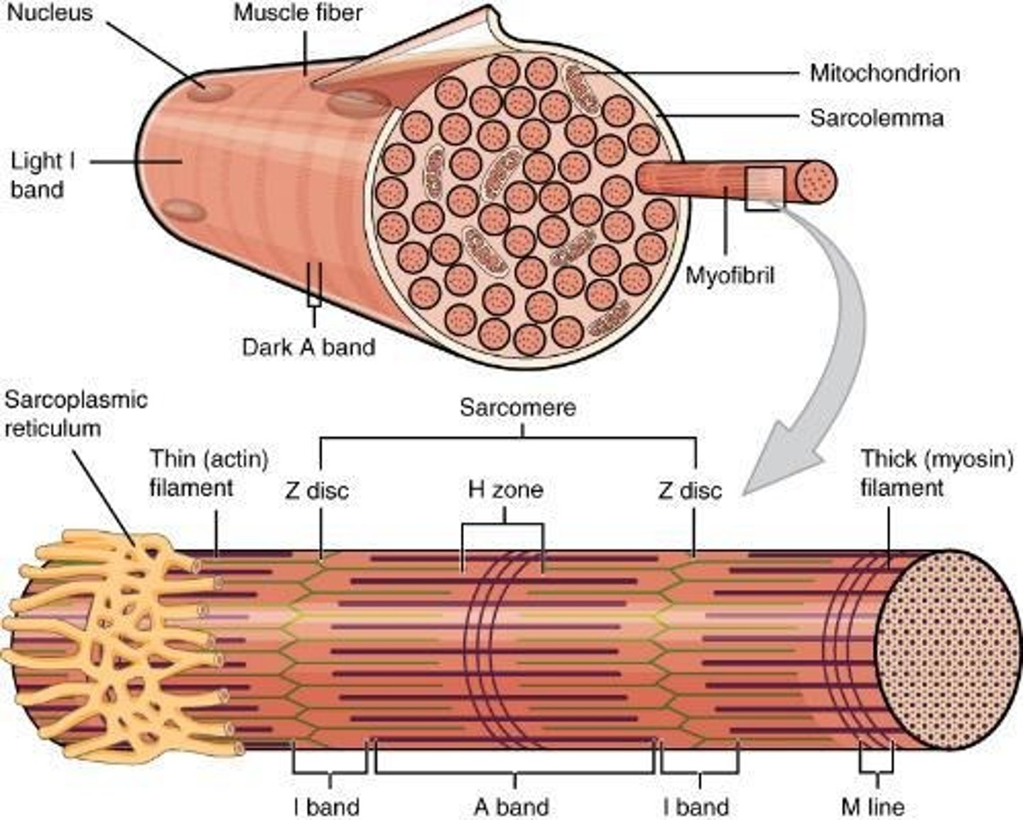
Sarcolemma
Cell membrane of a muscle cell.
Sarcoplasm
Cytoplasm of a muscle cell.
Sarcoplasmic Reticulum
Specialized Endoplasmic Reticulum of a muscle cell. It is specialized for calcium storage in muscle cells, so that it can release and retrieve calcium ions for muscle contraction.
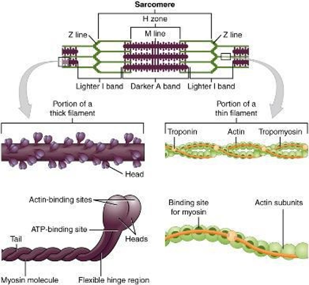
Sarcomere(always asked on the nursing tests!!!)
Functional contractile unit of the muscle cell.
It spans from one Z disc to the next Z disc("From Z to shining Z").
Z disc
Zig zag line that is the attachment point for actin filaments.
M line
Middle line attaching myosin filaments.
Middle of the sarcomere, line that attaches the myosin filament.
I band
Light stripe with only thin actin. It's light colored because it only has the thin actin.
Memory tip: "I saw the light"
A band
Dark stripe since it's where actin and myosin overlap.
Memory tip: "Afraid of the dark"
H zone
Region with only myosin, no actin overlap.
Membrane potential
Electrical gradient(difference in voltage) across a cell membrane.
The inside of the cell membrane is usually more negatively charged because of the 3:2 ratio of Na+ ions and K+ ions. 3 sodium ions are pumped out while 2 potassium ions are pumped in, so basically, there are more positive ions being pumped out than being pumped in. Therefore, there are more positive ions, and a more positive charge, outside the cell than inside the cell.
How do neurons and muscle cells use their membrane potentials?
They make electrical signals by manipulating the flow of ions across the membrane to change the charge by using special ion channels in the membrane.
What is the normal voltage of the inside of the cell?
about -70 millivolts
Action potential
Electrical signal traveling across a membrane as a wave.
It's quick and consistent.
Excitation-contraction coupling
Its the steps necessary for a muscle to contract. These are the simplified steps(there is a lot of steps for excitation):
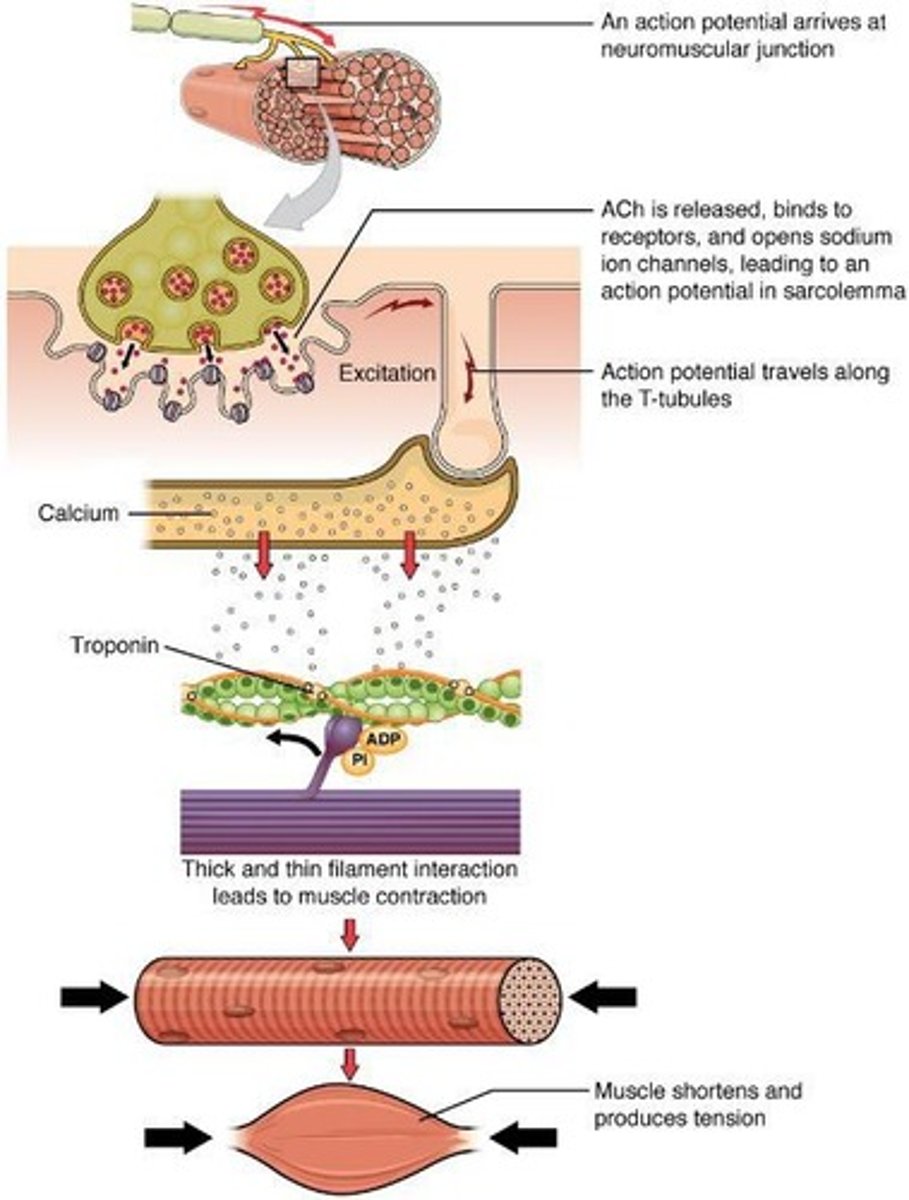
1. The muscle must be "excited"(stimulated to fire an action potential). For skeletal muscle cells, the "excitation" is always a nerve stimulus.
2. The excitation triggers the action potential.
3. Muscle action potential triggers calcium(Ca2+) to be released from the sarcoplasmic reticulum and the sarcolemma
4. Ca2+ binds to troponin
5. Troponin changes shape and allows tropomyosin to detach from the binding sites.
6. The myosin heads bind to the binding spots of the actin filament.
Neuromuscular Junction (NMJ)
Contact point between nerve and muscle.
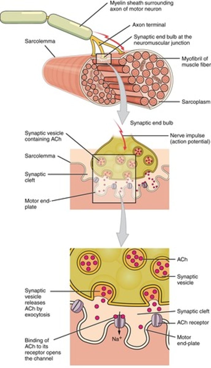
Motor Neuron
Nerve signaling muscle from spinal cord.
The nerve body is in the spinal cord, but the axon travels all the way from the spinal cord to the muscle.
Acetylcholine (ACh)
Neurotransmitter released at NMJ.
Synaptic cleft
Space between nerve and muscle fibers.
What are the steps of action potential in order to release calcium?
1. The action potential of the neuron travels from the cell body all the way down the axon and ends at the NMJ where it triggers the release of acetylcholine(ACh)
2. Acetylcholine (ACh) diffuses across the synaptic cleft (space between the nerve and the muscle) to bind to a receptor on the motor endplate of the muscle cell.
3. When ACh binds to its receptor, the receptor changes shape, opening a channel to allow positively charge ions to flood into the cell.• This equalizes the voltage on both sides of the membrane; thus we say the membrane has been depolarized since it is no longer electrically polar.
4. Depolarization triggers voltage-gated sodium channels to open all along the membrane, spreading the action potential to the whole muscle like a waterfall
5. The action potential passes through the cell membrane and through the T-Tubules.
6. The T-tubules are connected to the sarcoplasmic reticulum, so when the action potential moves from the T tubules to the terminal cisternae of the sarcoplasmic reticulum, the sarcoplasmic reticulum releases calcium into the cell.
7. (ACh in the synaptic cleft is then degraded to end the excitation)
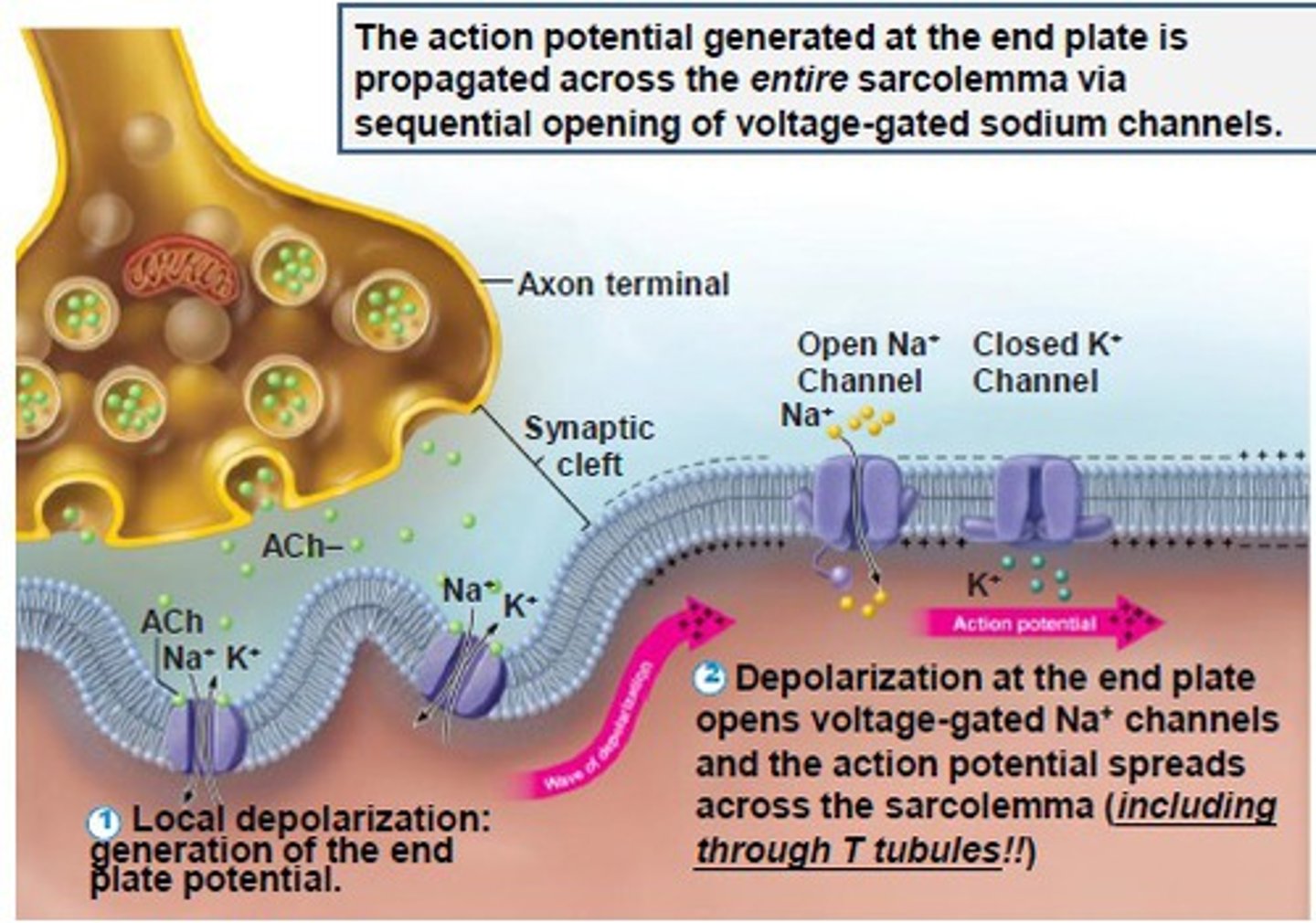
Depolarization
Loss of membrane potential, since it let positive ions into the cell. The membrane is no longer electrically polarized because the voltages on both sides of the membrane have been equalized because the channels allowed the positive ions to flow in.
Sarcoplasmic reticulum (SR)
The endoplasmic reticulum in muscle cells that specialize in Calcium storage, release, and retrieval in a muscle cell in order to control muscle contractions.
Voltage-gated sodium channels
Sodium Channels that open during depolarization in order to let positive sodium ions in, so that the action potential spreads like a wave.
Myofibrils
Contractile fibers within muscle cells. They have actin and myosin fibers.
T-Tubules
Narrow tubes conducting electrical impulses into muscle and connecting the cell membrane to the sarcoplasmic reticulum.
They pass the action potential all around the myofibrils into the terminal cisternae which then makes the sarcoplasmic reticulum release calcium.
Terminal Cisternae
The part of the sarcoplasmic reticulum that connects to the T-tubules. It
Triad
One T-tubule sandwiched between two terminal cisternae.
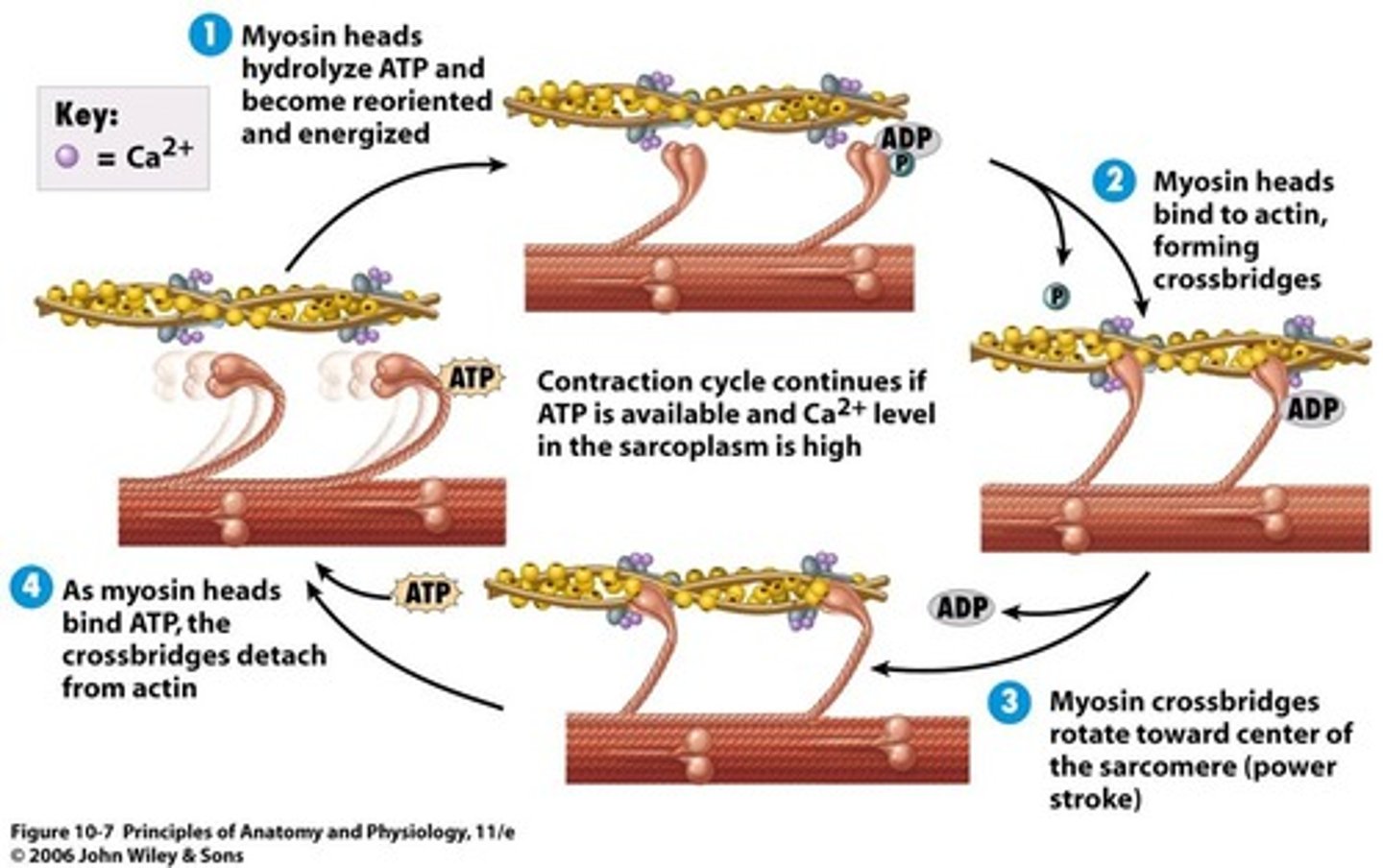
Cross-Bridge
Connection between actin and myosin heads which triggers contraction.
Troponin
attaches tropomyosin to actin so that the myosin can't bind to the actin.
Tropomyosin
Protein that hides actin binding sites.
Thin Filament
actin.
Clue: It has the thinnest name. Actin has a shorter name than myosin, so actin is the thin filament.
Thick Filament
Myosin
It has a tail(the rope looking part) and a head(which has the binding sites for actin and ATP).
Clue: Myosin has a thicker name than actin, so myosin is the longer filament.
Myosin Head
Contains binding sites for actin and ATP.
Power Stroke
Myosin head pivots, pulling actin during contraction.
ATP (Adenosine Triphosphate)
Energy source required for myosin head "re-cocking" or resetting. It's also used to relax the muscle cell.
Z Lines
Boundaries of a sarcomere, move closer during contraction.
I Band
Region of sarcomere that shortens during contraction.
A Band
Region that remains the same width during contraction because at full contraction, the thick and thin filaments overlap.
Steps for skeletal muscle contraction:
(a) The active site on actin is exposed as calcium binds to troponin.
(b) The myosin head is attracted to actin, and myosin binds actin at its actin-binding site, forming the cross-bridge.
(c) During the power stroke, the phosphate generated in the previous contraction cycle is released. The myosin head pulls the actin fiber toward the M line.
(d) A new molecule of ATP attaches to the myosin head, causing the cross-bridge to detach. (After the myosin pulls, it needs to let go of the rope(actin) so it needs to relax with ATP in order to grab more rope and pull later or to stop contracting.)
(e) The myosin head hydrolyzes ATP to ADP and phosphate, which returns the myosin to the cocked position which is ready to attach to the actin again.
Sarcoplasmic Calcium Pump
Pumps Ca2+ back into SR during relaxation.
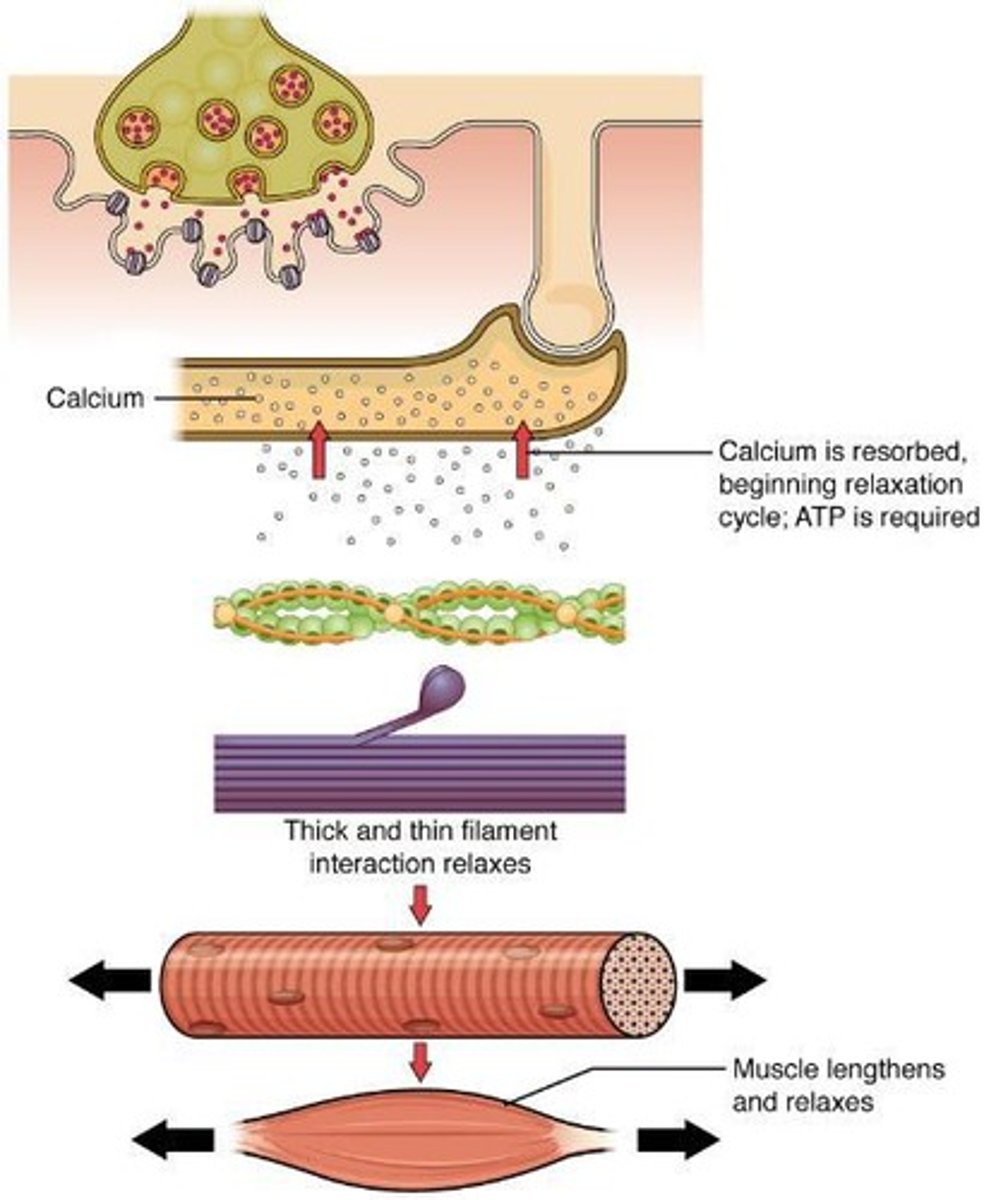
How does a muscle stop contracting?
It stops when the calcium ions are pumped back into the sarcoplasmic reticulum.
It also stops when it runs out of ATP and becomes fatigued.
Muscle relaxation steps
• Release of ACh ceases
• The membrane repolarizes
• Calcium pumps return calcium to the sarcoplasmic reticulum
• Binding sites on actin are "reshielded" as the troponin binds the tropomyosin to the binding sites once more.
• Muscle loses tension and returns to normal length
M-Line
Center of the sarcomere where filaments pull.
Muscle Fiber Shortening
Occurs as actin and myosin slide past each other during contraction.
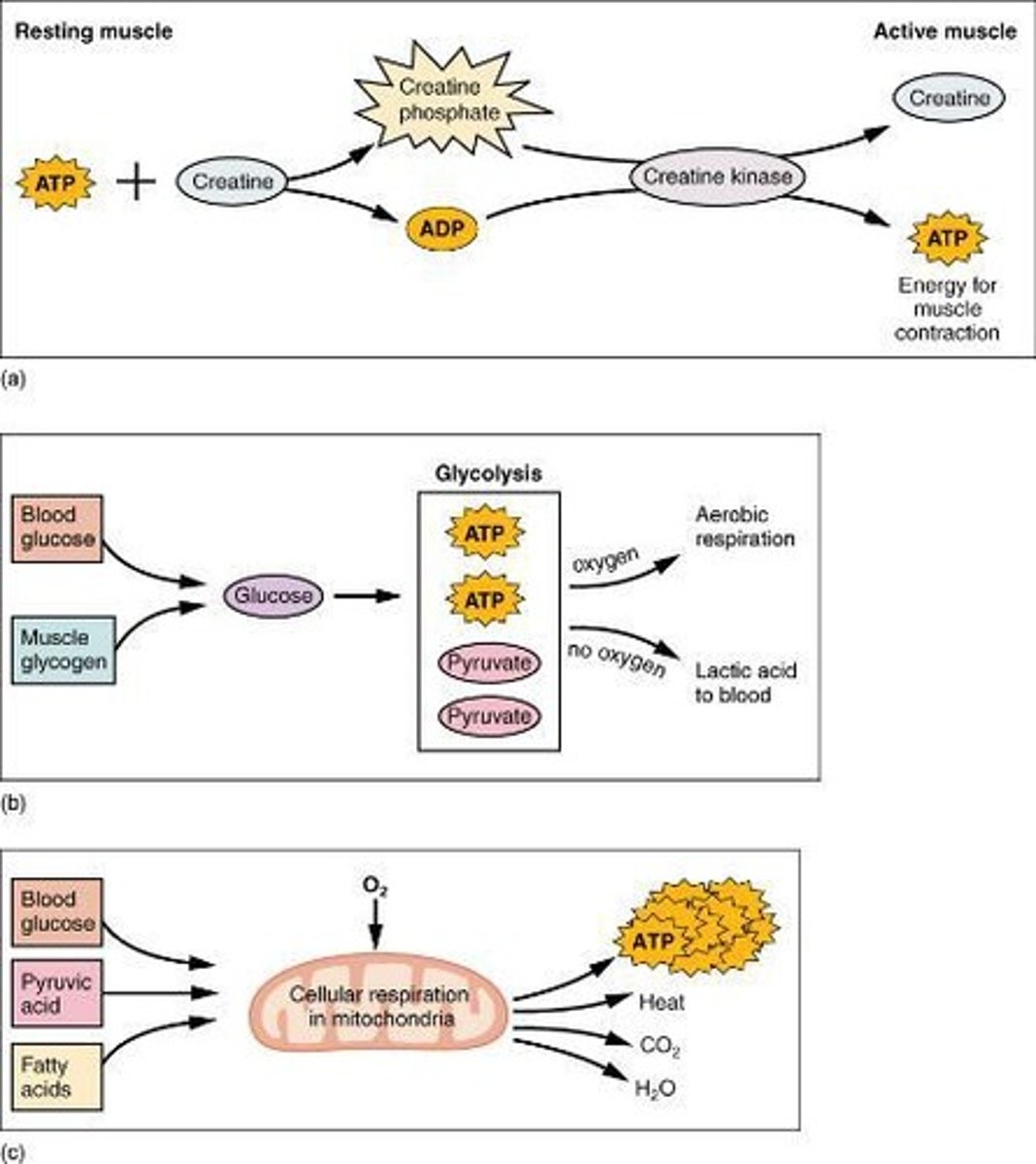
Ways muscle cells get energy
- Creatine phosphate(fastest)
- glycolysis(faster than aerobic respiration, but slower than creatine phosphate. Doesn't make a lot of ATP though, so it is just quick, short term energy)
- Aerobic respiration
Creatine Phosphate
Stores phosphates for immediate ATP regeneration whenever ATP is needed for really fast contractions like for jumping or for sprinting.
It's the fastest way for muscles to get energy.
Glycolysis
Anaerobic breakdown of one glucose molecule into two pyruvates which creates a net total of 2 ATP.
If there is enough oxygen, then the pyruvates will be used for aerobic respiration.
If there isn't enough oxygen, then the pyruvates will be converted into pyruvic acid which is converted to lactic acid which leads to muscle fatigue and soreness.
Aerobic Respiration
Converts pyruvic acid to ATP using oxygen. Water and CO2 are by products of this reaction.
It is very efficient and makes 36 ATPs from 1 glucose.
This process only happens in the mitochondria of the cell.
How much ATP from one glucose molecule that goes through aerobic respiration?
1 glucose produces 36 ATP in respiration.
Myoglobin
Oxygen-binding protein in muscle fibers that stores Oxygen for times when the muscle isn't getting enough Oxygen from the blood.
Rigor Mortis
3rd stage of death that happens 2-3 hours after death. It's when the sarcoplasmic reticulum disintegrates, which releases calcium so the muscles are tense. They stay tense because the sarcoplasmic reticulum isn't there to suck the calcium back up. But after 8 hours after death the myosin and actin breaks down and the muscle stiffness goes away.
What affects Muscle Strength?
- number of muscle fibers in cells(predetermined by genes and doesn't change unless lost due to damage)
- amount of myofibrils and sarcomeres
- hypertrophy
- atrophy
What affects the number of myofibrils and sarcomeres inside a single muscle fiber?
• Activity vs inactivity
• Hormones
• Thyroid
• Stress
• Steroids
Hypertrophy
Increase in muscle size/bulk due to activity. This is muscle growth. The increase in muscle size ISN'T an increase in muscle cells, but an increase in the number of myofibrils and sarcomeres in a single muscle fiber.
Atrophy
Decrease in muscle size due to disuse(from lazy lifestyle or from immobility from things like an injury or disease).
Duchenne muscular dystrophy (DMD)
Genetic disorder affecting muscle function, primarily in males since it is found on the X chromosome.
It's a condition where there is accumulated scar tissue in muscle since the dystrophin protein is made incorrectly(due to gene change) and the dystrophin rips the muscle every time the muscle is used. This leads to accumulation of scar tissue. The person usually loses muscle from the legs and the disease progresses upwards. They usually die from inability to breathe; their life expectancy is usually in their 20s.
Muscle tension
Force generated by shortening sarcomeres during contraction.
What are the two types of muscle tension?
isotonic and isometric
Isotonic contraction
Muscle tension remains constant while moving a load. The muscle length changes since the muscle is moving the load.
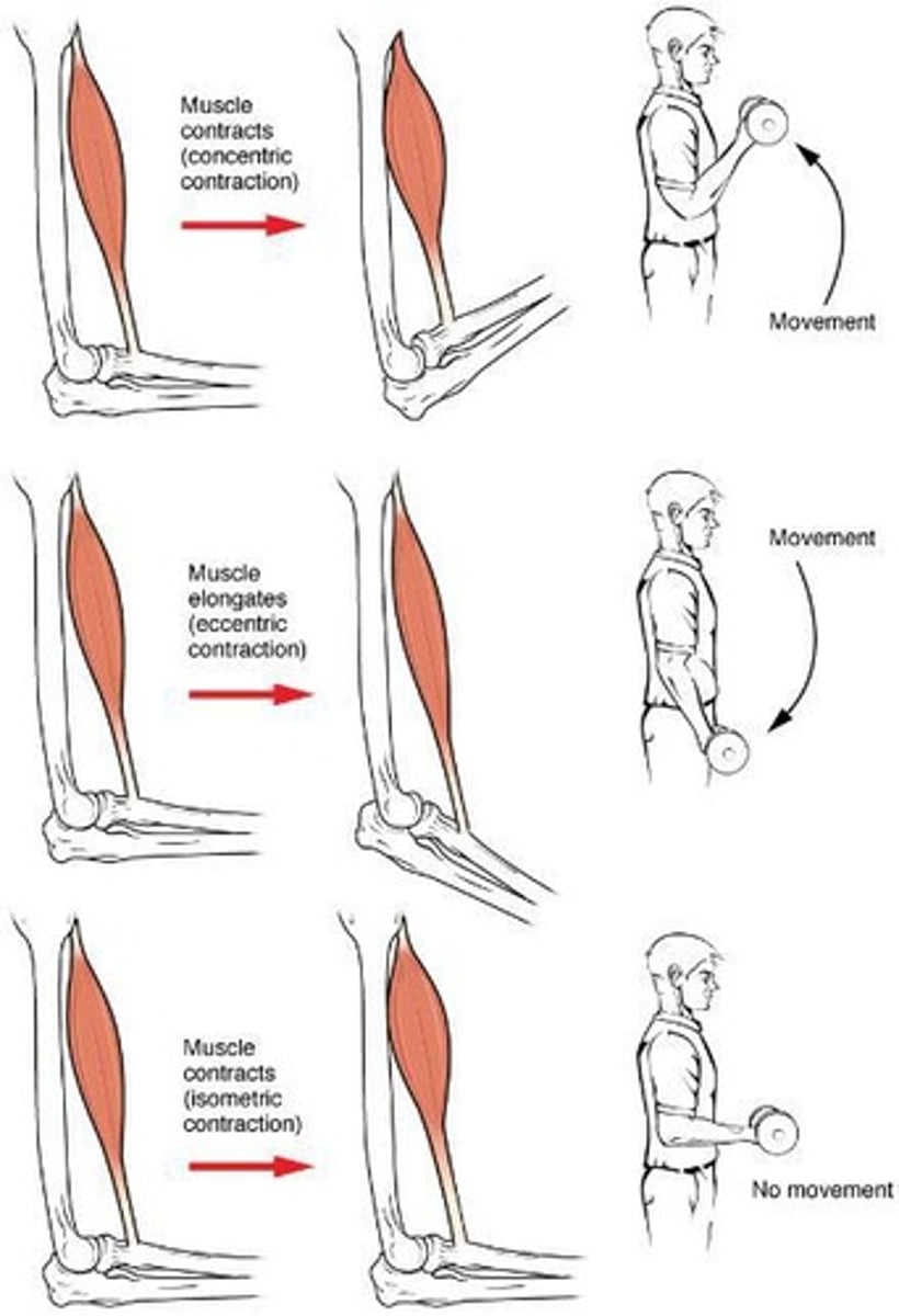
What are the two types of isotonic contraction?
concentric and eccentric
Concentric contraction
Muscle shortens, decreasing joint angle while moving a load.
Eccentric contraction
Muscle lengthens, increasing joint angle while moving a load.
Isometric contraction
Muscle tension remains constant, no movement or change in angle of the joint.
It's very important for maintaining posture and body position.
Motor Unit
A Motor neuron and all the muscle fibers that it controls.
Small Motor Unit
A Motor neuron that innervates a few fibers for fine motor control.
Ex: muscles in the eye have a ration of 1 motor neuron per 6 muscle fibers which allows for very precise and fine controlled movements.
Large Motor Unit
Motor neuron innervates many fibers(like thousands of fibers) for simple but powerful movements. gross motor control.
Ex: Muscles of the thigh like quadriceps.
Why do muscles have a large range of muscle unit sizes?
Because different size motor units are used for tasks of different sizes. Small motor units are used for light tasks to create small amounts of muscle tension. Larger motor units are used for more challenging tasks
• Example: Your bicep can lift a feather using a small motor unit or an incredibly heavy weight by recruiting larger motor units
Muscle Tone
Slight contraction is maintained in the muscle in order to keep the proteins regulated.
This is why skeletal muscle is almost never completely relaxed.
Hypotonia
Decreased muscle tone, often due to central nervous system(brain/spinal cord) damage or loss of innervation to the muscle from damage or disease.
People with this condition usually have low reflexes.
It can also mean when muscle is flaccid and not holding tone, like when sleeping
Hypertonia
Increased muscle tone, associated with upper motor neuron(brain) damage. This can be caused by Parkinson's disease, cerebral palsy, etc.
They usually also have hyperreflexia.
Tetany
involuntary, often sustained muscle contractions, spasms, cramps.
It's when all your muscles are contracting at once, like when you get electrocuted. Your heart can’t go into tetany since it is made different.
Some common causes are:
- overstimulated nerves
- issues of calcium(parathyroid or kidney problems)
- "Tetanus" caused by Clostridium tetani bacteria
Tetanus
Infection caused by Clostridium tetani, leading to muscle spasms.
How are muscle fibers categorized, and what are the categories?
They are categorized by how fast they contract and how they make ATP. Most muscles have a MIX of fiber types.
These are the categories:
- Slow Oxidative
- Fast Oxidative
- Fast Glycolytic
Oxidative fibers
They use Oxygen to make ATP(aerobic).
Clue: "Oxi" and "Oxy"gen.
Glycolytic fibers
make ATP without oxygen, so it's anaerobic