opportunistic fungi: zygomycetes
 Call Kai
 Learn
Learn Practice Test
 Spaced Repetition
 Match
Match Flashcards
 Knowt Play
1/20

There's no tags or description
Looks like no tags are added yet.
Name | Mastery | Learn | Test | Matching | Spaced | Call with Kai | Chat |
|---|
No analytics yet
Send a link to your students to track their progress
21 Terms
mucormycosis predisposing factors
diabetes, leukemia/lymphoma, malnutrition, barrier break, immunosuppression
rhinocerebral mucormycosis
caused by rhizopus or mucor. primary underlying condition: diabetes. hyperglycemia → enhanced growth. impaired neutrophil chemotaxis. causes convulsions. mortality >80%. treated with IV amphotericin B.
thoracic (pulmonary) mucormycosis
caused by any of the zygomycetes. primarily pulmonary: inhalation, aspiration (from rhinocerebral)
gastric mucormycosis
caused by any of the zygomycetes. primary underlying condition: malnourished children
cutaneous mucormycosis
caused by zygomycetes (mostly rhizopus). post trauma/barrier break or disseminated from other site. painful, necrotic cutaneous lesion
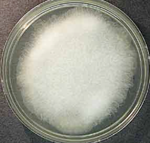
absidia spp colony appearance (SAB @ RT)
opportunistic infections of lungs (most common).
rapid growth, wooly, white-grayish-brown, reverse: uncolored
absidia spp microscopic appearance
hyphae (stolon): broad, aseptate, thin-walled.
sprangiophores: branched, columellas w/ swelling (apophysis) beneath sporangium, flask shaped.
sporangia: pyriform (pear shaped).
rhizoids: few, situated on stolons b/t sporangiophores

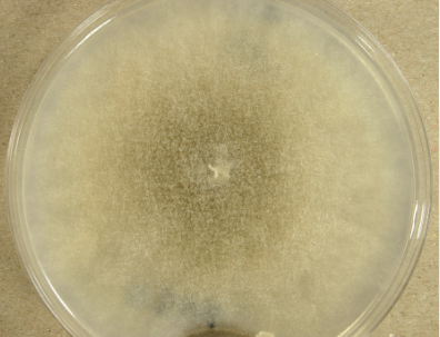
mucor spp colony appearance (SAB @ RT)
zygomycosis in severely debilitated patients, ear infections, allergies.
rapid growth, wooly, white at first then becomes gray, reverse: pale
mucor spp microscopic appearance
hyphae (stolon): broad, aseptate or sparsely septate, thick walled.
sprangiophores: branched, columnella w/o apophyses, some chlamydoconidia.
sproangia: globose (round).
rhizoids: absent

rhizopus spp colony appearance (SAB @ RT)
rapidly progressing agent of zygomycosis in brain, lungs, & intestines. patients w/ diabetic ketoacidosis, malnutrition, severe burns, immunocompromised.
rapid growth, wooly (“lid lifter”), white at first then becomes gray w/ dark tips (salt n pepper), pale reverse

rhizopus spp microscopic appearance
hyphae (stolon): broad, aseptate or sparsely septate, thin-walled
sprangophores: brown, unbranched. solitary or in tufts off stolen, diverging from point which rhozpoids form. apophysis absent or scarcely present.
sproangia: globose (round), columella
rhizoids: present (below sporangiophores

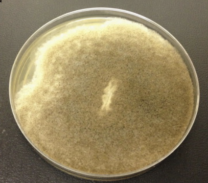
rhizomucor spp colony appearance (SAB @ RT)
occasionally agent of zygomycosis. seen in leukemia patients.
rapid growth (can grow at high temps, 54C), wooly, olive-gray to pale brown surface, white reverse.
rhizomucor spp microscopic appearance
hyphae (stolon): very broad, aseptate or sparsely, thin-walled.
sprangophores: solitary or branched (arranged in unbel), apophysis absent.
sproangia: globose (round), well developed columella.
rhizoids: rudimentary, stolons present but rare or hard to recognize

syncephalastrum spp colony appearance (SAB @ RT)
pathogenicity rare, dermatomycoses & onychomycoses.
rapid growth, wooly, pale-gray to dark gray surface, pale reverse

syncephalastrum spp microscopic appearance
hyphae (stolon): broad, aseptate or sparely (old colonies), thin-walled.
sprangophores: branched, terminate in a vesicle.
sproangia: finger-shaped (mesosporangioa), form around vesicle.
rhizoids: present

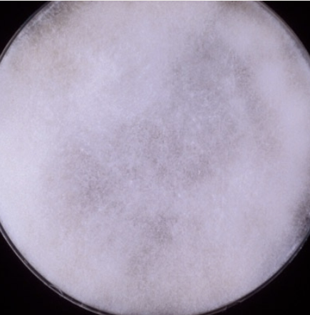
cunninghamella colony appearance (SAB @ RT)
occasional pathogen, pulmonary or disseminated zygomycosis in severely debilitated patients.
rapid growth, cottony, white to gray surface, pale reverse
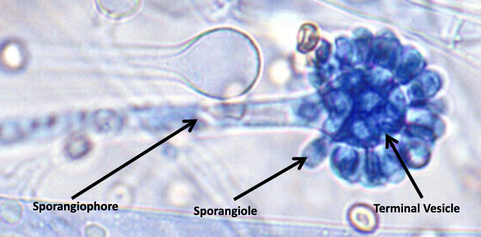
cunninghamella microscopic appearance
hyphae (stolon): broad, aseptate or sparsely
sprangophores: branched, terminate in vesicle
sproangia: one-spored sporangioles, form on vesicle surface
basidiobolus ranarum colony appearance (SAB @ RT)
localized to subcutaneous tissue: woody, hard, painless nodule. can cause infection in immunocompetent hosts, commensal in intestinal tract of frogs, lizards, & toads.
rapid growth at 30C, less rapid at 37C. glabrous becoming folded & granular. buff/yellowish gray front, white reverse

basidiobolus ranarum microscopic appearance
have 2 lateral appendages that resemble beaks

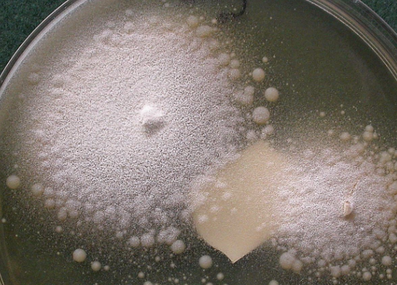
conidiobolus coronatus colony appearance (SAB @ RT)
tropical chronic inflammatory disease isolated to nasal mucosa.
rapid growth at 37C, glabrous becoming powdery, buff/yellowish gray front, white reverse. white satellite colonies form with age, spores forcibly discharged by sporangiophores fill sides of inoculated tubes & plates
conidiobolus coronatus microscopic appearance
hyphae (stolon): broad, few septa (inc w age)
sprangophores: unbranched, bear single spores
sproangia: can produce hair-like appendages called villae or protuberances called papillae