Chapter 7: Urinary System
 Call Kai
 Learn
Learn Practice Test
 Spaced Repetition
 Match
Match Flashcards
 Knowt Play
1/203

There's no tags or description
Looks like no tags are added yet.
Name | Mastery | Learn | Test | Matching | Spaced | Call with Kai | Chat |
|---|
No analytics yet
Send a link to your students to track their progress
204 Terms
nitrogenous wastes
urea
creatinine
uric acid
metabolic waste products filtered from bloodstream by kidney
creatinine
byproduct of muscle energy metabolism (creatine breakdown); nitrogenous waste execreted in urine
urea
created bye liver as byproduct of protein metabolism
uric acid
waste product formed from breakdown of purines, found in Cody’s cells and in certain foods
functions of kidney
filter nitrogenous wastes to form urine; about 200 quarts of blood are filtered every day to form 2 quarts of urine
maintain proper balance of water, electrolytes (sodium, potassium), and acids
release hormones: renin, erythroprotein(EPO), calciferol
degrade and eliminate hormones from the bloodstream
renin
enzyme important in adjusting blood pressure
erythropoietin (EPO)
hormone that stimulates RBC production in bone marrow; secreted by kidney
calciferol
active form of vitamin D necessary for the absorption of calcium from the intestine
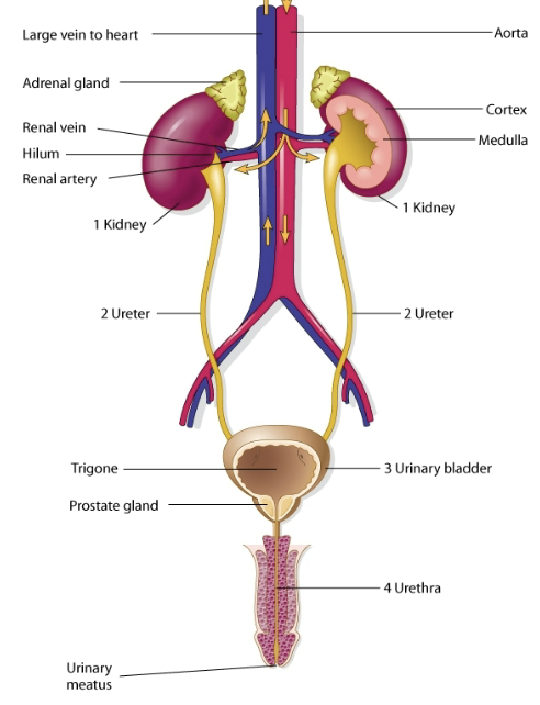
organs of male urinary system
kidney
ureter
urinary bladder
urethra
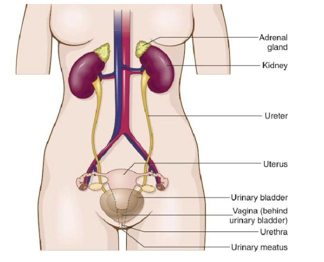
organs of female urinary system
kidney
ureter
urinary bladder
urethra
voiding
urination
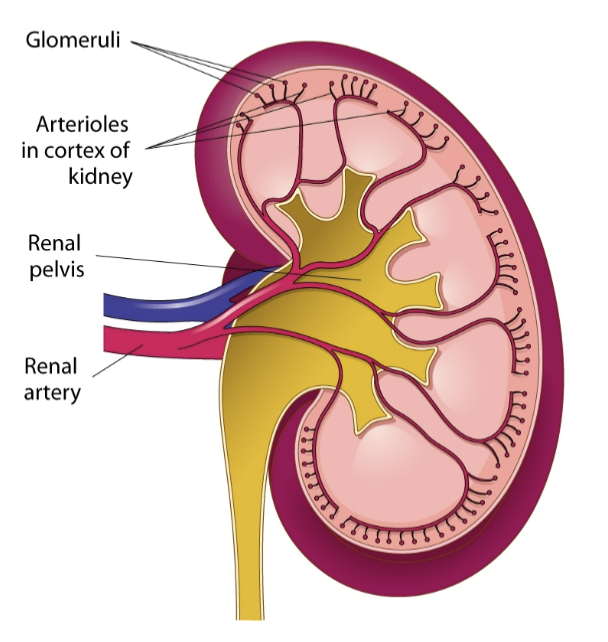
how kidneys produce urine
blood enters the kidneys through the right and left renal arteries
arteries carry blood to the capillaries
glomeruli filter the blood
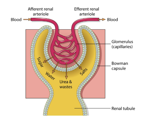
glomerulus and glomerular/Bowman capsule
blood passes through the glomeruli
glomerular (Bowman) capsule surrounds each glomerulus
renal tube is attached to each Bowman capsule
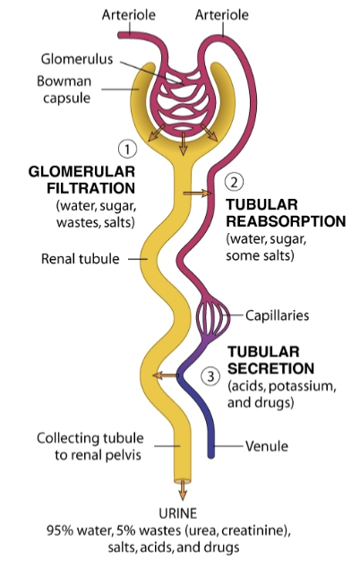
3 steps in formation of urine
glomerular filtration (water, sugar, wastes, salts)
tubular reabsorption (water, sugar, sodium)
tubular secretion (acids, potassium, drugs)
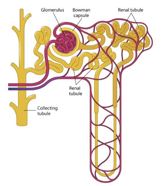
the glomerulus and a renal tubule combine o form a unit called a
nephron
approx. how many nephrons are in a kidney
1,000,000
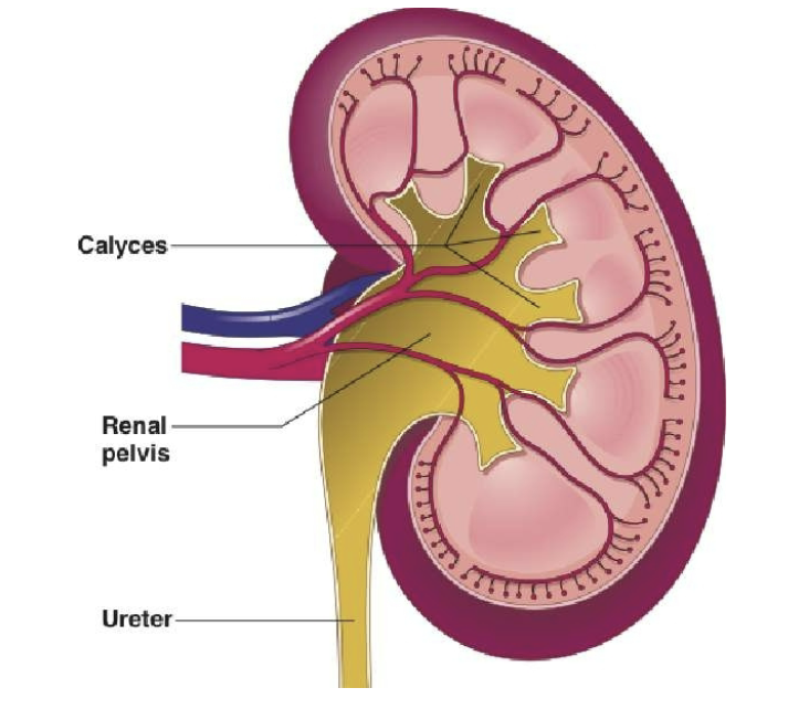
all collecting tubules lead to the
renal pelvis
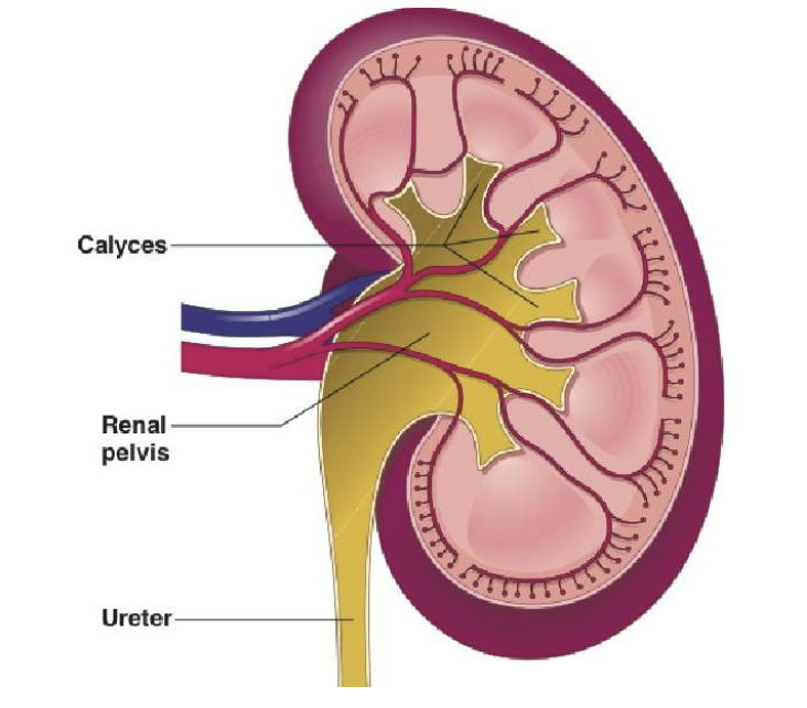
calyces or calices
small, cuplike regions of renal pelvis; the term comes from the Greek, kalux, meaning a cup or case surrounding a flower bud
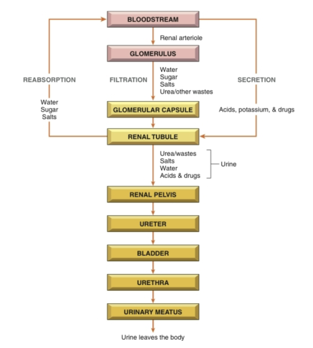
functions of the kidneys
remove nitrogenous wastes
balance water and electrolytes
release hormones
degrade and eliminate hormones
arteriole
small artery
catheter
tube for injecting or removing fluids
cortex
outer region of an organ
electrolyte
chemical element that carries an electrical charge when dissolved in water
-poietin
substance that forms
filtration
process whereby some substances pass through a filter
glomerular capsule
enclosing structure surrounding each glomerulus; also called Bowman capsule
glomerulus
tiny ball of capillaries in the kidney
hilum
depression in the part of an organ where blood vessels and nerves enter an leave; comes from Latin meaning a small thing
kidney
one of two bean-shaped organs that filter nitrogenous waste from the bloodstream to form urine
meatus
opening or canal
medulla
inner region of an organ; the term comes from the Latin, medulla, meaning marrow
nephron
functional unit of kidney where filtration, reabsorption, and secretion take place
nitrogenous waste
substance containing nitrogen and excreted in urine, ex. urea, uric acid, creatinine
potassium (K+)
an electrolyte regulated by the kidney
reabsorption
renal tubules return material necessary to the body back into the bloodstream
renal artery
blood vessel that carries blood to the kidney
renal pelvis
central collection region in the kidney
renal tubules
microscopic tubes in the kidney where urine is formed after filtration
renal vein
blood vessel that carries blood away from the kidney
renin
enzyme secreted by the kidney that raised bp
sodium (Na+)
electrolyte regulated in the blood and urine by the kidneys; a common form of sodium is sodium chloride (salt)
trigone
triangular area in the urinary bladder
urea
major nitrogenous waste excreted in urine
ureter
one of two tubes leading from the kidneys to the urinary bladder
urethra
tube leading from the urinary bladder to the outside of the body
uric acid
nitrogenous waste product excreted in the urine
urinary bladder
hollow, muscular sac that holds and stores urine
urination (voiding)
process of expelling urine; aka micturition
cali/o, calic/o
calyx (calix); cup shaped
cyst/o
urinary bladder
glomerul/o
glomerulus
meat/o
meatus
nephr/o
kidney
pyel/o
renal pelvis
ren/o
kidney
trigon/o
trigone
ureter/o
ureter
urethr/o
urethra
vesic/o
urinary bladder
albumin/o
albumin
azot/o
nitrogen
bacteri/o
bacteria
dips/o
thrist
kal/o
potassium
ket/o, keton/o
ketone bodies
litho/o
stone
natr/o
sodium
noct/o
night
olig/o
scanty
-poietin
substance that forms
py/o
pus
-tripsy
to crush
ur/o
urea
-uria
urination; urine condition
urin/o
urine
tests in urinalysis
color
appearance
pH
protein
glucose
specific gravity
ketone bodies
sediment
phenylketonuria
bilirubin
in urinalysis, what does test of specific gravity reflect
amount of wastes, minerals, and solids in the urine
glomerulonephritis
inflammation of the glomeruli, the tiny filtering units of kidneys; it can reduce the kidneys’ ability to filter blood properly
interstitial nephritis
inflammation of the kidney’s interstitial tissue (tissue surrounding the nephrons), often caused by medications, infections, or autoimmune diseases
nephrolithiasis
formation or presence of kidney stones in kidneys
nephrotic syndrome
kidney disorder in which the kidneys leak large amounts of protein into urine, causing swelling (edema), low blood protein, and high cholesterol
polycystic kidney disease (PKD)
kidneys contain masses of cysts; typically polycystic kidneys weigh 20 times more than their usual weight
pyelonephritis
inflammation or infection of the kidney and renal pelvis, usually caused by a bacterial urinary tract infection (UTI) that spreads to the kidney
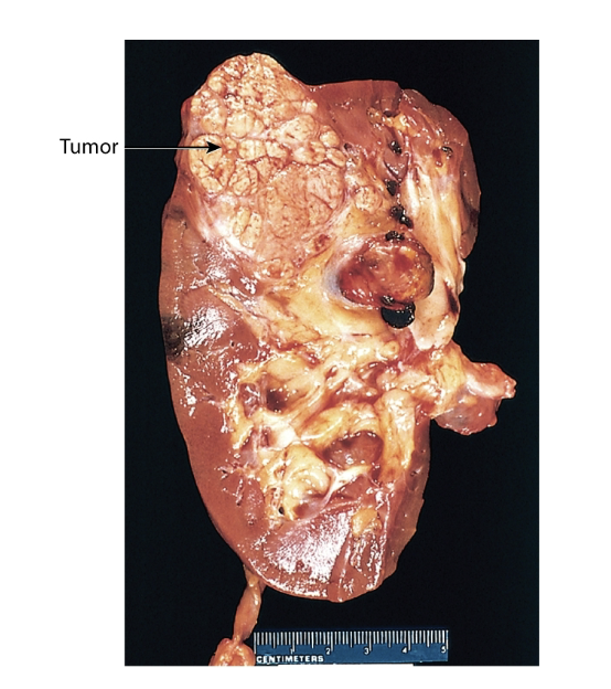
renal cell carcinoma
most common type of kidney cancer in adults, begins in the cells lining the kidney tubules
renal failure
condition in which kidneys lose their ability to filter wastes and excess fluid from the blood, may be acute (sudden) or chronic (long term)
renal hypertension
high blood pressure caused by kidney disease or narrowing of the arteries supplying the kidneys (renal artery stenosis)
Wilms tumor
a malignant kidney tumor that occurs primarily in young children (also called nephroblastoma)
bladder cancer
malignant tumor that develops in the tissues of the urinary bladder, most commonly in the bladder lining (urothelium)
diabetes insipidus (DI)
disorder in which the body cannot properly regulate water balance, causing large amounts of dilute urine and excessive thirst; its is usually due to lack of antidiuretic hormone (ADH) or the kidneys not responding to ADH
diabetes mellitus (DM)
disorder in which the body cannot properly regulate blood glucose (sugar) because of problems with insulin production or insulin action, resulting in high blood sugar
frequent voluntary urination at night
nocturia
blood urea nitrogen (BUN)
a blood test that measures the amount of urea nitrogen in the blood; it helps evaluate kidney function because healthy kidneys remove urea from the bloodstream
creatinine clearance test
test that measures how efficiently the kidneys filter creatinine from the blood; estimate the glomerular filtration rate (GFR), which reflects kidney function
CT urography x-ray
CT scan of urinary tract that uses contrast dye to produce detailed images of the kidneys, ureters, and bladder; helps detect kidney stones, tumors, blockages, and other urinary tract abnormalities
kidneys, ureters, and bladder (KUB) x-ray
plain x-ray of the kidneys, ureters and bladder; commonly used to detect kidney stones, bowel obstruction, or abnormal calcifications
renal angiography x-ray
x-ray study in which contrast dye is injected into the renal arteries to visualize the blood vessels of the kidneys; helps diagnose narrowed arteries, aneurysms, or other vascular problems
retrograde pyelogram (RP) x-ray
x-ray procedure in which contrast dye is injected through the urethra and bladder into the ureters and renal pelvis to visualize the urinary tract; used to detect blockages, stones, or tumors
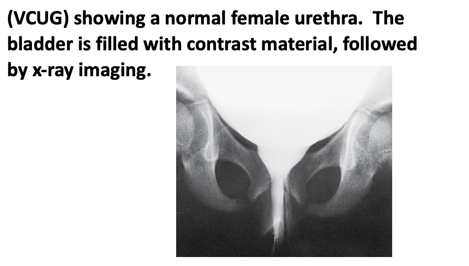
voiding cystourethrogram (VCUG) x-ray
x-ray study of the bladder and urethra performed while the patient urinates (voids); contrast dye is placed into the bladder through a catheter to detect vesicoureteral reflux (backward flow of urine) or structural abnormalities
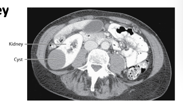
CT computed tomorgrpahy
scan with contrast shows a benign cyst on the kidney
ultrasonography
imaging urinary tract structures using high frequency sound waves