rabbit muscles
1/38
There's no tags or description
Looks like no tags are added yet.
Name | Mastery | Learn | Test | Matching | Spaced | Call with Kai |
|---|
No analytics yet
Send a link to your students to track their progress
39 Terms
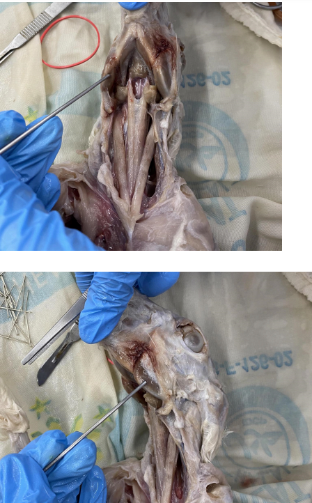
Masseter
O: Zygomatic Arch
I: Angular process of the mandible
A: Elevates the lower jaw
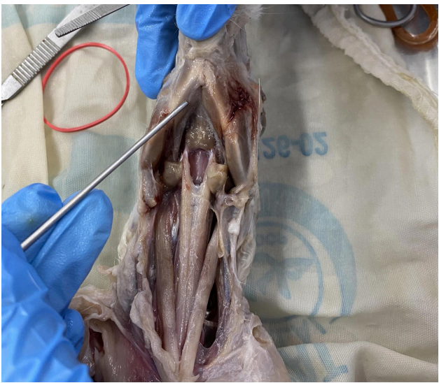
Digastric
O: Jugular and mastoid process of the skull
I: Mandible
A: Depresses the lower jaw
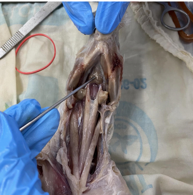
Mylohyoid
O: Mandible
I: Median raphe
A: Raise the floor of the mouth; bring hyoid forward
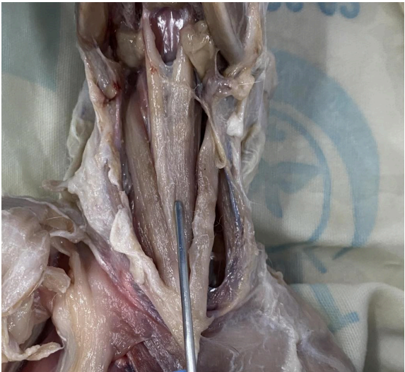
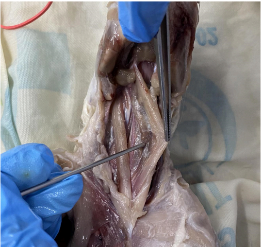
Sternohyoid
O: First costal cartilage in median ventral
line; Posterior surface of manubrium of the sternum
I: Body of hyoid; Lower margin of the hyoid bone
A: Draws the hyoid posteriorly
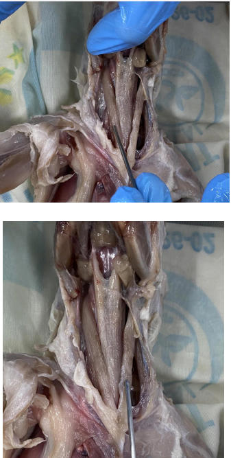
Sternothyroid
O: Sternum (with sternohyoid)
I: Thyroid cartilage of the larynx
A: Pull larynx posteriorly
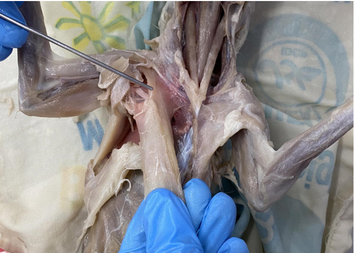
Sternomastoid
O: Median raphe; manubrium
I: Mastoid process of the skull
A: Turn the head; depress head on the neck
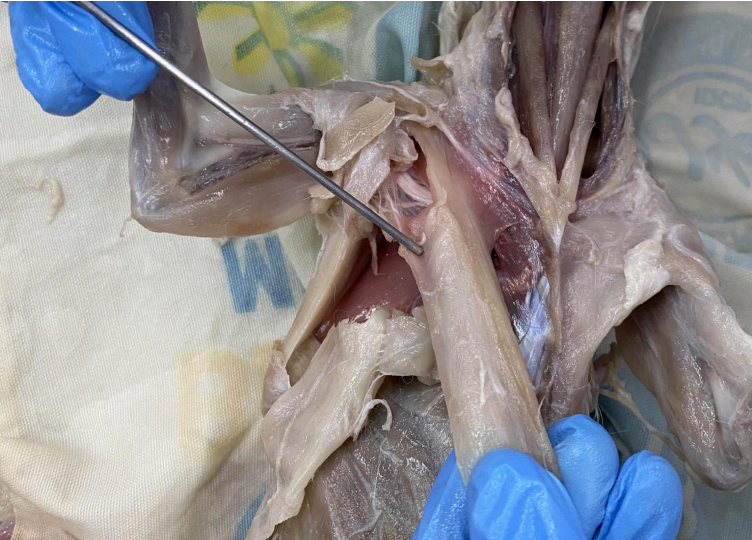
Pectoralis Major
O: Sternum; median ventral raphe
I: Pectoral ridge on ventral side of humerus
A: Adduction of the arm (pulling toward the chest) Posterior fibers: Retraction (pulling the arm back)
Pectoralis Minor
O: Sternum
I: Ventral side of the humerus (proximal to origin of p. major)
A: Stabilizes scapula by drawing it inferiorly and anteriorly against the thoracic wall
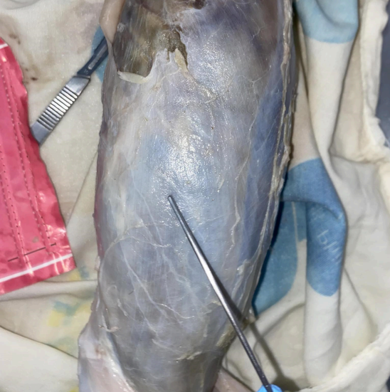
External Oblique
O: Lumbodorsal fascia; posterior ribs
I: Linea alba white line located at the midline of the trunk region pinakagitna na line
(medial)
A: Constrict the abdomen
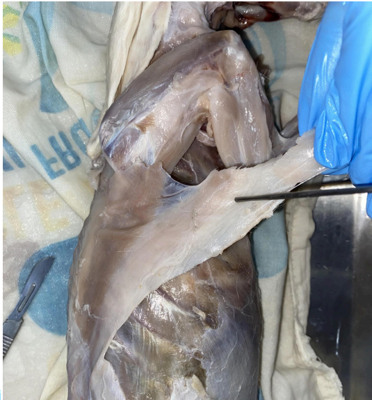
Latissimus dorsi
O: Neural spines of the last thoracic and most lumbar vertebrae; lumbodorsal fascia some thoracic to lumbar
I: Tendon on the medial surface of the humerus
A: Arm adduction, extension
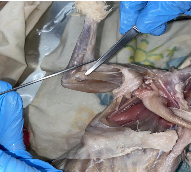
Epitrochlearis
O: Latissimus dorsi
I: Olecranon
A: Extend the forearm (with the triceps); rotate the ulna
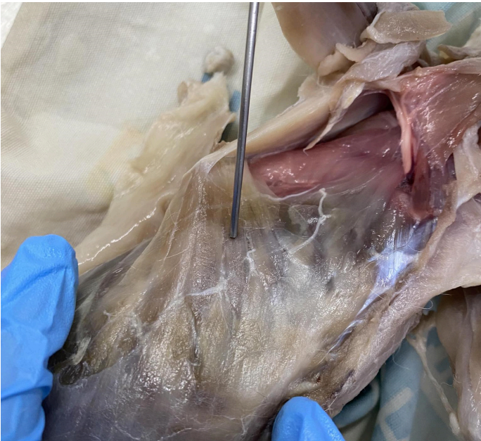
serratus anterior/ventralis
O: First nine or ten ribs or their cartilages
I: medial border of Scapula
A: draws the scapula forward; Protracts and rotates scapula, assists in arm elevation
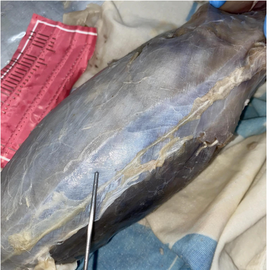
Rectus Abdominis
O: pubis
I: Sternum; costal cartilages
A: Constrict the abdomen; retract the sternum and
ribs curl ups or sit ups constrict the abdomen (exercise)
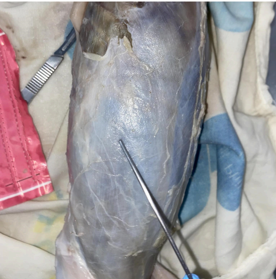
External Oblique
O: Lumbodorsal fascia; posterior ribs
I: Linea alba (white line located at the midline of the trunk region pinakagitna na line (medial))
A: constrict the abdomen; flexes and rotates vertebral column
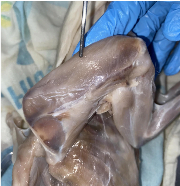
Acromiotrapezius
O: Neural spines of cervical and first thoracic vertebrae
I: Metacromion process and spine of scapula; fascia of spinotrapezius
A: Draw scapula dorsad and hold two
scapulae together
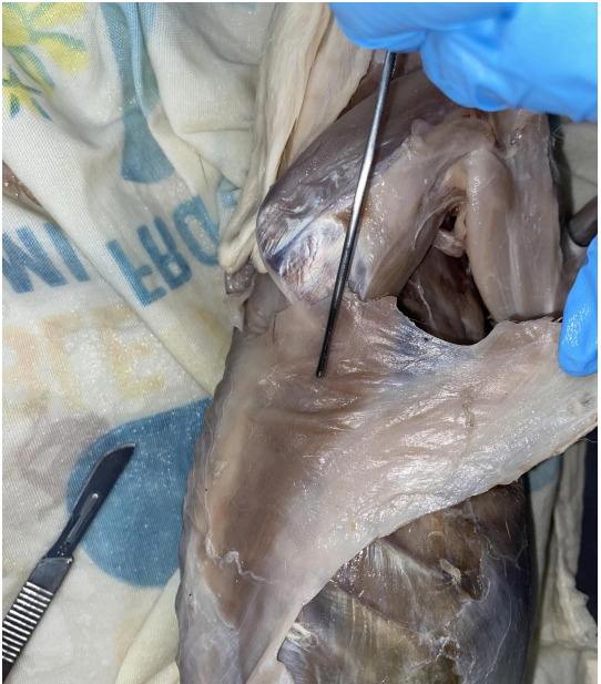
Spinotrapezius
O: Spines of thoracic vertebrae
I: Fascia of scapula
A: Stabilize and move the scapula dorsally and caudally
acromiodeltoid
O: Acromion process
I: Spinodeltoid; brachialis muscle; deltoid ridge of the humerus
A: Retraction and abduction of humerus
spinodeltoid
O: Spine of scapula
I: Deltoid ridge of the humerus (with the
acromiodeltoid)
A: Retract and abduct the humerus (with the acromiodeltoid)
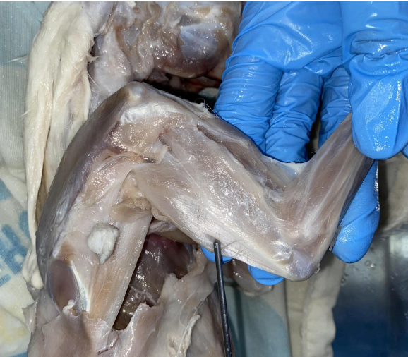
Triceps brachii long head
O: Glenoid border of the scapula
I: Olecranon process of the ulna
A: Extend the forearm
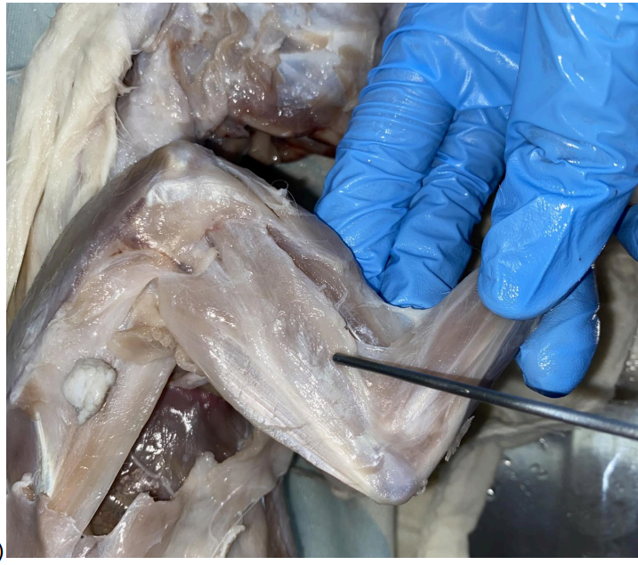
Triceps brachii lateral head
O: Greater tuberosity and deltoid ridge of the humerus
I: Olecranon process of the ulna
A: Extend the forearm
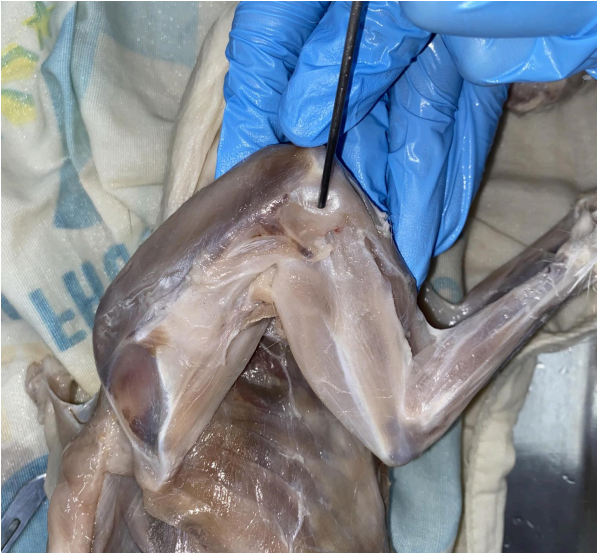
Levator scapulae ventralis
O: Transverse process of the atlas; occipital bone
I: Metacromion process; neighboring fascia
A: draw scapula craniad
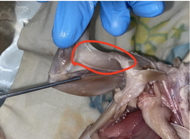
Biceps brachii
O: Supraglenoid tubercle of the scapula
I: Bicipital tuberosity of the radius
A: Flex forearm; assist in supination of the forearm
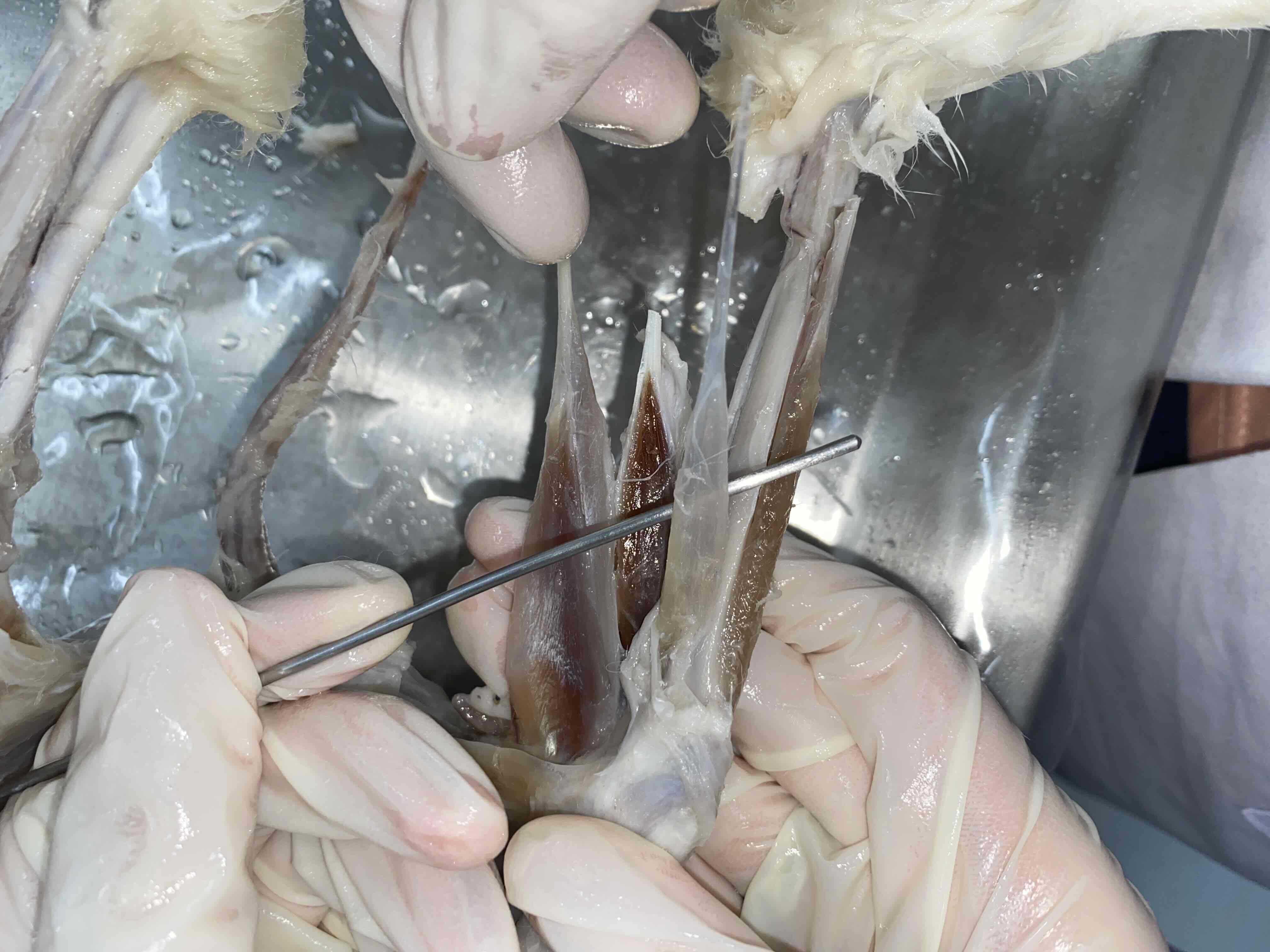
Extensor digitorum lateralis
Extensor digitorum longus/lateralis
O: Lateral surface of the femur/humerus, above the lateral epicondyle
I: Digits (via tendons passing internal to the wrist ligaments and splitting into 3 or 4 tendons underlying those of the extensor digitorum communis)
A: Extend digits
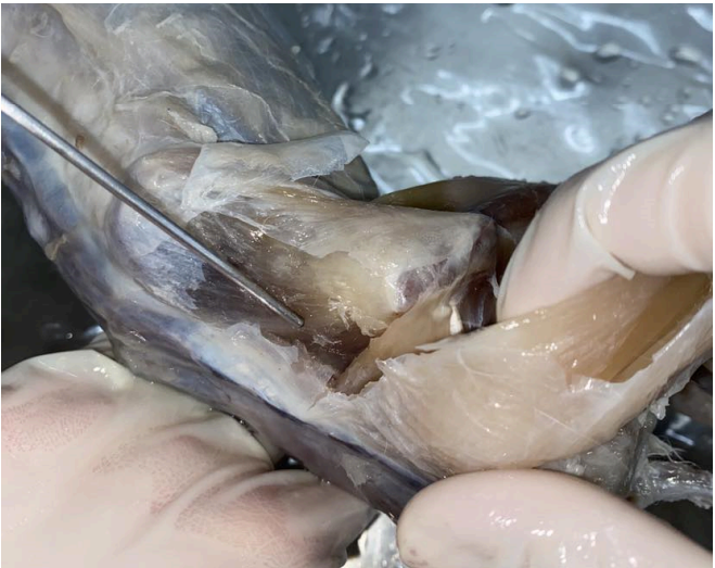
Gluteus medius
O: Adjacent fascia; crest of the ilium; lateral surface of the ilium; transverse process of the last sacral and first caudal vertebrae
I: Strong tendon on the great trochanter of the
femur
A: abducts thigh
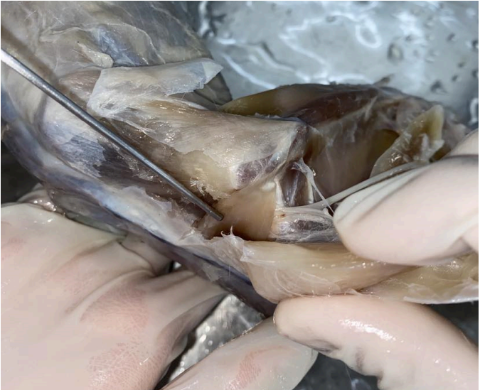
Gluteus maximus
O: Fascia; transverse process of the last sacral and first caudal vertebrae
I: Fascia lata; greater trochanter of the femur
A: Abducts thigh (In common with gluteus medius)
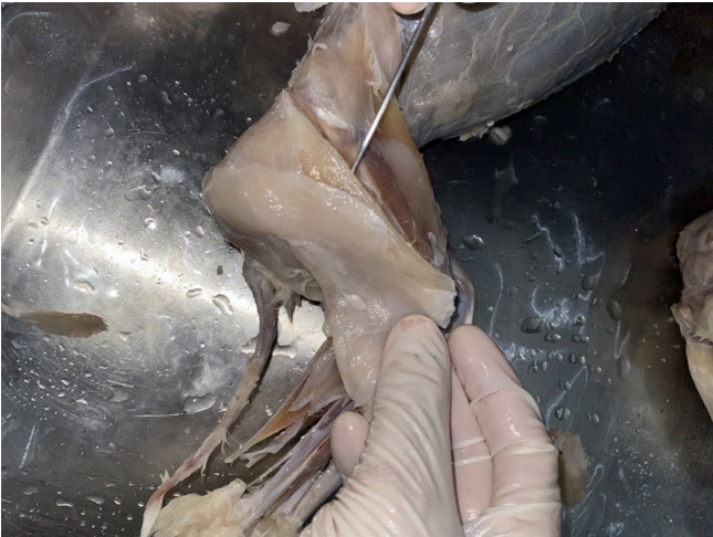
Biceps femoris
O: the Ischium
I: Patella and tibia (via a tendon); fascia of the shank
A: Abducts thigh; flex the shank
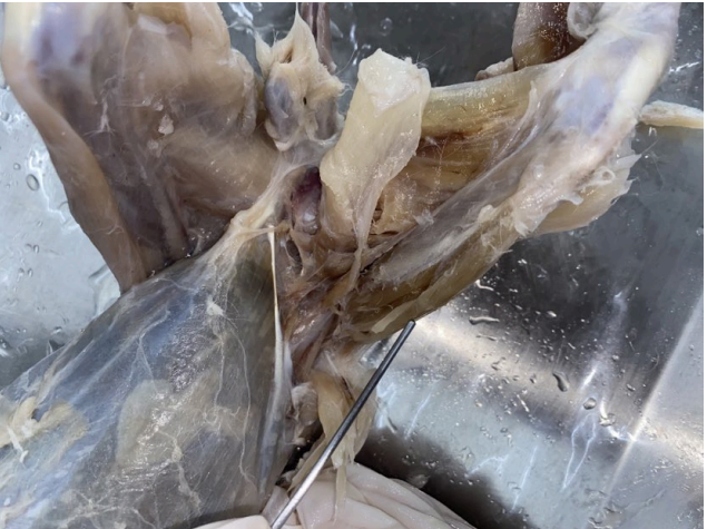
Vastus lateralis
O: Greater trochanter and surface of the femur
I: Patella and adjacent ligaments
A: Extend the shank
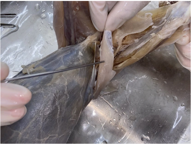
Tensor fascia latae
O: iliium and neighboring fascia
I: fascia lata
A: tightens the fascia lata

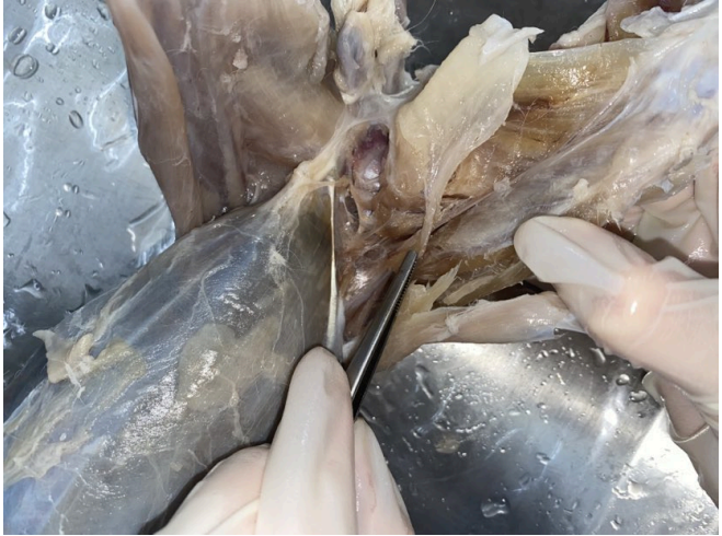
Sartorius
O: Crest and ventral border of the ilium
I: Proximal end of the tibia; patella; fascia and
ligaments in between
A: Adduct and rotate thigh; extend shank
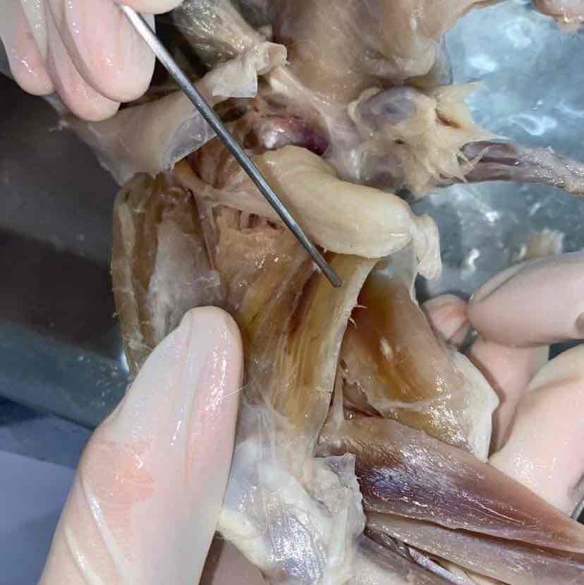
semimembranosus
semimembranosus
O: ischium, lower or back part of the hip bone
I: Medial epicondyle of femur; proximal end of tibia
A: Extend the thigh
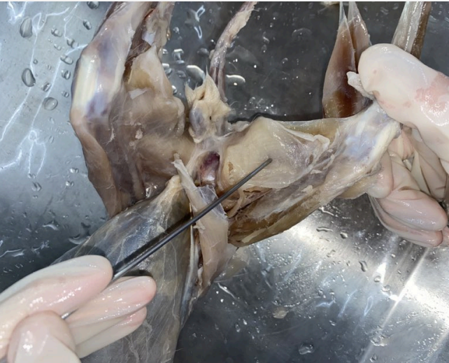
Gracilis
O: Ischial and pubic symphyses
I: Tibia (via an aponeurosis) long bone
A: adduct leg
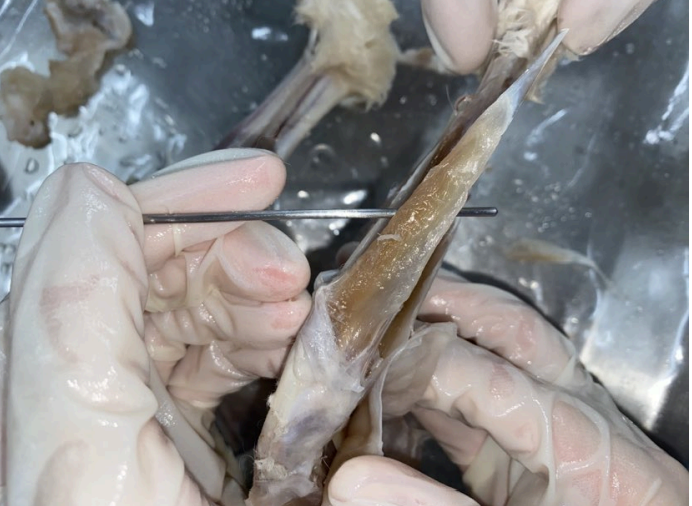
Tibialis anterior
O: Proximal parts of tibia fibula
I: First metatarsal via a strong tendon
A: Extend (dorsiflex) foot
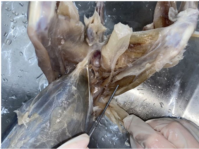
Rectus femoris
O: Ilium, near the acetabulum
I: Patella and adjacent ligaments
A: Extend the shank
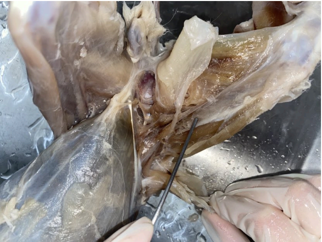
Vastus medialis
O: Femur
I: Patella and adjacent ligaments
A: Extend the shank
Vastus intermedius
O: surface of the femur
I: Patella and adjacent ligaments
A: Extend the shank
gastrocnemius
O: Surface fascia; femur; tendon and fascia of the plantaris muscle
I: Calcaneus (via the Achilles' tendon)
A: Ventroflex the foot
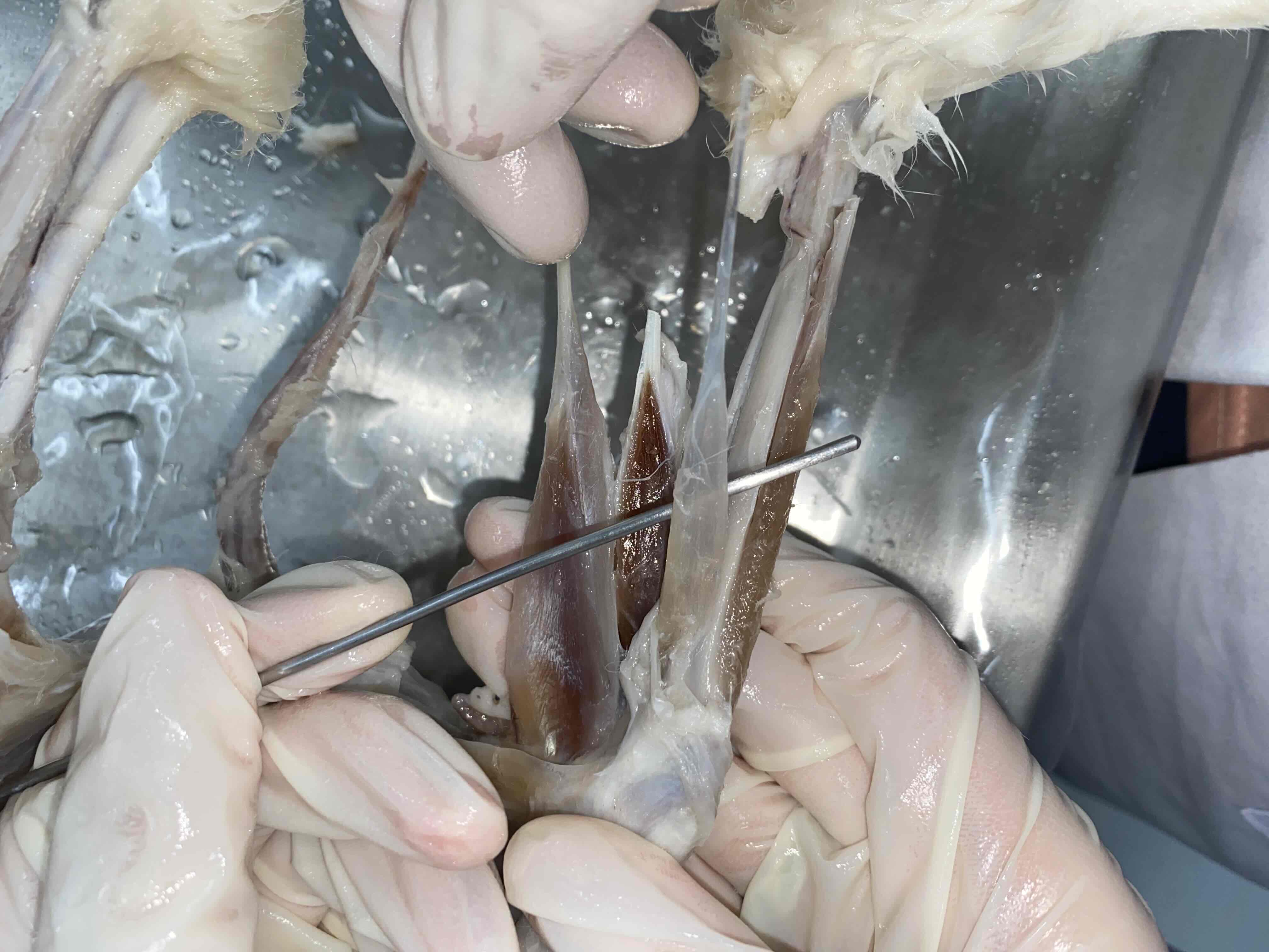
plantaris
O: Patella and femur
I: Digits (via a tendon divided into four)
A: Flex digits; extend ankle
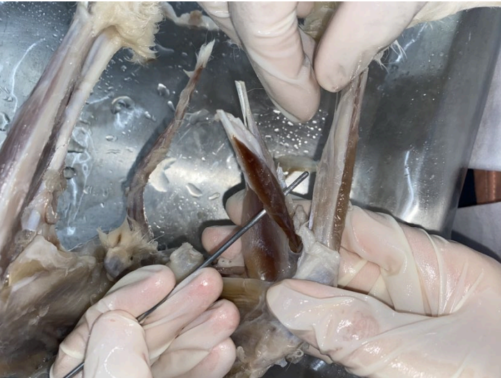
Soleus
O: fibula
I: Calcaneus (joins tendon of the gastrocnemius)
A: Ventroflex the foot
(with the gastrocnemius)
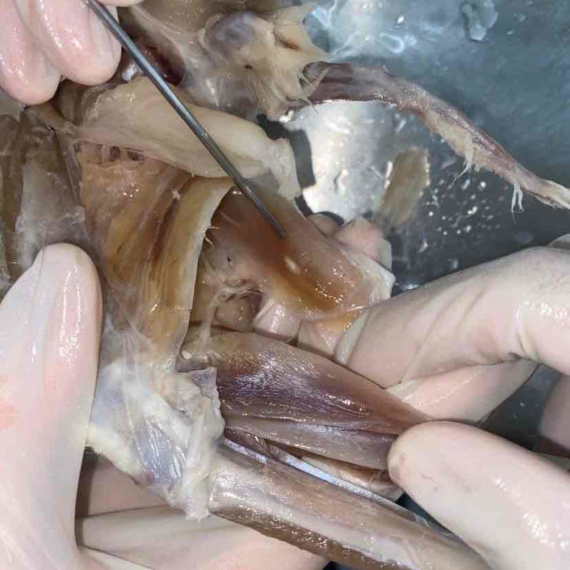
semitendinosus