Voice Disorders: Anatomy and Physiology
 Call Kai
 Learn
Learn Practice Test
 Spaced Repetition
 Match
Match Flashcards
 Knowt Play
1/48

Earn XP
Description and Tags
Comprehensive vocabulary flashcards covering sub-systems of speech, laryngeal anatomy, muscle actions, innervation, vocal fold histology, and phonatory mechanics from Voice Disorders Exam 1 notes.
Name | Mastery | Learn | Test | Matching | Spaced | Call with Kai | Chat |
|---|
No analytics yet
Send a link to your students to track their progress
49 Terms
Subsystems of Voice and Speech
Respiration
Phonation
Resonance
Respiration (Subsystem)
The speech subsystem encompassing the upper and lower respiratory tract; primary function is gas exchange (O2 intake and CO2 release), and secondary function is providing airflow for speech.
Phonation (Subsystem)
The speech subsystem centered in the larynx that creates the source sound for voice production.
Resonance (Subsystem)
The speech subsystem consisting of the vocal tract that modifies the shape and acoustic quality of the source sound.
Checking Action
A respiratory control process that maintains constant airflow during speech by resisting the passive recoil forces of inspiratory muscles and expanded ribs.
Normal Subglottic Pressure
The phonatory pressure needed underneath the vocal folds for normal voicing, typically 4–6 cm H2O (can be as low as 3 cm H2O).
Laryngeal Framework
One bone: hyoid bone. The six types of cartilage structuring the larynx: Epiglottis (1), Thyroid cartilage (1), Cricoid cartilage (1), Arytenoid cartilage (2), Cuneiform cartilage (2), and Corniculate cartilage (2).

Cuneiform and Corniculate Cartilages
Paired accessory cartilages situated in the posterior larynx atop and within the aryepiglottic folds above the arytenoids.
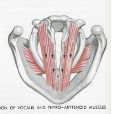
Thyroarytenoid Muscle (TA) Action
An intrinsic laryngeal muscle that causes the arytenoid cartilages to ROCK inward during phonation.

Interarytenoid Muscle (IA) Action
An intrinsic laryngeal muscle (consisting of transverse and oblique arytenoids) that causes arytenoids to GLIDE up and inward for vocal fold adduction during phonation.

Lateral Cricoarytenoid Muscle (LCA) Action
An intrinsic laryngeal muscle that ROTATE the vocal process of the arytenoids INward during phonation to adduct the vocal folds.

Posterior Cricoarytenoid Muscle (PCA) Action
The sole intrinsic vocal fold abductor muscle that ROTATE arytenoids OUTward to pull vocal folds apart during respiration.
Intrinsic Muscles
Lateral cricoarytenoid
Interarytenoid
Transverse arytenoid
Oblique arytenoid
Posterior cricoarytenoid (PCA – pulls cords apart)
Medial thyroarytenoid/Thyrovocalis
Cricothyroid
Lateral thyroarytenoid/Thyromuscularis
Vocal Fold Adductors
Lateral cricoarytenoid
Interarytenoid
Transverse arytenoid
Oblique arytenoid
Vocal Fold Abductor
Posterior cricoarytenoid (PCA – pulls cords apart) Rotates Out
Vocal Fold Tensors - Pitch Control
Intrinsic muscles that increase vocal fold tension for pitch control, comprising the Medial Thyroarytenoid (Thyrovocalis) and Cricothyroid muscles.
Vocal Fold Relaxer - Pitch Control
The Lateral Thyroarytenoid (Thyromuscularis) muscle, which acts to reduce vocal fold tension.
Suprahyoid Muscles - elevators
Extrinsic laryngeal elevation muscles located above the hyoid bone, including the digastric, mylohyoid, geniohyoid, stylohyoid, genioglossus, and hyoglossus muscles.
Infrahyoid Muscles - depressors
Ribbon-like 'strap' muscles located below the hyoid bone that act as laryngeal depressors, including the sternohyoid, sternothyroid, omohyoid, and thyrohyoid muscles.
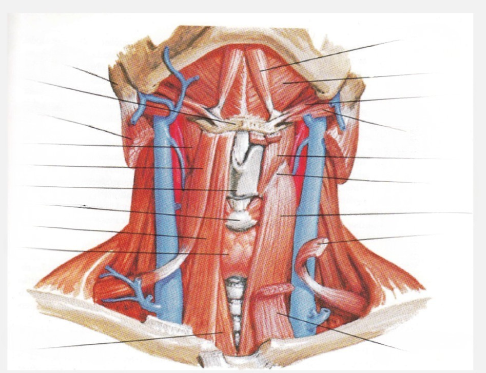
Sternohyoid
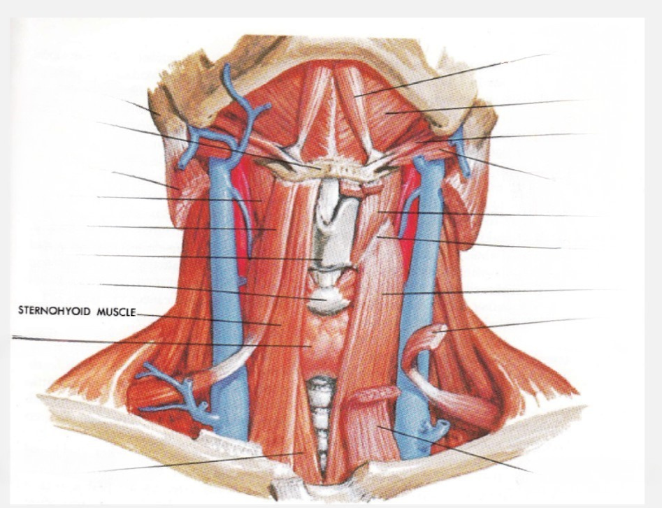

Sternothyroid
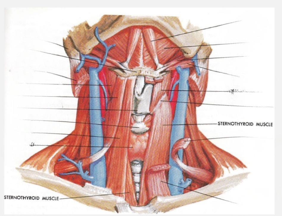

Omohyoid
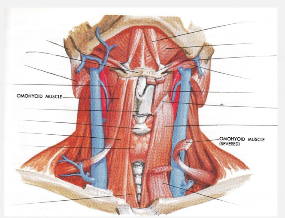

Stylohyoid


Thyrohyoid

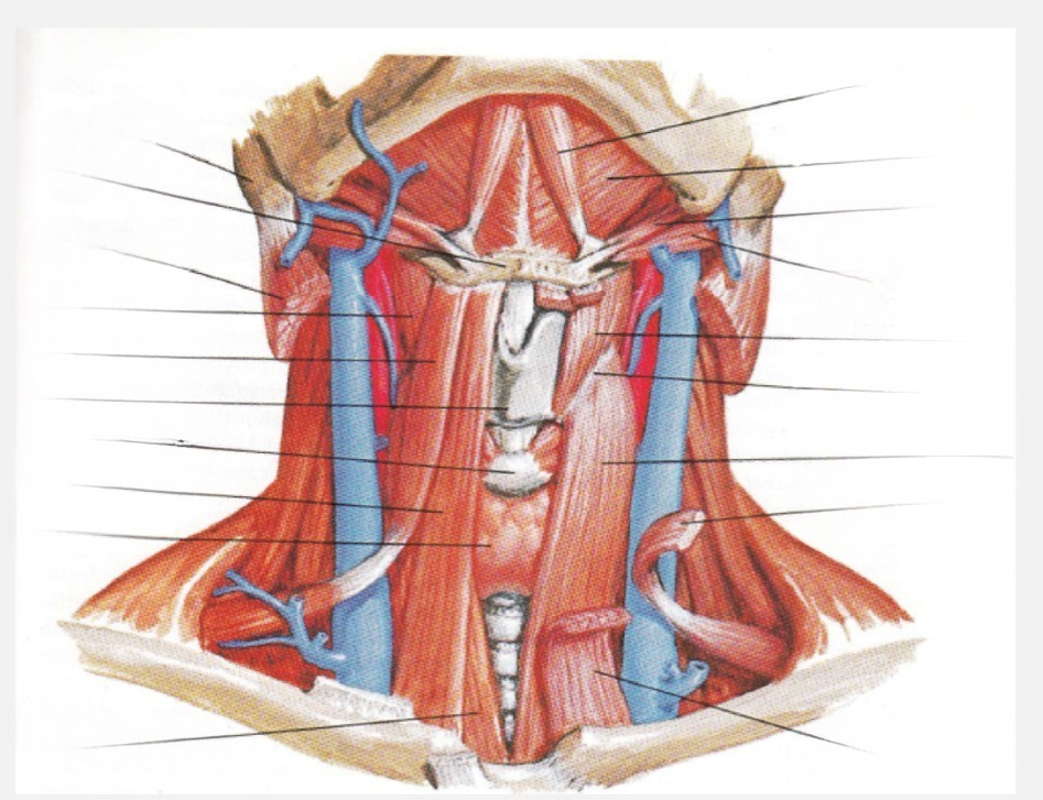
Mylohyoid


Digastric (anterior and posterior belly)

Genioglossus

Geniohyoid
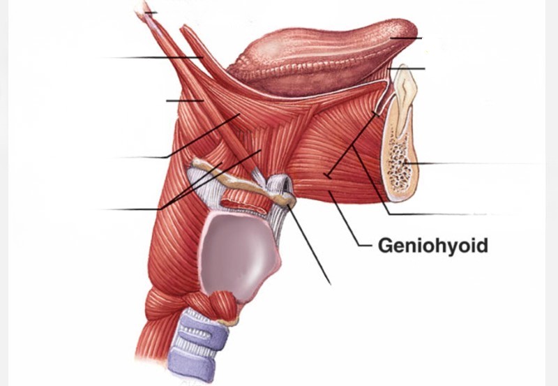
Hyoglossus

Superior Laryngeal Nerve - Internal Branch
A branch of CN X providing sensory innervation to the area above the vocal folds. SENSORY ABOVE
Superior Laryngeal Nerve - External Branch
A branch of CN X providing motor innervation specifically to the cricothyroid muscle. MOTOR to CT
Recurrent Laryngeal Nerve (RLN)
A branch of CN X that provides motor innervation to all intrinsic laryngeal muscles except the cricothyroid muscle, and sensory innervation from mucosa in the laryngeal vestibule below the vocal folds. SENSORY BELOW
Left Recurrent Laryngeal Nerve Course
Path of the left RLN looping under the AORTIC ARCH near the heart, rendering it vulnerable to damage during cardiac procedures leading to left vocal fold paralysis.
Right Recurrent Laryngeal Nerve Course
Path of the right RLN looping around the RIGHT SUBCLAVIAN ARTERY.

Superior and Recurrent Laryngeal Nerves Diagram
Anatomical schematic showing the right lateral course of the superior laryngeal nerve branches and the recurrent laryngeal nerve along the larynx.
Superior Laryngeal Nerve Damage Consequences
Paralysis of cricothyroid muscle causing lower pitch, inability to produce high notes or falsetto, rapid vocal fatigue, loss of vocal projection, voice asymmetry, reduced supraglottic sensation, and aspiration risk.
Unilateral Recurrent Laryngeal Nerve Damage
Paralysis of one vocal fold in a PARAMEDIAN position, causing breathy voice quality, hoarseness, roughness, reduced loudness, vocal fatigue, and shortened maximum phonation time.
Bilateral Recurrent Laryngeal Nerve Damage
Paralysis of both vocal folds near the MIDLINE, causing severe airway compromise (stridor, breathing distress) and a weak or whispery voice.
Epithelium (Vocal Fold)
The outermost, thin, stiff tissue layer of the vocal fold mucosa.
Superficial Layer of Lamina Propria (Reinke's Space)
A loose mucosal layer beneath the basement membrane that behaves mechanically like soft gelatin.
Intermediate Layer of Lamina Propria
An elastic component of the vocal ligament that behaves mechanically like a bundle of soft rubber bands.
Deep Layer of Lamina Propria
A fibrous component of the vocal ligament that behaves mechanically like a bundle of cotton threads.
Thyroarytenoid Muscle (Vocal Fold Layer)
The muscular body (vocalis) of the vocal fold, characterized mechanically as a bundle of stiff rubber bands.

Vocal Fold Tissue Layers Diagram
Cross-sectional visualization showing the stratified architecture of the vocal fold: epithelium, basement membrane, superficial lamina propria, intermediate lamina propria, deep lamina propria, and vocalis muscle.
Myoelastic-Aerodynamic Theory
Theory stating vocal fold vibration is driven by tracheal airflow and elastic properties of fold tissues: subglottic pressure forces adducted folds open, and tissue elasticity alongside the Bernoulli effect draws them closed.
Bernoulli Effect in Phonation
Principle where airflow velocity increases as it passes through the narrow glottis, dropping local air pressure and pulling the inferior borders of the vocal folds together.
Passive Tissue (Cover-Body Theory)
The non-muscular mucosal cover (epithelium, lamina propria) that is set in motion passively by airflow and modified by intrinsic muscle contraction.
Active Tissue (Cover-Body Theory)
The thyroarytenoid (vocalis) muscle body that possesses active contractile properties to alter vocal fold shape and stiffness.
Pitch Control Mechanics
Modulation of fundamental frequency (F0) where increased tissue stiffness raises F0, while increased vibrating mass or length lowers F0.