Neural Processing of Vision I : Retina (and Formation of Receptive Fields)
1/29
There's no tags or description
Looks like no tags are added yet.
Name | Mastery | Learn | Test | Matching | Spaced | Call with Kai |
|---|
No analytics yet
Send a link to your students to track their progress
30 Terms
why is the retinal pigment epithelium important
it absorbs photons missed by photoreceptors, minimizing image scatter
it is where discs of the outer segment are digested for waste disposal
explain and describe the retinal pathway
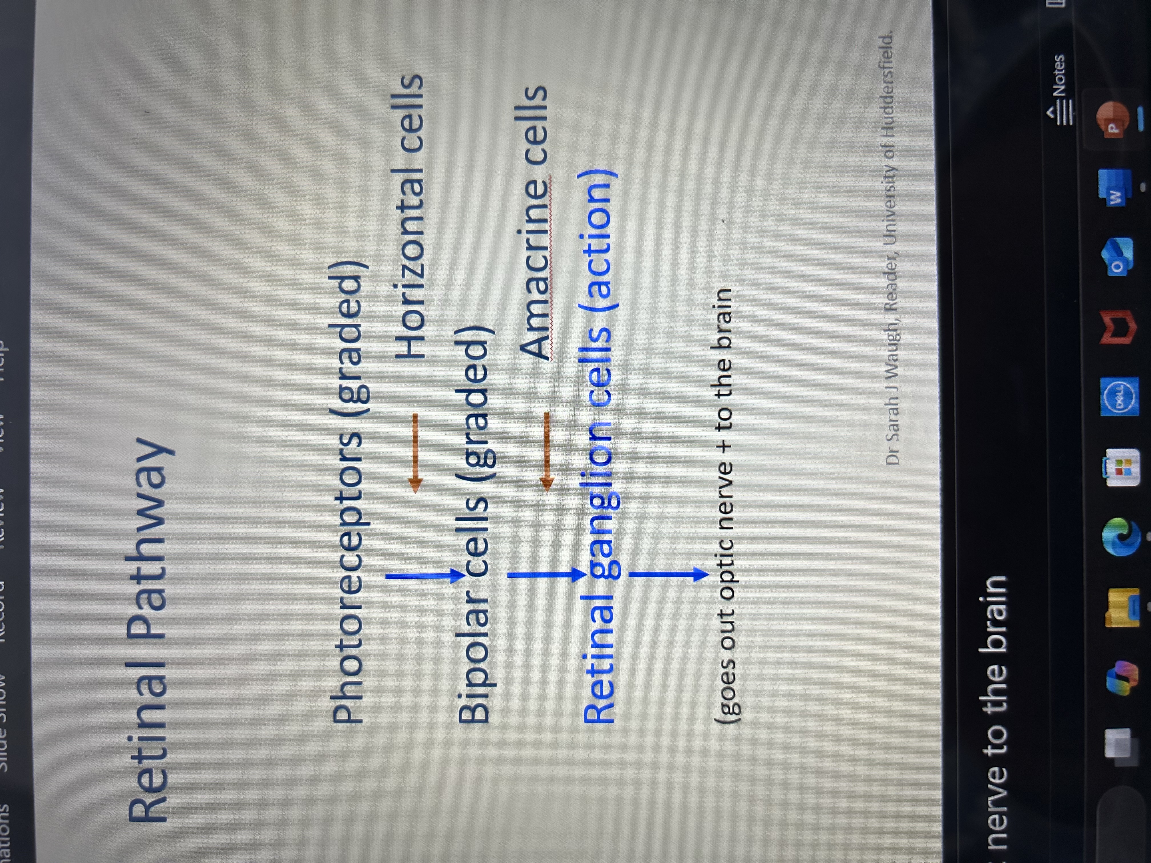
what are retinal ganglion cells - rGCs
Do not absorb light but receive input from other retinal cells - Not light sensitive – only photoreceptors do
Their output is in form of generating action potentials or neural impulses (brief electrical discharges) carried to the brain
Scientists can record changes in electrical activity occurring near single neurones (single cell recordings) and monitor the number of action potentials /second occurring in response to different visual stimuli.
explain how single cell recording for rGCs works
rGC has a “baseline level” or spontaneous activity without any visual stimulus
Move small spot around screen to find region of stimulated retina for which the spot causes an increase in number of action potentials /s - a “burst” of impulses (ON response)
turn light off, returns to spontaneous activity level
Move position of stimulus to neighbouring area of retina. Turn light on, notice decrease in firing rate
In same position, turn light off and see an increase in spontaneous activity (OFF response)
If you map out ON and OFF regions you are mapping out a “Receptive Field” for that neuron.
essentially: can target a single GC and Can map out on and off regions of the rGCs - by listening to the firing rate of the Aps
define receptive field
•the region of the visual field in which presentation of a stimulus evokes responses (changes in firing rate) from a given sensory neuron
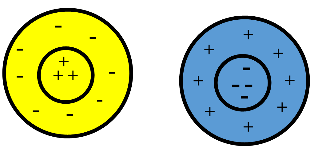
explain ON and OFF center responses and receptive fields at the rGCs (4)
Every ganglion cell has a receptive field – either ON centre receptive field or OFF centre receptive field – all the way across the retina
Centre-surround Receptive Field: has concentrically organized regions of excitation and inhibition.
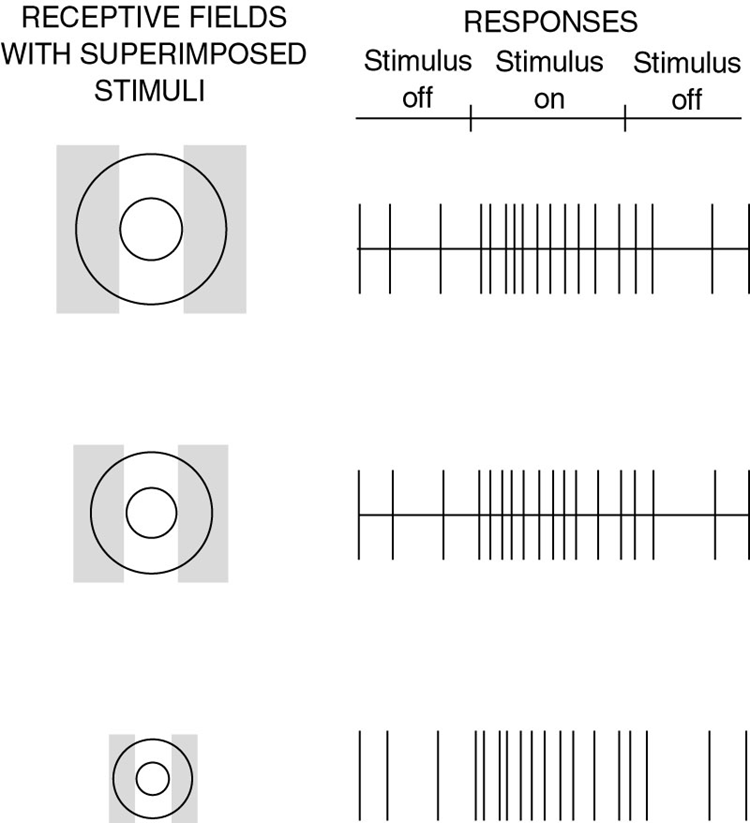
Centre-surround antagonism (through lateral inhibition - surround influences center response) allows size to be encoded. Stimuli that fill the central excitatory area, but do not encroach on the inhibitory surround, lead to the strongest response
circular !! important point - receptive fields at the level of rGCs are said to have concentric circular ON and OFF regions
Cell Type | Light in center does what? | Light in surround does what? |
|---|
ON-center | Turns the cell on (fires more) | Turns the cell off (fires less) |
OFF-center | Turns the cell off | Turns the cell on |
explain relation of size and GC receptive fields (5)
RF size determines size of most effective stimulus
Different size receptive fields can code for different sized images - can code for size at retina and edges !!
Size of center is related to the dendritic field of the anatomy of that ganglion cell
If you put white stripe/region so that it is over whole receptive field – the ON and OFF cancel each other out - wipes out – as if there is no stimulus at all – we do not want this
Thus analysis of object size is already becoming important at the retinal level
can we code for orientation at the retina and why
no
because RFs are round/circular - doesn’t matter how you position the stripes it will fire the same - so cannot code for orientation
why are rCGs said to be linear
In general, the cell’s response is determined by adding responses from excitatory and inhibitory influences within the cell’s receptive field
what is spontaneous discharge
The weights of excitatory and inhibitory regions are approximately equal, so that if they receive equal stimulation, they will cancel each other out and response = spontaneous discharge
how many ON and OFF center rGCs are there (and distinguishing between)
There are approximately equal numbers of ON centre and OFF centre rGCs
They form mosaics covering entire retina but information from them remains segregated (so that they don’t cancel each other out!)
Can code dark things on a light background and VV – don’t cancel each other out
How do neurones between photoreceptors and ganglion cells contribute to creation of ON and OFF centre receptive fields?
via bipolar cells
need 2 types of BCs for coding - one for ON-center and one for OFF-center responses, connecting photoreceptors to their respective rGCs
essentially need: ON bipolar cells and OFF bipolar cells
explain the 2 types of bipolar cells (ON and OFF) and how they are activated
ON cone bipolar: is activated with increased photon catch. It depolarises, leading to an increase in neurotransmitter release (to rGC).
OFF cone bipolar: is activated with decreased photon catch leading to depolarization and an increase in neurotransmitter release (to rGC)
They react to same signal coming from the cones in 2 different ways and feed that on to the rGCs !!!
where does the antagonist surrounds come from and what is this
the outer part of a retinal cell’s receptive field that responds in the opposite way to the center — it helps the eye detect contrast and edges
this comes from horizontal cells
what are horizontal cells
Every photoreceptor contacts both HCs and BPs
Horizontal cells (HCs) are spread over wide retinal areas over numbers of receptors, so have large receptive fields
They act laterally across cone responses and so are thought to provide the surround of the rGC RF
explain what lateral inhibition is in terms of horizontal cells
inhibition that travels across a neural network to impede the forward flow of activity
HCs modify photo receptor responses to create the surround
what do HCs do (3)
•HCs modify the strength of the signal coming from neighbouring photoreceptors before BPs
•They attenuate (turn down) the signals from the photoreceptors with weakest responses thereby heightening lightness differences in image
•They provide a fundamental role in creating the surround (through lateral inhibition) of rGC receptive fields
HCs and flicker
The signal coming form HCs in formation of the surround response is slightly delayed in time compared to centre response (this is important when we consider how the eye responds to flicker in Semester 2!)
describe and explain amacrine cells (4)
They outnumber both HCs and GCs
Amacrine cells may influence the temporal responses of ganglion cells, i.e., how vigorously they respond over time
They may enhance responses to RF surround in primates
AII (A2) amacrine cells convey rod signals to cone bipolars (have a special role in scotopic vision)
name and describe the 4 types of rGCs
1)M cells (Parasol): project to cells in magnocellular layers - good for colour vision R/G – fine details – a lot near fovea
2)P cells (Midget): project to cells in parvocellular layers - parvo cells of LGN - analyzing motion and flicker – no colour – located everywhere
3)K cells (Bistratified) : project to koniocells between magno- and parvo-cellular layers of LGN - blue/yellow colour vision - appear to be excited by blue light and inhibited by yellow light
4) Biplexiform cells: connect directly to photoreceptors - the function of biplexiform cells is not clear, but they may be involved in pupil control and diurnal rhythm. (Mather)
characteristics of M, P and K cells - cell body size, dendritic field area, conduction velocity, %cell population, spatial + temporal resolution, contrast + spectral sensitivity
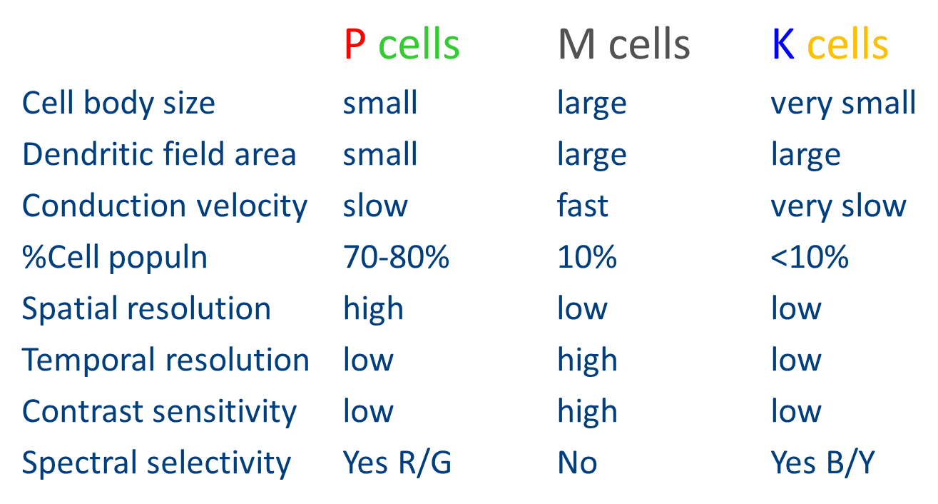
which rGCs are involved in spatial vision
•RFs of P cells are smaller suggesting that they support fine spatial detail
•M cells respond better to small differences in light level, i.e. higher contrast sensitivity at low contrasts
what rGCs are involved in temporal vision and motion perception
•M cells respond well to transient changes in stimulus, faster at responding; P cells respond poorly and prefer sustained stimuli
what rGCs are involved in colour vision
•P cells are sensitive to wavelength (colour - R/G); M cells are sensitive only to luminance changes – cannot do colour vision at all
K cells are sensitive to B/Y wavelengths
explain colour opponency in rGCs (4)
Whether a GC responds to different wavelengths depends on which cone classes contribute to its receptive field
If both excitatory and inhibitory regions have inputs from same cone classes, there will be no spectral selectivity (M cells)
If there is input from different cone classes to centre and surround then “spectral opponency” will be exhibited, e.g., cell will be excited by one portion of the spectrum and inhibited by another
basically - Need to have different amount of cones classes going into center than surround in order to do colour vision
what can receptive fields at level of retinal ganglion cells code for/do
can already code for size and colour
sensitivity vs resolution - degree of convergence
Degree of convergence depends on retinal location so that at fovea there is about 2 midget-ganglion cells serving 1 cone and in periphery there are several hundred receptors served by a single ganglion cell (“pooling”)
WHY? To design an eye capable of both high sensitivity and good spatial acuity
The eye varies how many photoreceptors connect to each ganglion cell — using low convergence in the fovea for sharp vision, and high convergence in the periphery for better night vision — to balance both detail and light sensitivity
visual illusions like Hermann grid and Mach bands are thought to be due to what?
lateral inhibition effects present in rGC receptive fields due to horizontal cells
lateral inhibition effects in LGN RFs
These illusions happen because horizontal cells send inhibitory signals across photoreceptors, shaping the center-surround receptive fields of ganglion cells — and that lateral inhibition sometimes tricks the brain into seeing contrast that isn’t really there
explain the Hermann grid
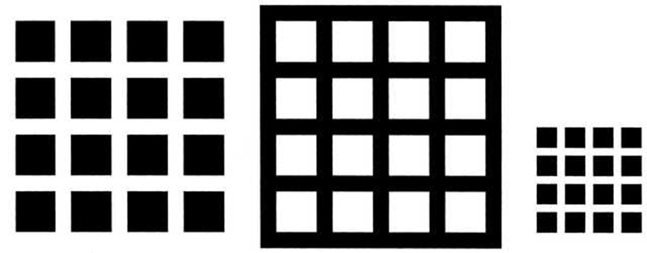
(You see gray dots at the intersections of white lines)
The intersections stimulate more surround inhibition than the straight parts of the lines.
So the ganglion cells fire less there, making the intersections look dimmer.
But when you look directly at one — no gray dot! Because your fovea has no surround inhibition there.
✅ Cause: Strong lateral inhibition from receptive fields in the peripheral retina
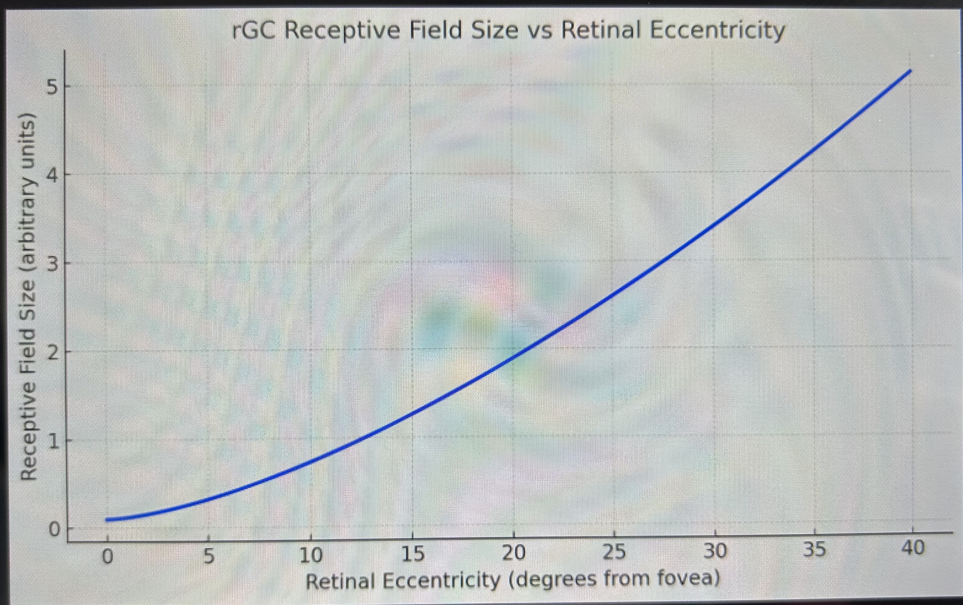
Draw a graph depicting how sizes of rGC receptive fields change with increasing retinal eccentricity - and explain
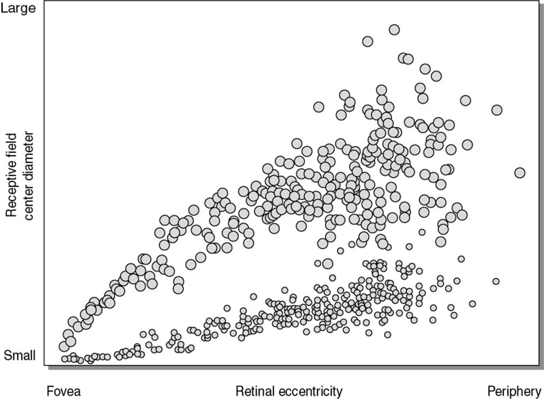
retinal ganglion cell (rGC) receptive field sizes increase with retinal eccentricity:
🔵 At the fovea (0°): very small receptive fields → high detail (resolution).
➡ As you move into the periphery: receptive fields get larger → better light sensitivity, but lower detail.
This visual helps explain why central vision is sharp and peripheral vision is better in low light but blurrier