Systolic Function & 3D/4D
1/21
There's no tags or description
Looks like no tags are added yet.
Name | Mastery | Learn | Test | Matching | Spaced |
|---|
No study sessions yet.
22 Terms
Where on an ECG does IVCT & Systole Occur
Right after the QRS complex
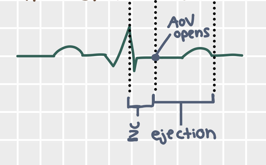
Where on an ECG does mechanical systole occur
After IVCT (after QRS) to after the T wave

Measures of cardiac function
SV
EF
CO
SW
GLS
GRS
E/e’
Dp/dt
Time constant of ventricular relaxation
Stroke volume equations
EDV - ESV
0.785 LVOT Diameter² * LVOT VTI
Normal Stroke Volume
70 - 110 mL/beat
CO equation & normal value
SV * HR
4 - 8 L/min
Measures of regional LV function
visual
Strain
TDI Strain rate
Measures of LV global systolic function
EF
GLS Average
TEI index/LIMP
Visual
EF Equations
(EDV - ESV) / EDV
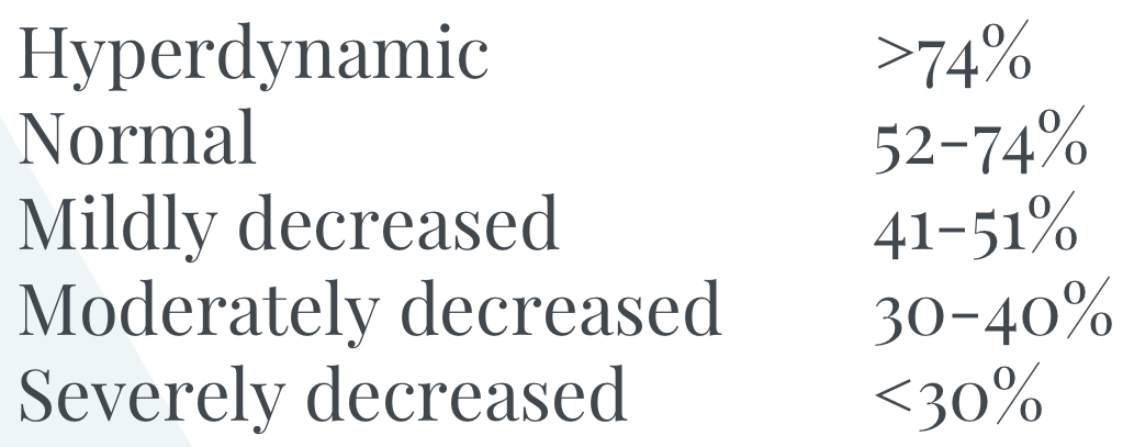
Normal LVEF with biplane
54 - 74
Normal EF Values
Fractional Shortening equation & normal value
(LVIDd - LVIDs)*100 / LVIDd
25-45%
Is it ok for the LV to be foreshortened when obtaining 3D volume?
Yes, as long as the whole LV is in view!
What is LV mass?
The total weight of the myocardium; obtained by (myocardium volume * density of cardiac muscle)
Normal LV mass value
66 - 200g
Draw the LV regional function bullseye
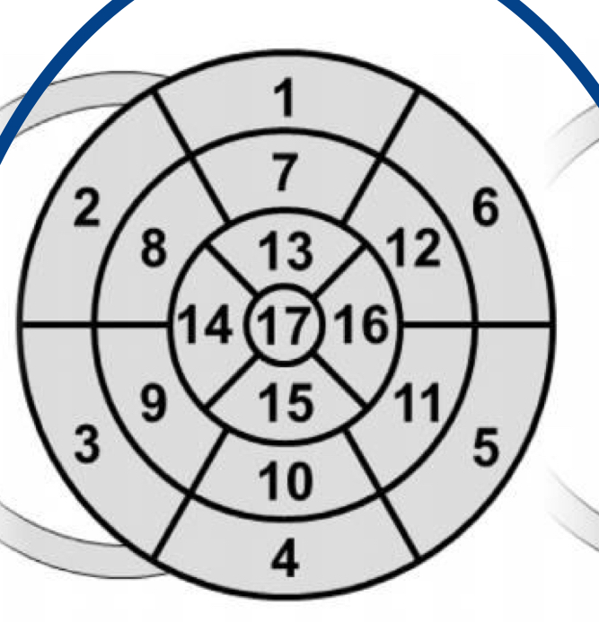
Grading of LV regional dysfunction
1 = normal/hyperkinetic
2 = hypokinetic
3 = a kinetic
4 = dyskinetic
5 = aneurysmal
Stress echo may reveal coronary artery stenosis depending on the regional _________.
Coronary flow reserve
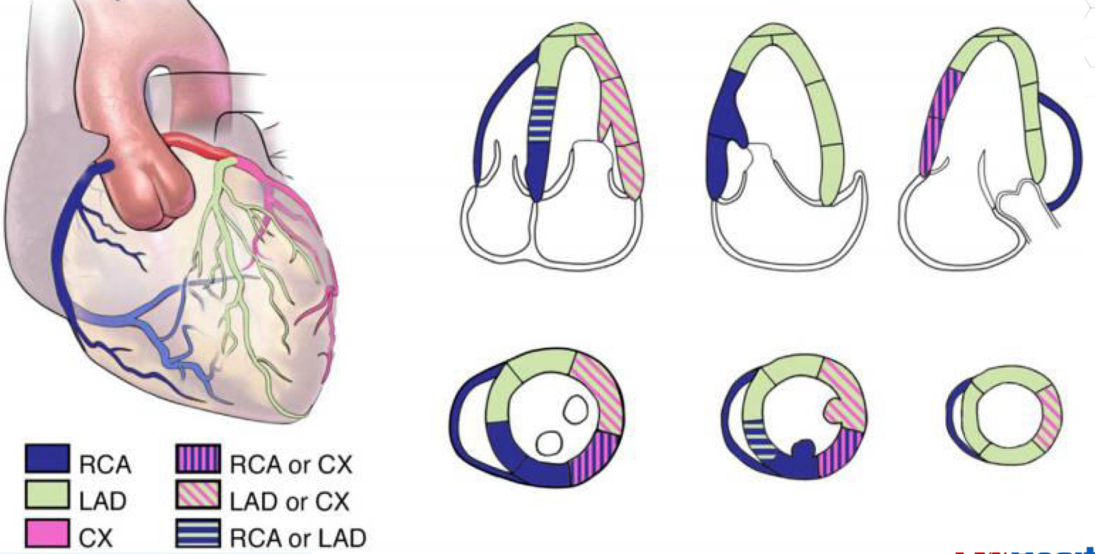
Walls perfused by the LAD
A4C:
IVS (inferoseptal)
Apex
Anterolateral
A3C
Anteroseptal
Apex
Apical inferolateral
A2C
Anterior
Apex
Apical inferior

Walls perfused by the RCA
RV
A4C
Base & mid IVS (inferoseptal)
A3C
base & mid inferolateral
A2C
Base & mid inferior

Walls perfused by the CX
A4C
Anterolateral wall
A3C
Base & mid inferolateral