Synaptic Transmission Objectives
 Call Kai
 Learn
Learn Practice Test
 Spaced Repetition
 Match
Match Flashcards
 Knowt Play
1/69

There's no tags or description
Looks like no tags are added yet.
Name | Mastery | Learn | Test | Matching | Spaced | Call with Kai | Chat |
|---|
No analytics yet
Send a link to your students to track their progress
70 Terms
electrical synapse
gap junction consisting of a field of connexin pores that pass ions and signaling molecules directly from one cell to another without passing through the extracellular fluid
gap junctions
aggregates of intercellular channels that permit direct cell-cell transfer of ions and small molecules (i.e. ATP & 2nd messengers)
Gap junctions are formed by?
two connexons of adjacent cells docking together
Connexons
6 subunits (connexins) with an aqueous central pore that allows exchange of electrical current → direct cytoplasmic continuity
electrical synapse flow of information
- virtually instantaneous
- bidirectional
chemical synapse
gap between two neurons or a neuron and a non-neuronal cell where information passes chemically, in the form of neurotransmitter molecules
neurotransmitters
chemical substance released at the end of a nerve fiber and transmit messengers between neurons or from neurons to some other structure (i.e. muscle fiber)
neurotransmitters are packed in ______ and released into the _____ where they can act on ______
membrane-bound vesicles; synaptic cleft; postsynaptic receptors
chemical vs electrical synapse speed
Chemical synapses are slower than electrical synapses because they depend on the release of NT molecules
o This results in a delay between when the action potential reaches the presynaptic terminal and when the NT leads opening of postsynaptic ion channels
▪ This mechanism also means the flow of information is unidirectional
chemical synapse mechanism
1) NTs are synthesized in soma (cell body) and then stored in vesicles
2) An action potential invades the presynaptic terminal
3) Depolarization of presynaptic terminal causes opening of voltage-gated Ca2+ channels
4) Influx of Ca2+ through channels
5) Ca2+ causes vesicles to fuse with presynaptic membrane
6) NT is released into synaptic cleft via exocytosis
7) NT binds to receptors on postsynaptic membrane
8) Opening or closing of postsynaptic channels
9) EPSP or IPSP changes excitability of postsynaptic cell
10) Retrieval of vesicular membrane from plasma membrane
role of Ca2+ channels in chemical synapses
Ca2+ is required for NT release → when an action potential reaches the nerve terminal, voltage-gated Ca2+ channels open and Ca2+ rushes into the neuron terminal (due to greater extracellular [Ca2+]
chemical synapses in low extracellular Ca2+
→ 0 to a few NT-containing vesicles are released → only reaches subthreshold in postsynaptic membrane
chemical synapses in high extracellular Ca2+
multiple NT-containing vesicles are released → reaches suprathreshold in postsynaptic membrane
Quantal NT Release
NTs are released into a synapse in packaged vesicles called quanta (1 quantum = 1 vesicles worth of NT)
Quantum
smallest measurable unit, generates a miniature end plate potential (mEPP)
miniature end plate potential (mEPP)
smallest amount of stimulation that one neuron can send
mEPP size based on vesicles
• 1 vesicle → mEPP (w/ small variation in size)
• 2 vesicles → double mEPP size
• 3 vesicles → triple mEPP size
end plate potential (EPP)
aggregate sum of many mEPPs
If the EPP depolarizes the postsynaptic membrane to the threshold level, it will?
fully activate voltage-gated Na+ channels and produce the action potential
types of synaptic potentials
o End Plate Potential (EPP)
o Evoked Potential
o Spontaneous Potential
Presynaptic vesicles are formed by?
budding off an endosome via Clathrin-dependent exocytosis
In order for Clathrin to enclose a vesicle what must be present?
12 pentagons must be present in the lattice
Ca2+ influx is sensed by?
synaptotagmin
Synaptic Vesicle Recycling
1) Presynaptic vesicles are formed by budding off an endosome via Clathrin-dependent exocytosis
a. Clathrin coat falls off the vesicle once it buds off the endosome
2) Synapsin then attaches the vesicle to the cytoskeleton and docks it at the membrane, moving it closer to the active zone
3) SNAREs are involved in priming which tucks the vesicle very snuggly against the plasma membrane at the active zone (next to voltage-gated Ca2+ channels)
4) Ca2+ influx is sensed by synaptotagmin which causes the vesicle to fuse with the membrane for NT release
5) The vesicle is then recaptured via Clathrin-dependent endocytosis → the vesicle buds inward back into the presynaptic membrane
a. Clathrin coat falls off the vesicle once it buds off the plasma membrane
6) Vesicle refuses with endosome and can be recycled
active zone
region of presynaptic membrane that mediates NT release
Synapsin
tethers synaptic vesicles to the actin cytoskeleton within the presynaptic terminal and docks it at the membrane
SNARE complex
proteins involved in the priming of vesicles for fusion by tucking them snuggly against the membrane → mediates vesicle fusions
SNARE Complex proteins
▪ SNAP-25: attached to plasma membrane
▪ Syntaxin: attached to plasma membrane
▪ Synaptobrevin: attacked to vesicle membrane
Synaptotagmin
serves as a calcium sensor for NT release → Ca2+ (4 total) binding to synaptotagmin triggers vesicle fusion and release
Each synaptotagmin molecules has ______ sites (how many total?)
two Ca2+ binding; 4 total since there are two synaptotagmin molecules
Clathrin
involved in coating membranes that are exocytosed from the endosome and endocytosed from the plasma membrane (does not link to membrane directly)
criteria for classification as a neurotransmitter
I. Present in the nerve terminal
II. Released by electrical stimulation of neuron
III. Specific receptors exist for said NT
IV. Direct application of substance mimics neuron stimulation
V. There is a mechanism for terminating the action
how can NT action be terminated?
I. Diffusion away from the site of action (i.e. peptide NTs)
II. Re-uptake into cells (i.e. amino acids, amines)
III. Enzymatic cleavage (i.e. acetylcholinesterase; peptidase)
who discovered neurotransmitters
Otto Loewi
otto loewi experiment
1) Using two frog hearts → heart #1 was placed in a chamber filled with saline which was connected to another chamber that contained heart #2
a. Fluid from chamber #1 was allowed to flow into chamber #2
2) Electrical stimulation of the vagus nerve (attached to heart #1) caused heart #1 to slow down
3) After a delay, heart #2 also slowed down
4) Loewi hypothesized that electrical stimulation of the vagus nerve release a chemical into the fluid of chamber #1 that flowed into chamber #2 → he called this chemical "Vagusstoff"
"Vagusstoff" is now known as?
acetylcholine
Small Molecule NTs Synthesis
Synthesized locally within the axon terminal
Peptide NT synthesis
Follows synthesis of any secretory protein (transcription starts in nucleus, translation occurs in cytoplasm)
Small Molecule NTs post modifications
none
Peptide NT post-modifications
Enzymes modify pre-peptides to produce peptide NTs
Small Molecule NT packaging
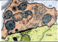
Packaged via vesicular transporters into small clear-core vesicles
Peptide NT packaging
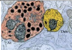
Packaged by Golgi apparatus into large dense-core vesicles
Small Molecule NT route of transport
axon terminal → vesicle → release into synaptic cleft
Peptide NT route of transport
Nucleus → vesicle → transported down microtubules to axon terminal → released out the sides of terminal
Small Molecule NT histology
Peptide NT histology
Small Molecule NT Ca2+ channels
N-type → opens & closes quickly
Peptide NT Ca2+ channels
L-type → stays open longer (& N-type)
Small Molecule NT Ca2+
Requires localized increase in [Ca2+] → single action potential is enough to cause NT release from vesicles
Peptide NT Ca2+
Requires large increase in [Ca2+] since vesicles are farther from synaptic cleft → larger increase in [Ca2+] allows the ions to diffuse farther away to reach theses vesicles → requires train of APs (not just one)
Small Molecule NT recycling
yes, can be
Peptide NT recycling
degraded, can not be recycled
Acetylcholine (ACh)
used at neuromuscular junction
ACh mechanism of transmission
1) Acetyl CoA + Choline → Acetylcholine (by Choline Acetyltransferase)
2) ACh is packaged into vesicles by ACh vesicular transporters
3) ACh is released into synaptic cleft
4) Two routes:
a. ACh runs into ACh esterase which breaks it down into acetate + choline
b. ACh binds to its receptors on postsynaptic membrane to exert its effect (can eventually get broken down by ACh esterase as well once it leaves the receptor)
5) Choline can be transported back into presynaptic membrane via transporter to be recycled
Glutamate
major excitatory NT in CNS
glutamate mechanism of transmission
1) Glutamine is converted to glutamate (by Glutaminase)
2) Glutamate is packaged into vesicles by glutamate vesicular transporters
3) Glutamate is released into synaptic cleft
4) Glutamate can bind to its receptors on postsynaptic membrane, or can get transported back (2 routes)
a. Glutamate can be transported back to the presynaptic membrane to be recycled
b. Glutamate can be transported into a glial cell:
i. In glial cell → glutamate is converted back to glutamine (by Glutamine Synthetase)
ii. Glutamine can be transported out of the glial cell and transported back into the presynaptic membrane to be recycled
GABA
one of the major inhibitory NTs in CNS
GABA mechanism of transmission
1) Glutamate is converted to GABA (by Glutamic Acid Decarboxylase)
2) GABA is packaged into vesicles by GABA vesicular transporters
3) GABA is released into synaptic cleft
4) GABA can bind to its receptors on postsynaptic membrane, or it can be transported back into presynaptic membrane to be recycled
a. They may also just diffuse away from the synaptic cleft
Glutamate and GABA will never be released by the?
same neuron → if glutamic acid decarboxylase is present then GABA will be the specific NT that is being released
Glycine
second major inhibitory NT in CNS
glycine mechanism of transmission
1) Glucose is converted to serine (do not need to know how)
2) Serine is converted to glycine (by Serine Transhydroxymethylase)
3) Glycine is packaged into vesicles by glycine vesicular transporters
4) Glycine is released into synaptic cleft
5) Glycine can bind to its receptors on postsynaptic membrane, or it can be transported back into presynaptic membrane to be recycled
a. They may also just diffuse away form the synaptic cleft
Dopamine mechanism of transmission
1 of 3 catecholamines
1) Tyrosine is converted to L-DOPA (by Tyrosine Hydroxylase)
2) L-DOPA is converted to dopamine (by Dopa Decarboxylase)
3) Dopamine is packaged into vesicles by dopamine vesicular transporters
4) Dopamine is released into synaptic cleft
5) Dopamine can bind to its receptors on postsynaptic membrane, or it can be degraded by MAO-B or COMT
a. Can also be re-uptaken into presynaptic membrane
Norepinephrine (NE)
1 or 3 catecholamines
NE mechanism of transmission
1) Dopamine is converted to NE (by Dopamine β-hydroxylase)
2) NE is packaged into vesicles by NE vesicular transporters
3) NE is released into synaptic cleft
4) NE can bind to its receptors on postsynaptic membrane, or it can be degraded by MAO-A or COMT
a. Can also be re-uptaken into presynaptic membrane
if Dopamine β-hydroxylase is present in the cell, dopamine will?
NOT be released and instead converted to NE
Epinephrine (Epi)
1 or 3 catecholamines
epi mechanism of transmission
1) NE is converted to Epi (by PMNT)
a. If this enzyme is present in the cell → NE will NOT be released and instead converted to Epi
2) Epi is packaged into vesicles by Epi vesicular transporters
3) Epi is released into synaptic cleft
4) Epi can bind to its receptors on postsynaptic membrane, or it can be degraded by MAO-A or COMT
a. Can also be re-uptaken into presynaptic membrane
if PMNT is present in the cell, NE will NOT?
be released and instead converted to Epi
Serotonin (5-HT)
Indoleamine
serotonin mechanism of transmission
1) Tryptophan is converted to 5-hydroxy Tryptophan (by Tryptophan hydroxylase)
2) 5-hydroxy Tryptophan is converted to 5-HT (by Aromatic Amino Acid Decarboxylase)
3) 5-HT is packed into vesicles by 5-HT vesicular transporters
4) 5-HT is released into synaptic cleft
5) 5-HT can bind to its receptors on postsynaptic membrane, or it can be re-uptaken into presynaptic cleft (by SERT)