Articulations, joints and adaptations
1/16
There's no tags or description
Looks like no tags are added yet.
Name | Mastery | Learn | Test | Matching | Spaced | Call with Kai |
|---|
No analytics yet
Send a link to your students to track their progress
17 Terms
types of joint
can be classified based on structure or the type of movement that the structure supports
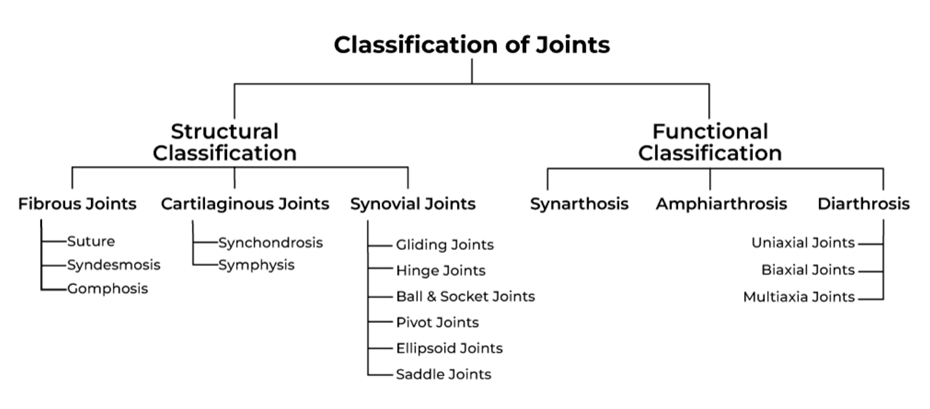
classification by movement
CLASSIFIED BY MOVEMENT
some in the skull do not move (synarthrosis) but are still interface between muscular skeletal tissues
some in the spine are slightly moveable (amphiarthrosis)
diarthrosis - highly moveable

classified by structure
CLASSIFICATION BY STRUCTURE
FIBROUS
no synovial cavity held together by collagen fibres
suture - interlocking between skull and bones - some fuse
syndesmoses - band/ligaments - tibiofibular joints - slight movement allowed
gomphoses - bolt or nail - E.g tooth cone shaped into a socket
CARTILAGENOUS
no synovial cavity held together by cartilage
synchondroses - connected by cartilage E.g: epiphysial plates of hyaline cartilage
symphyses - growing together - flat disc of fibrocartilage - vertebrate disks
SYNOVIAL
synovial cavity, united by dense irregular capsule tissue - more complex movements, synovial cavity filled with synovial fluid
synovial fluid is an ultrafiltrate from the capillaries feeding the cartilage
fluid enriched with hyaluronan - gives viscolubricative properties to the fluid and also prvenets drainage of the cavity
held together by accessory ligaments to support more complex movement
human defining joint
the hip is a human defining joint, although joints in the hand and the thumbs are unique among species
gives us the ability to become bipedal, free up our hands for holding tools etc - thought to be an evolutionary weak point as is prone to disease/osteoarthritis
pelvis starts to form between 6-8 weeks of pregancy but are not bone at this point - ossification is stalled genetically so it can remain able to change shape
compared to the pelvic blades in chimpanzees and gorillas, ours are shorter and wider to allow us to walk upright - also increase in size of birth canal
understanding evolution of the human hip
by understanding the genetic origin for bipedalism we can understand the genetic architecture and therefore link this to the underlying cause of disease
illuminate targets to prevent diseases
articulations - scorpion tails
unique to scorpions
tubes move against each other to maneuver the stinger with speed and accuracy
articulations - pistol shrimp
snap their claws so fast - generate heat that is as hot as the surface of the sun for a small period of time
speed of movement is beyond the capabilities of muscle to bone connections
due to this “cocking mechanism” which acts as an energy store
this ability appears twice throughout evolution - characterised by ridges which can release energy when over a certain threshold
attempts to sequence the genes responsible - turns out that there are conserved genes with these shrimp and the TMJ joint in jaw
development of srticulations
cartilage in the knee is prone to tearing or failing
attempts to understand where these joints fail and how this changes shape
also studies on how in uterine kicking can cause dysplasia through shape change
adaptation of cartilage
cartilage has evolved certain properties to adapt to different shapes of joints
cartilage one of the most adaptable tissues
post mortem biopsy samples of knee joint taken to explore how old the collagen was through matrix turnover
done by measuring the carbon 14 levels incorporated into the matrix (carbon 14 present due to the ammount of nuclear testing present in the 1960s-1970s where there was a peak in atmospheric levels)
collagen turnover
collagen in skin turns over quite frequently - few days ago to few weeks
collagen formation in knee joint taken place in early teens - slightly later in medial side than lateral side (few years)
tells us that the information required to build articulate cartilage doesnt arrive until adolescence
movement that took place in preteen - years are extremely informative in forming cartilage in teenage years
also nothing that you can do about it after its formed - although this is not true for all organisms (axolotls growing new limbs)
adaptation to walking
humans adapted very well to walking
are we outliving the evolutionary design of our system?
important to get young kids moving in the way that we think humans should
limb loss
abystoma mexicanum also known as axolotl can regenrate limbs without any tissue damage and with every tissue being able to be replaced
blastema: a tissue formed after tissue injury comprising progenitor cells or differentiated cells that can differentiate into various cell types during regeneration
limb regeneration
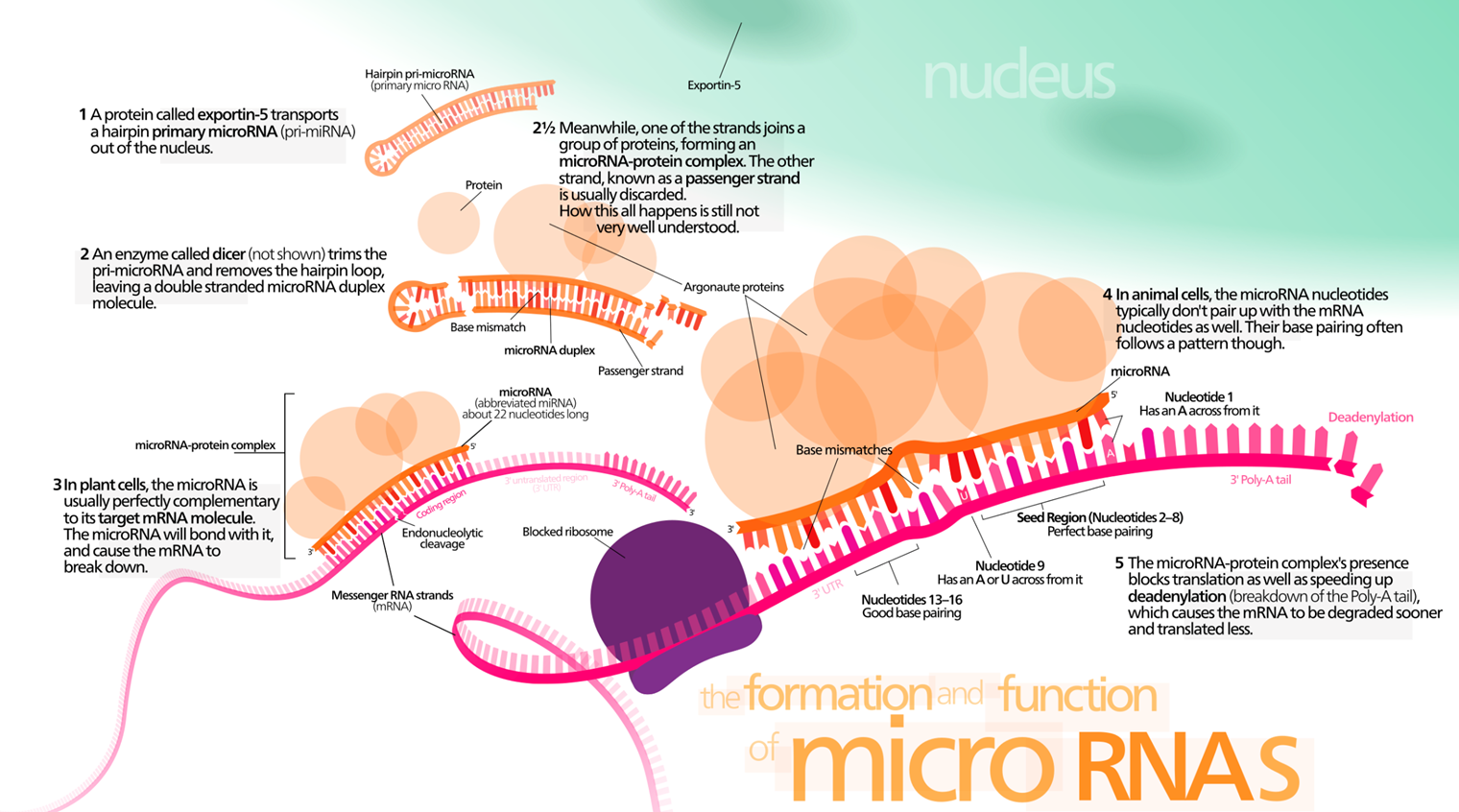
a protein called exportin 5 transports a hairpin primary microRNA (pri-miRNA) out of the nucleus
an enzyme called dicer trims the pri-microRNA and removes the hairpin loop, leaving the double stranded microRNA duplex molecule
Meanwhile, one of the strands joins a group of proteins forming a micro-RNA protein complex. The other strand, known as a passenger strand is usually discarded. How all this happens is still not well understood
in plant cells, the microRNA is usually perfectly complementary to its target RNA molecule. The micro mRNA will bond with it and cause the mRNA to break down
in animal cells the microRNA nucleotides typically dont pair up with teh mRNA nucleotides as well. Their base pairing often follows a pattern though
The microRNA protein complex’s prescence blocks translation as well as speeding up deadenylation, which causes the mRNA to be degraded sooner
molecules of skeletal regenerations
introns have characteristic sizes in axolotl —> particularly the genes associated with development
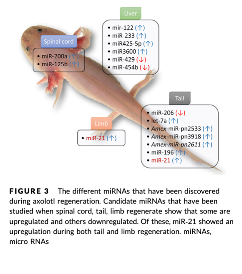
these ncRNAs particularly miRNA are dynamically regulated during regeneration
297 putative mature miRNA
352 conserves miRNA
play key roles in timing ad control of gene expression order and organise processes behind blastema creation
roles of miRNA
de-differentiation after injury
ability to beccome any cell type
hypothesised that human cartilage may share some of teh evolutionarily conserved miRNA circuits of teh blastema in highly regenerative animals
molecular protein clocks in human skeletal tissue
The spontaneous post-translational modification deamidation (removal of amide groups on side chains of asparagine and glutamine) can be used to assess protein ‘age’ .
Non-deamidated proteins and young – deamidated are old
Mass spectroscopy – proteomics can quantify
Gradients of protein turnover reflecting an innate tissue repair capacity (joint site, tissue depth)
highest tissue turnover was measured in ankle, superficial zones of cartilage in OA cartilage
also enriched for miR-21
maladaptation
is when a previously advantageous trait becomes a maladaptive one due to the change in environment
temporal - change in existing environment (E.g: climate change)
spatial - E.g: a population mixing
as evolution is gradual whilst these other changes are relatively rapid —> causes a period of disequilibrium or mismatch that either results in loss of adaptation or extinction
With humans, we dont evolve to be healthy but we evolve to have large amounts of offspring to counter challenging conditions
Examples: the neolithic revolution caused change to agricultural lifestyle —> began storing energy as fat, the “thrifty” gene now works against us with modern abundance of food