module 2- cells of the nervous system
1/34
There's no tags or description
Looks like no tags are added yet.
Name | Mastery | Learn | Test | Matching | Spaced |
|---|
No study sessions yet.
35 Terms
hierarchical structure of the brain (bottom-top)
molecules
cells
circuits/systems
behaviour
cognition
Golgi
invented the silver stain
allowed him to pick out individual cells & see them in detail
reticular network/theory: saw that neurons had processes & that they were fused together
THEORY IS WRONG
Cajal
used Golgi’s silver stain
made drawings of what he saw
neuron doctrine: each neuron is a discrete cell (not joined together in a continuous network)
THEORY IS CORRECT
principle of dynamic polarisation: neurons transmit info in a particular direction
principle of connectional specificity: structure of the nervous system isn’t random
electron microscopy
electron microscope
highest resolution (1mil x more than human eye)
can see cell ultrastructure (can see synapses)
limitation of using electron microscopes
can’t use living specimens
4 steps for immunofluorescence labelling methods
get an antibody/drug that has a fluorescent label which is selective for the part of the cell you want to look at
add the antibody/drug to the tissue (allow it to bind)
wash off any free antibody/drug
shine light on the wavelength that excites the fluorophore to see where fluorescence is coming from
strength of immunofluorescence labelling methods
can do it on living cells
limitation of immunofluorescence labelling methods
limited by the range of antibodies available
what antibody are microglial cells stained with
antibody for coronin (glial-specific protein)
what is confocal microscopy
combining fluorescence techniques with lasers
3 steps for confocal microscopy
laser is focused on different levels of a tissue labelled with fluorescent probe
see where fluorescence is coming from
use high sensitivity cameras + imaging software to create a 3D images of cells
2 strengths of confocal microscopy
can use in living cells
can see how the distribution of a molecule may change during a process
limitation of confocal microscopy
only has modest resolution (same as light microscope)
what is the brainbow technique
genetically modifying an animal so that its cells produce combinations of up to 4 fluorescent dyes
cells will end up being different colours
used to trace the pathways of individual neurons
2 major types of cells in the nervous system
neurons & glial cells
why are glial cells needed
the nervous system doesn’t have internal connective tissue for support
function of glial cells
structural/physiological support for the nervous system
mediate some signalling in the brain
each type of glial cell has a different function
6 types of glial cells
ependymal
oligodendrocytes
schwann cells
astrocytes
microglia
satellite
difference between glial cells & neurons
glial cells can divide
astrocytes (star-shaped)
majority of glia
fills space between neurons
regulates composition of extracellular fluids
role in directing the multiplication & differentiation of neural stem cells
oligodendrocytes/schwann cells
myelinates neurons
oligodendrocytes = CNS, provides insulation for many axons
schwann cells = PNS, each schwann cell insulates a single axon
microglia
brain scavengers
phagocytic cells that have an immune function
they are mobile (move to different tissues)
ependymal
found in brain ventricles
produce CSF
during development = direct the migration of cells in the brain
can turn into neurons so act as a backup of cells for regeneration
huntington’s disease
autosomal dominant → due to genetic abnormality in huntingtin gene
if polyQ repeat = >40 person will get huntingtons
27-35 repeats = unstable during cell replication so more copies can be added
mutant huntingtin protein isn’t broken down properly (accumulates in neurons & kills them)
what areas of the brain are sensitive to the effects of huntingtin gene
basal ganglia
astrocytes (changes cause neuroinflammation & neuronal death)
microglia (changes cause neuroinflammation & neuronal death)
what types of glial cells become activated in alzheimers + how
astrocytes & microglia → attaches to amyloid plaques
difference between neurons & nerves
neurons = cell
nerves = bundles of axons from different neurons
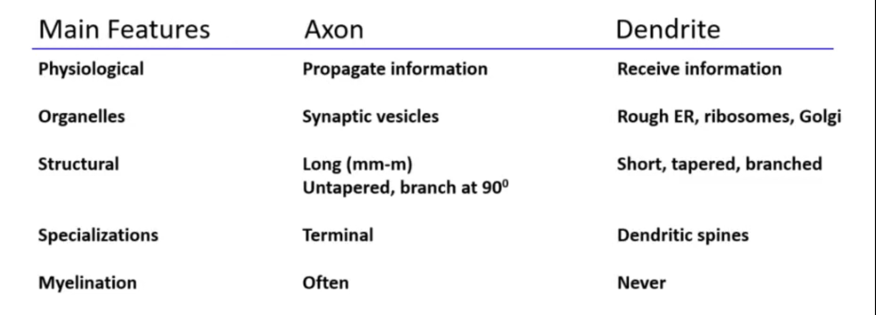
neural polarity/differences table (draw)
8 organelles found in the cytosol & dendtrites
peroxisomes
mitochondria
nucleus
ribosomes
ER
vesicles
golgi
lysosomes
3 organelles found in the axon hillock
synaptic vesicles
mitocondria
smooth ER
ion/ion channel differences in axons & dendrites
axon: Na & K channels, Ca channels (terminals), G-protein coupled receptors (terminals)
dendrites: Ca channels, ligand-gated ion channels (glutamate receptors), G-protein coupled receptors
3 functions of neuronal cytoskeleton
structural support (shape/diameter of axons & dendrites)
transports cargo to & from axons/dendrites
tethering (anchor) of components at the membrane surface
3 components of the neuronal cytoskeleton
microtubules: role structure via (de)polymerisation & transport, run longitudinally down axons/dendrites
neurofilaments: mechanical strength, filamentous
actin (microfilaments): mediate shape change, tethered to the membrane
why are neurons only rarely repaired in the CNS
damaged areas are colonised by glial cells to stop regrowth of axons
what do schwann cells do when theres damage in the PNS
promote regrowth of axons