final exam (images)
 Call Kai
 Learn
Learn Practice Test
 Spaced Repetition
 Match
Match Flashcards
 Knowt Play
1/36

There's no tags or description
Looks like no tags are added yet.
Name | Mastery | Learn | Test | Matching | Spaced | Call with Kai | Chat |
|---|
No analytics yet
Send a link to your students to track their progress
37 Terms
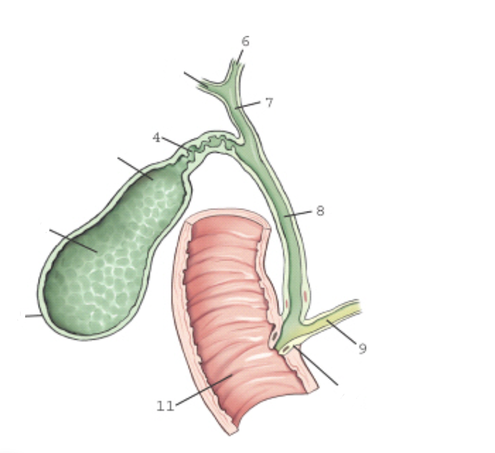
label this image
1 - fundus
4 - cystic duct
6 - left hepatic duct
7 - common hepatic duct
8 - common bile duct
9 - pancreatic duct
11 - duodenum
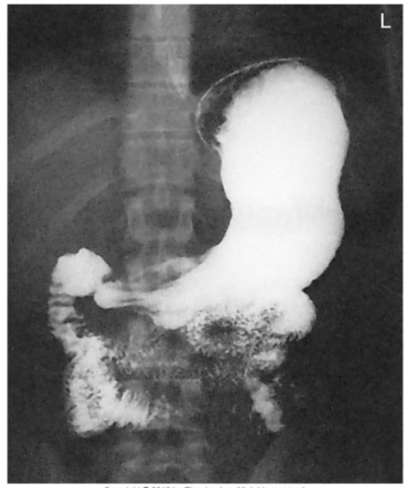
The position seen on this image is
PA stomach

The position seen on this image is
LPO
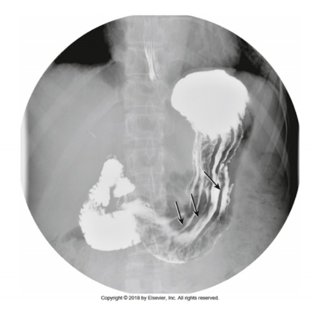
The position seen on this image is
AP
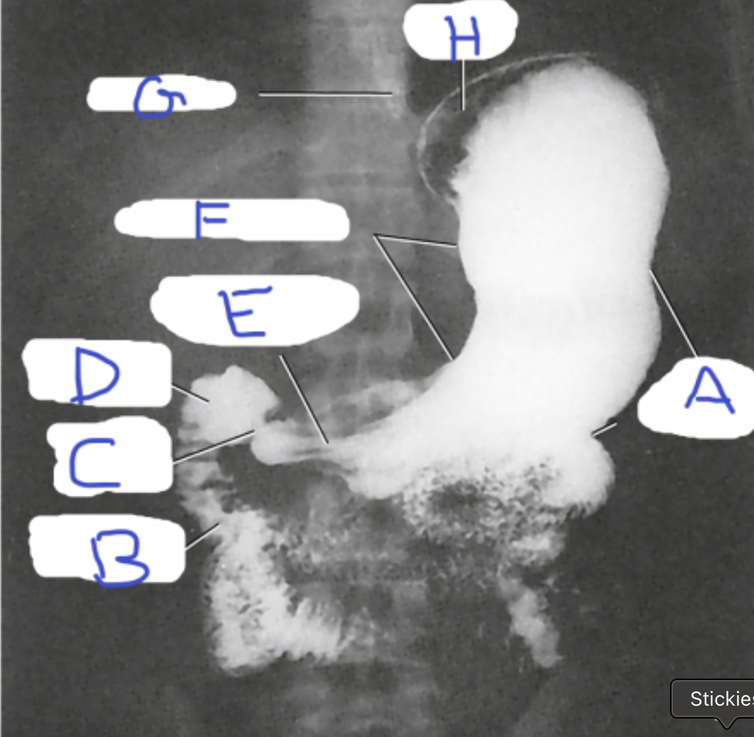
Label this radiograph A-H. For I- state the position J- state how you know
A- greater curvature of stomach
B- duodenum
C- pyloric sphincter
D- duodenal bulb
E- pyloric canal
F- lesser curvature of stomach
G- esophagus
H- fundus of stomach
I- PA prone
J- Air is in the fundus. Barium is in the pylorus and body of the stomach. The spinous processes of the vertebral column are running straight down the image.
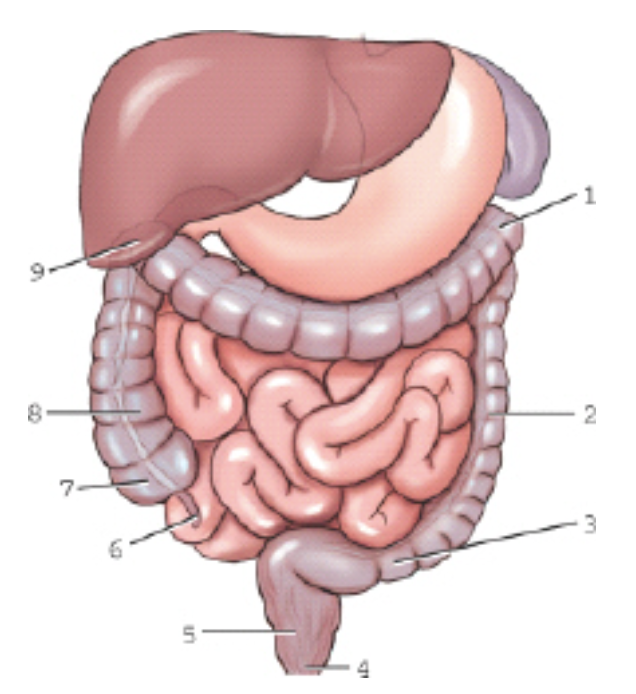
Label this image 1-9.
1 - left colic flexure
2 - descending colon
3 - sigmoid colon
4 - anus
5 - rectum
6 - appendix
7 - cecum
8 - ascending colon
9 - right colic flexure
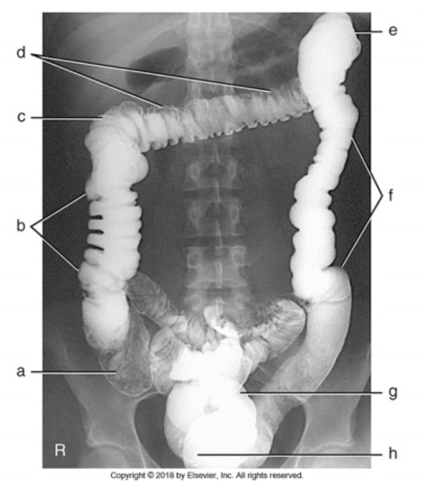
Label this image A-H. For I- what is the projection? For bonus point- how do you know?
a - cecum
b - ascending colon
c - right colic flexure
d - transverse colon
e - left colic flexure
f - descending colon
g - sigmoid colon
h - rectum
i - ap barium enema
*Barium is found in the ascending, transverse, descending, and sigmoid colons. These parts are retroperitoneal. Barium sinks. This would have to mean that the patient is laying supine.
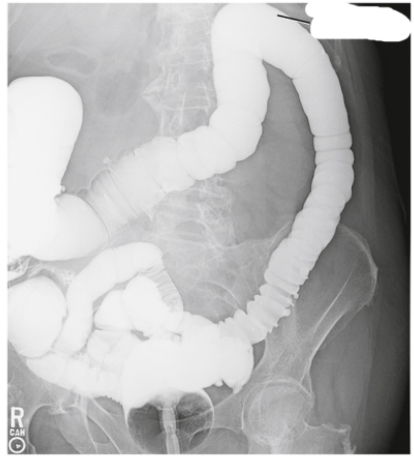
The image shown is? How do you know?
It is shot AP; the patient is in an RPO position. Barium is found in the retroperitoneal parts of the GI tract. This indicates the patient is supine. The left colic flexure and descending colon are best demonstrated here. This indicates the patient is in an RPO position.
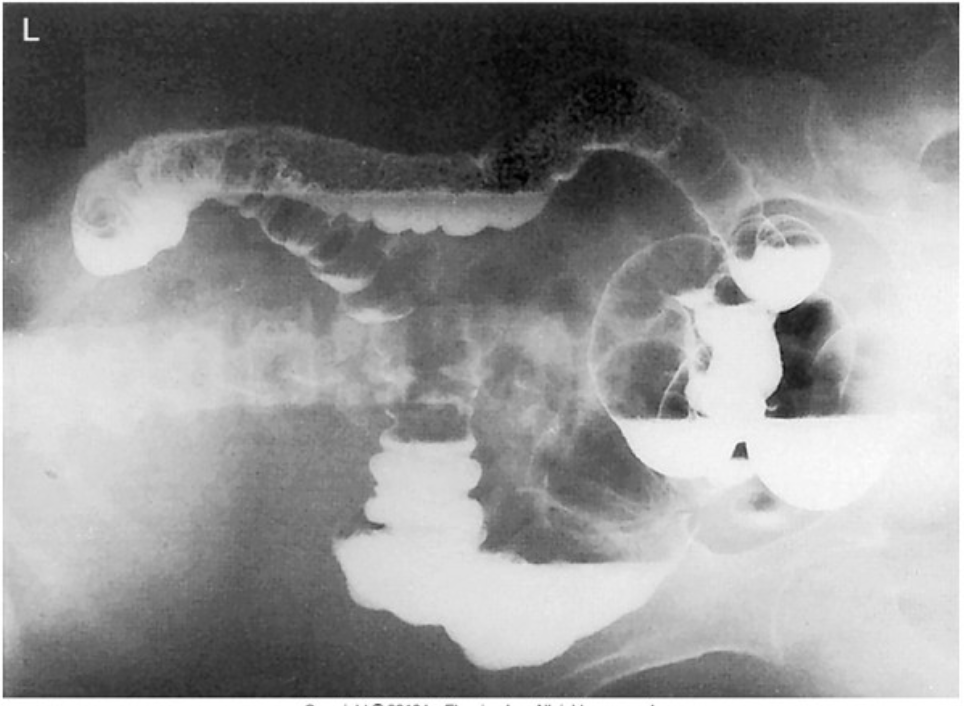
The image shown is? How do you know?
The patient is in a right lateral decubitus position. The left colic flexure typically sits higher than the right. Due to the air in that area, it can be determined that the patient's left side up. Barium will always sink. This indicates that the patient's right side is down (since it is filled with barium).
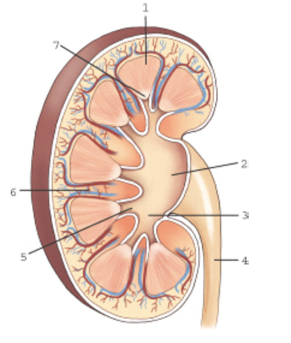
label this image
1 - medulla
2 - renal pelvis
3 - major calyx
4 - ureter
5 - minor calyx
6 - renal sinuses
7 - renal papilla
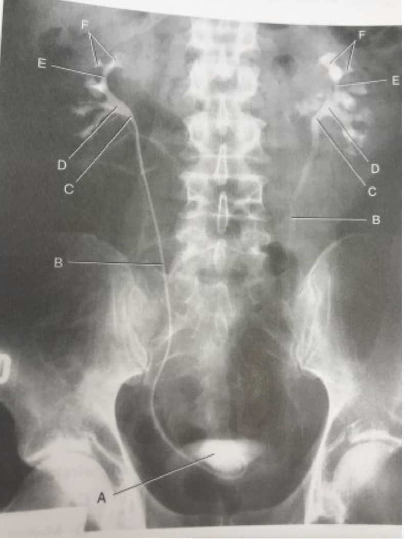
label this image
a- urinary bladder
b- midureter
c- UPJ
d- renal pelvis
e- major calyces
f- minor calyces

What position is shown in the image below?
RPO
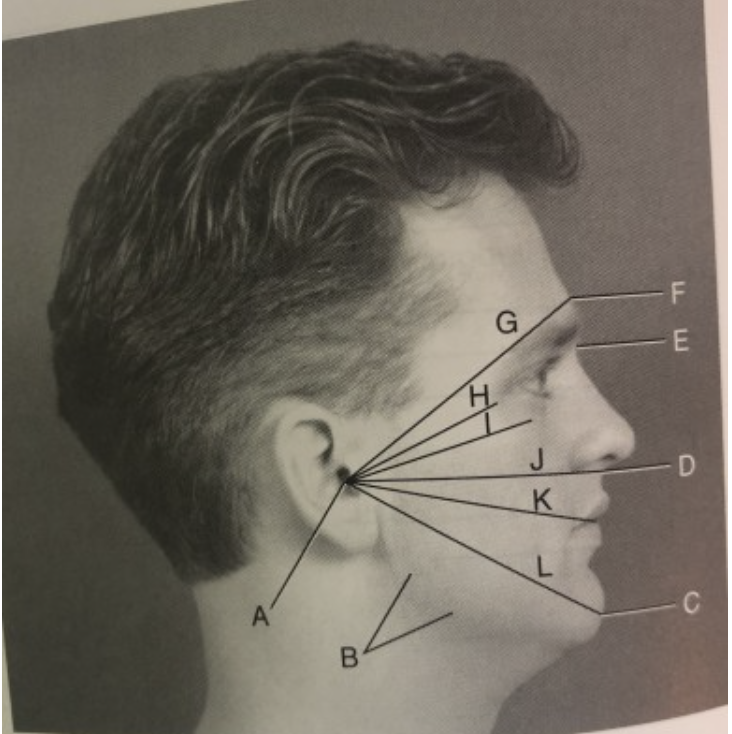
Identify the labeled landmarks and positioning lines used in skull and facial bone positioning, as shown in Fig 11.30 (including abbreviations)
a- EAM
b- gonion
c- mentum
d- acanthion
e- nasion
f- glabella
g- GML
h- OML
i- IOML
j- AML
k- LML
l- MML

Which cranial bone is labeled vi in this figure?
Greater wing of sphenoid
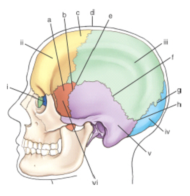
Which bony structure is labeled v?
Mastoid process

Which cranial suture is labeled g?
Lambdoidal
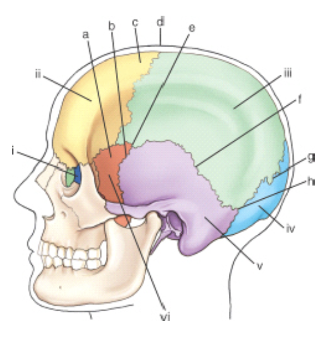
Which cranial suture is labeled f?
Squamosal
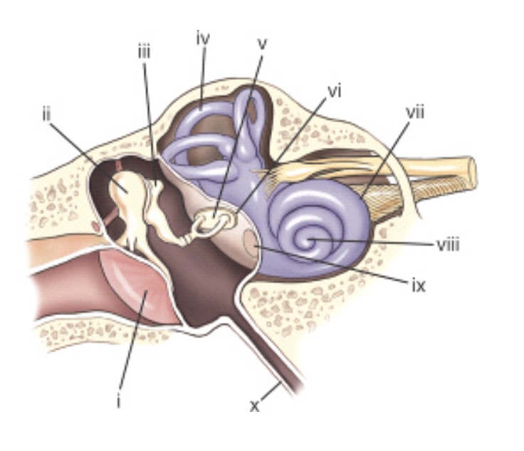
The cochlea is labeled:
viii
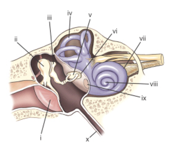
The part labeled ii is the:
malleus
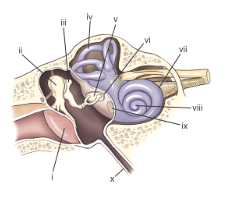
The part labeled i is the:
tympanic membrane.
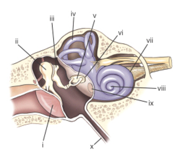
The part labeled x is the:
eustachian tube.
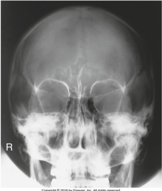
The radiograph shown is
PA Axial Caldwell
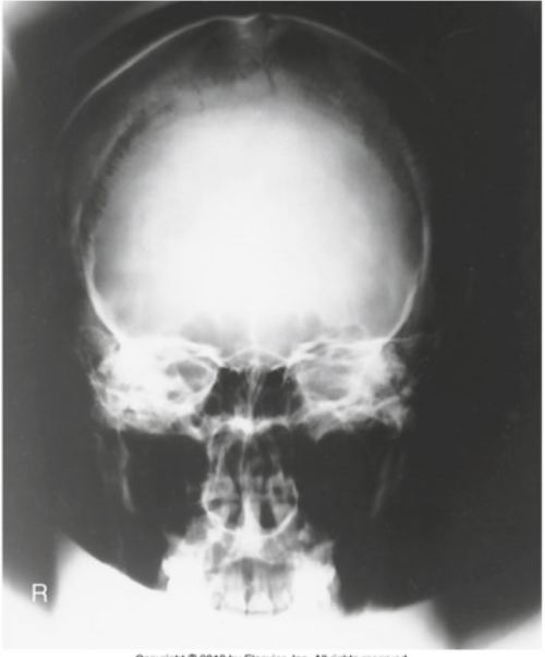
The radiograph shown is
PA

The radiograph shown is
Towne's
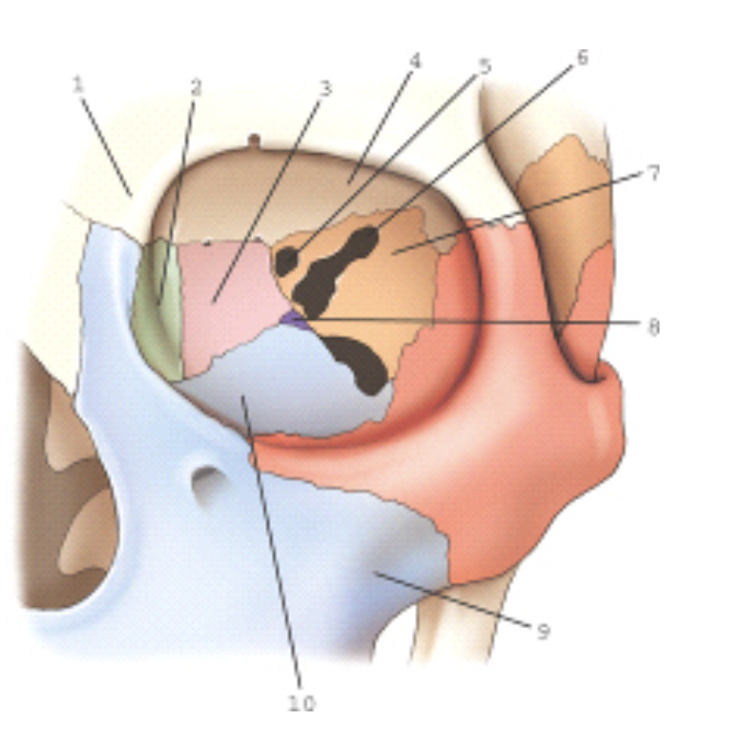
The bone labeled 2 is the:
lacrimal
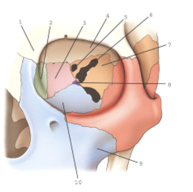
Part 10 is part of which bone?
Maxilla

Part 7 is part of which bone?
Sphenoid

Part 3 is part of which bone?
Ethmoid
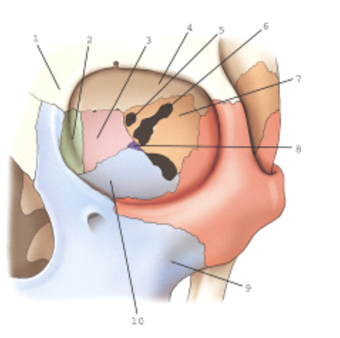
Number 6 is which of the following?
Superior orbital fissure
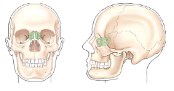
Which group of sinuses is shaded (screened) in the frontal and lateral views of the illustration below?
Ethmoid
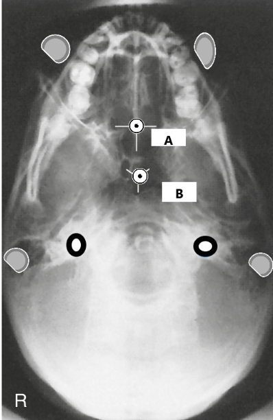
The letter A represents what anatomy?
ethmoid sinuses
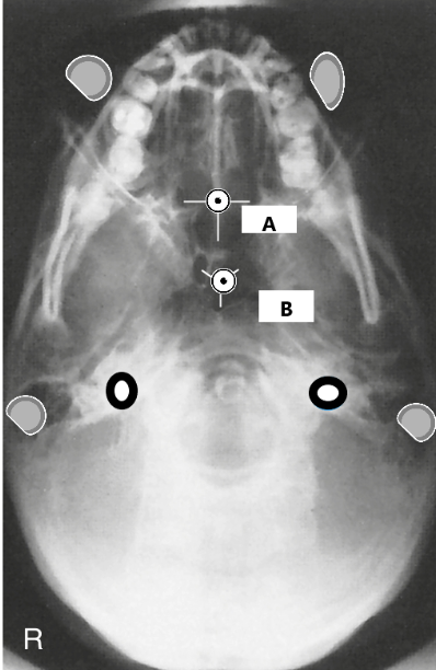
The letter B represents what anatomy?
sphenoid sinuses
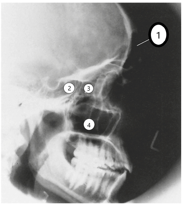
label this image
1- frontal sinus
2- sphenoid sinus
3- ethmoid sinus
4- maxillary sinus
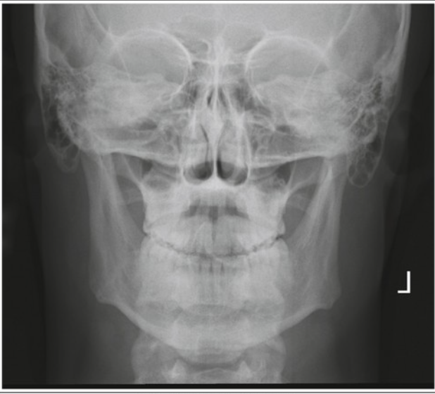
The below image is a PA mandible, pick the suitable critique.
Unacceptable; chin is insufficiently tucked

Is this radiograph of the lateral face shown acceptable or not?
unacceptable
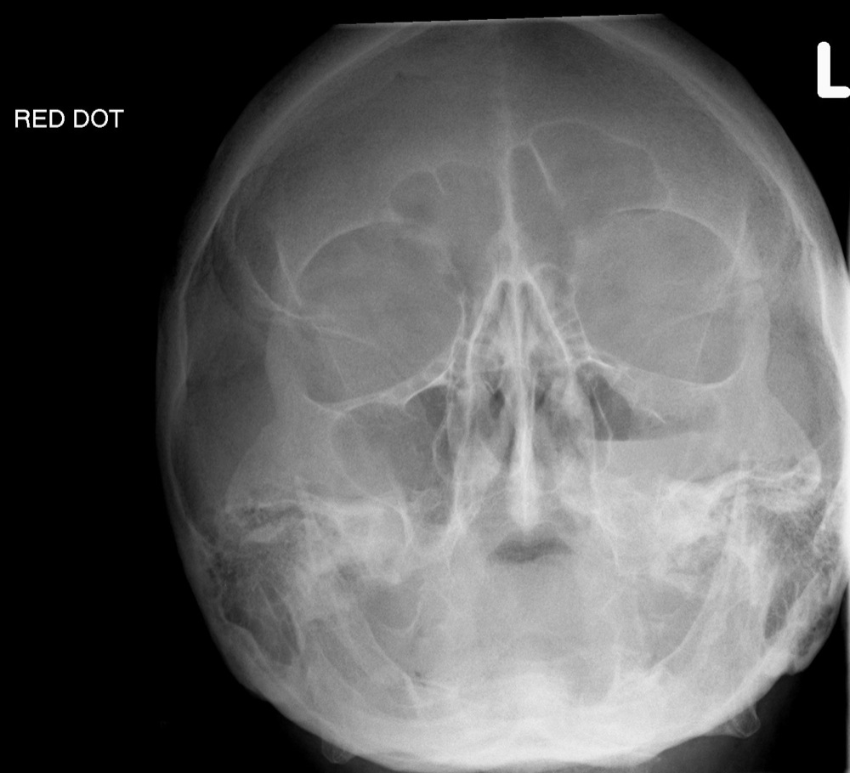
1. What is this image of 2. Is it acceptable or unacceptable? 3. Defend your answer
1. modified waters projection
2. acceptable, but not optimal
3. The petrous ridges in the correct place. The orbital floors are well demonstrated. However, they are a little bit tilted and rotated towards their right.
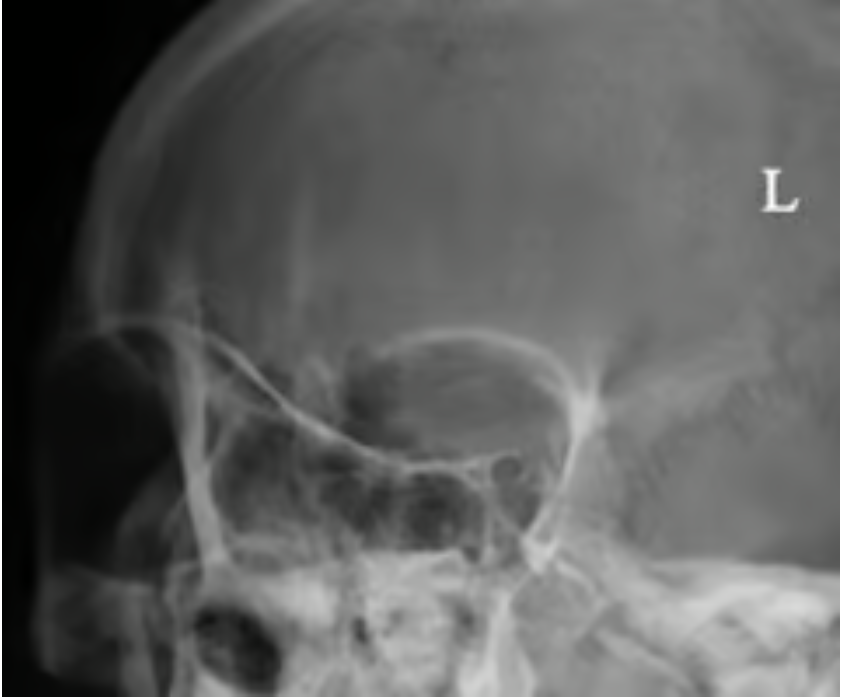
1. What is this projection? 2. Is it acceptable or unacceptable? 3. How do you know?
1. Rhese method
2. unacceptable
3. The patient is too rotated; the skull is almost at a lateral. The AML should be perpendicular to the IR for a proper Rhese.