TEAS 7- Science
 Call Kai
 Learn
Learn Practice Test
 Spaced Repetition
 Match
Match Flashcards
 Knowt Play
1/222

There's no tags or description
Looks like no tags are added yet.
Name | Mastery | Learn | Test | Matching | Spaced | Call with Kai | Chat |
|---|
No analytics yet
Send a link to your students to track their progress
223 Terms
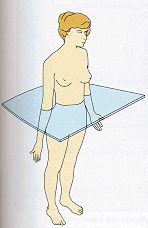
sagittal plane
vertical division of the body into right and left portions
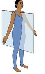
coronal plane
divides body into front and back
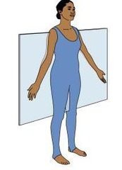
transverse plane
divides the body into superior and inferior parts
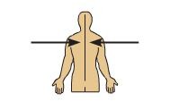
Medial
Toward the midline of the body
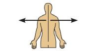
Lateral position
Away from the midline of the body
Proximal
Closer to the origin of the body part or the point of attachment of a limb to the body trunk
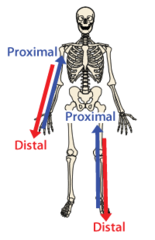
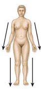
Distal
farther from the origin of a body part or the point of attachment of a limb to the body trunk
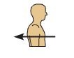
Anterior
toward the front
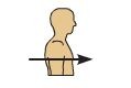
Posterior
back of body
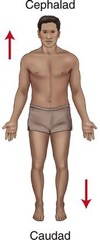
Cephalad/Cephalic
(adverbs) head
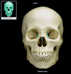
Cranial
(adjective) skull
Caudad
(adverb) toward the tail or posterior
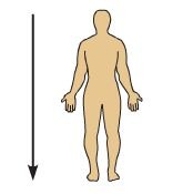
Caudal
(adjective) hindquarters
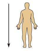
Superior
above; toward the head
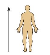
Inferior
Below; at a lower level
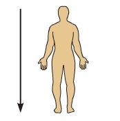
A: right hypochondriac
right kidney, liver, gallbladder, and small intestine
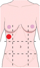
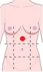
B: Epigastric (epi= on, above)
stomach, liver, adrenal glands, pancreas, spleen, small intestine
C: Left Hypochondriac
left kidney, spleen, pancreas, and colon
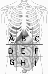
D: Right Lumbar
ascending colon, liver, gallbladder
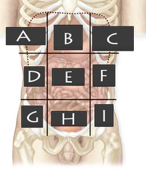
E: Umbilical
small intestine, duodenum, umbilicus
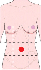
F: Left lumbar
descending colon, left kidney
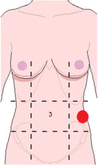
G: Right iliac
cecum, appendix
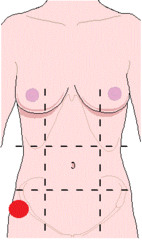
H - Hypogastric (hypo=below; gastric=stomach)
bladder, female internal reproductive organs, sigmoid colon
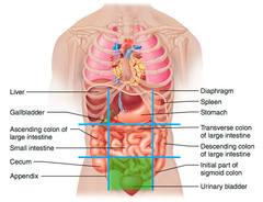
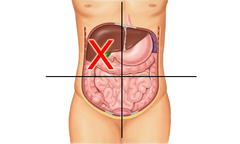
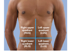
Right Upper Quadrant (RUQ)
duodenum, part of the ascending and transverse colon, hepatic flexure, liver, gallbladder, pancreas (head), right kidney, and right adrenal gland
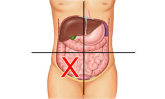
Right Lower Quadrant (RLQ)
cecum, appendix, right ovary and tube, right ureter, right spermatic cord
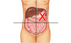
Left Upper Quadrant (LUQ)
stomach, liver (left lobe), pancreas (body), left kidney, left adrenal gland, splenic flexure, spleen, part of transverse and descending colon
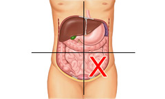
Left Lower Quadrant (LLQ)
left ureter, left fallopian tube, left ovary, part of descending colon, and sigmoid colon
Quadrants
four regions with the umbilicus (navel) at the center, helping to identify positions of organs
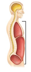
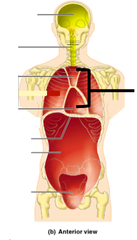
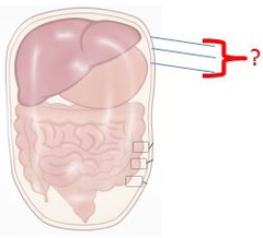
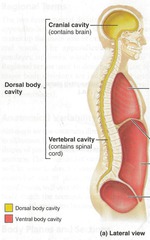
ventral cavity
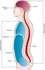
located along the front of the body, contains the body organs that sustain homeostasis; separated by the diaphragm
viscera
internal organs
thoracic cavity
above the diaphragm; contains the lungs and mediastinum, which separates the cavity into a right and left compartments
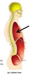
Mediastinum
area between the lungs containing the heart, thymus gland, esophagus, and trachea
abdominoplevic cavity
extends below the diaphragm to the pelvic floor; contains the upper abdominal cavity and lower pelvic cavity (not physically separated)
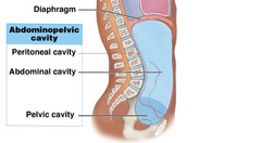
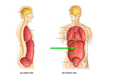
abdominal cavity
Contains stomach, large and small intestines, spleen, liver, gallbladder, pancreas
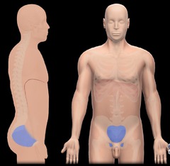
pelvic cavity
contains internal reproductive organs, bladder, and distal part of colon
Cavities are lined with
serous membranes
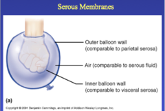
serous membrane
secretes fluid that separates the parietal layer from the visceral layer
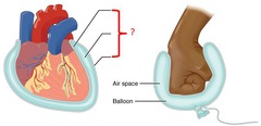
parietal pleura
lines the thoracic cavity
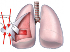
pericardial membrane
surrounds the heart
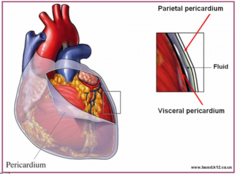
peritoneal membrane
lines the abdominal cavity
dorsal cavity
contains the brain and spinal cord
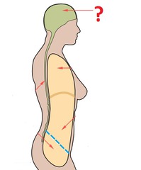
cranial cavity
contains the brain, 12 cranial nerves, and pituitary gland
Meninges
comprised of the dura mater, arachnoid mater, and pia mater; lines the cavity and surround the brain and spinal cord; contains cerebrospinal fluid between the arachnoid mater and pia mater in the subarachnoid space
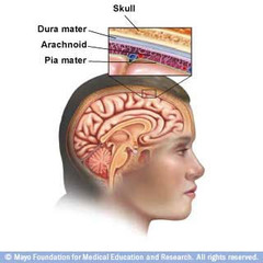
protects and cushions the dorsal cavity
meninges and cerebrospinal fluid
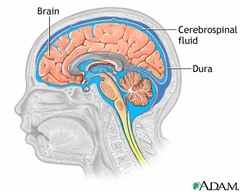
respiratory system functions
-pulmonary ventilation (breathing)
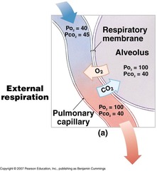
-gas exchange (external respiration)
-gas conditioning (internal respiration- warm, humidify, and cleanse air)
-sound production
-olfaction (smell)
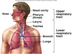
upper respiratory tract
nose, nasal cavity, mouth, pharynx, larynx
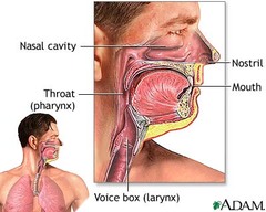
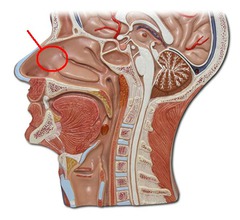
nose in nasal cavity
governs sense of smell
Trachea connects _____ and ____ to the_____.
connects pharynx and larynx to the lungs
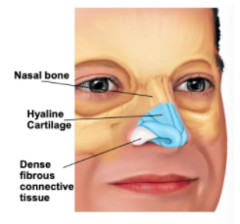
External nose structures
root, bridge, dorsum nasi, frontal maxillary bones, and septal alar cartilages
internal nose (nasal cavity)
lined with hairs and mucous membrane that act as a filter
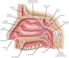
internal nose function
warms, moisturizes, and filters air that enters the body before it reaches the lungs
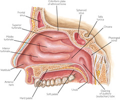
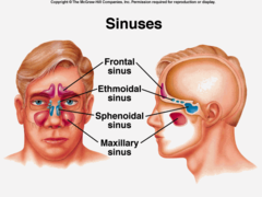
sinus system function
produce mucus to moisturize the inside more and protects us from dirt, dust, etc.
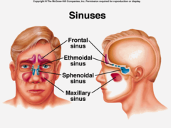
sinus stystem structure
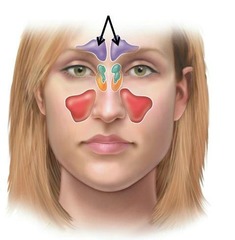
two maxillary sinuses and ethmoid sinus
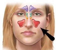
maxillary sinus
collectively are the largest sinus that can cause some pain; drains into ostium
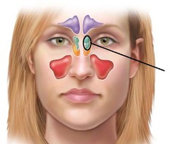
ethmoid sinus location
between the nose and the eyes
frontal sinus
located above each eye
Pharynx consists of
nasopharynx, oropharynx, laryngopharynx
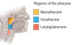
nasopharynx
lets you breathe through the nose
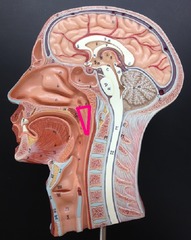
oropharynx
connects nose and back of mouth to the esophagus
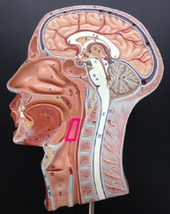
Hypopharynx
passageway for food
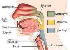
Larynx (voice box) function
-allows air to pass through the respiratory tract
-protects trachea by ensuring food and drink don't block airway
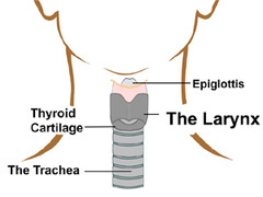
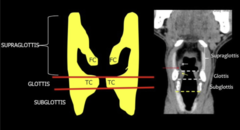
larynx sections
Supraglottis, Glottis, Subglottis
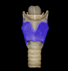
Larynx formation
nine cartilages connected by muscle and ligaments
lower respiratory tract components
trachea, lungs, bronchi, diaphragm
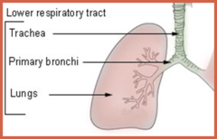
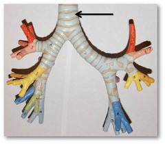
trachea (windpipe)
main airway to the lungs
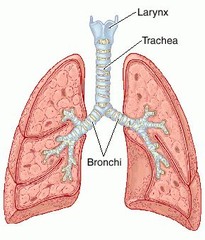
lungs
pull in O2 and push out CO2
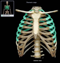
lungs are protected by
sternum, rib cage, and vertebrae
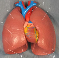
lung connects to
trachea via the left and right bronchi
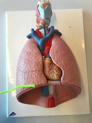
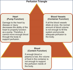
perfusion
helps fluid pass to an organ or tissue
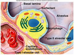
alveoli produces
surfactant (fluid)
Surfactant
prevent the lungs from collapsing
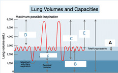
tidal volume
amount of air inhaled or exhaled with each breath under resting conditions
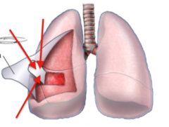
pleura
double-layered membrane surrounding each lung that secretes a fluid that lets the lungs move against the rib cage when we breathe
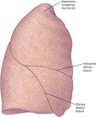
The right lung has how many lobes?
3 lobes
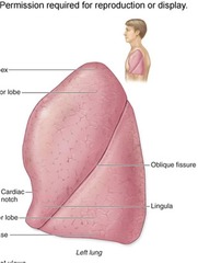
Left lung has how many lobes?
2 lobes, to make room for the heart
lung lobes function
bring O2 into and remove CO2 from the blood stream
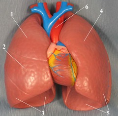
bronchus
one of the two tubes that connect the lungs with the trachea
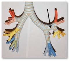
Bronchi function
help push air to and from the lungs
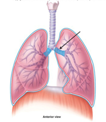
diaphragm function
primary muscle used in breathing
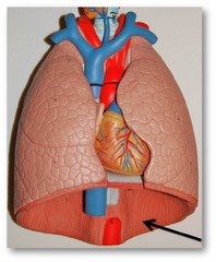
Diaphragm loaction
below heart and lungs
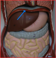
The diaphragm opening
lets other structures pass between the chest and abdomen
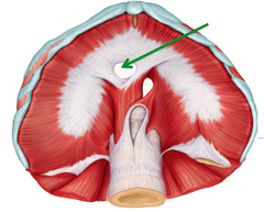
esophageal opening
provides passageway for esophagus and vagus nerve
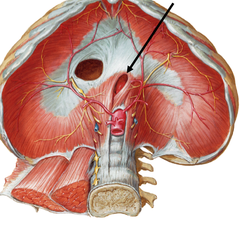
aortic opening
allows the aorta and thoracic duct to pass through
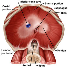
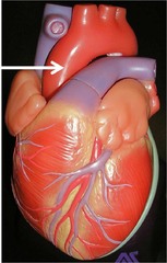
Aorta
transports blood from the heart and is the body's main artery
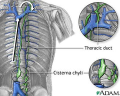
thoarcic duct
lymphatic system's main vessel
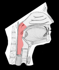
conducting zone (air)
provides a continuous passageway for incoming and outgoing air to travel
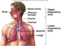
conducting zone structures
nose, pharynx, larynx, trachea, bronchi, and bronchioles,
respiratory zone
only involved with O2 and CO2 exchange (gas exchange)
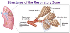
respiratory system
conducting and respiratory zone
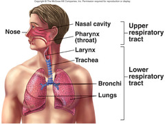
respiratory zone structures
bronchioles, alveoli, alveolar ducts
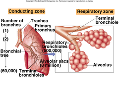
conducting zone (pic)
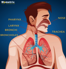
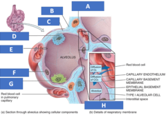
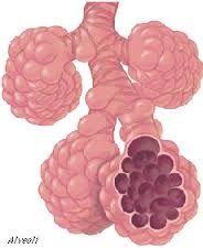
alveoli
very small air sacs in the site of the exchange of oxygen and carbon dioxide
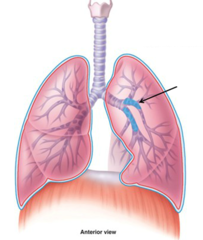
bronchi
main passageway attached to the lungs
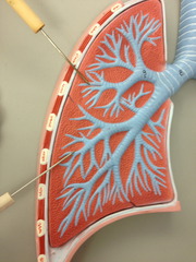
bronchiole
connects bronchi to the alveoli
Ventilation
movement of air in and out of the lungs
inhale
diaphragm moves downward, ribs push out and lungs are filled with air
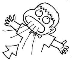
exhale
diaphragm is moved up, ribs relax, and air leaves lungs
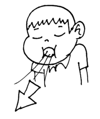
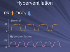
hyperventilation
the condition of taking abnormally fast, deep breaths
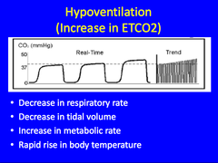
hypoventilation
decreased rate or depth of air movement into the lungs; too much CO2 in the blood stream
external respiration
exchange of gases between lungs and blood