nephrology and urology
 Call Kai
 Learn
Learn Practice Test
 Spaced Repetition
 Match
Match Flashcards
 Knowt Play
1/74

There's no tags or description
Looks like no tags are added yet.
Name | Mastery | Learn | Test | Matching | Spaced | Call with Kai | Chat |
|---|
No analytics yet
Send a link to your students to track their progress
75 Terms
acute kidney injury
rapid-onset disease of the kidneys resulting in a failure to produce urine
-BBB disruption from uremic toxins and inflammatory mediators
-HTN, depleted cardiac ATP
-impaired monocyte and cytokine production
short term effects of pediatric AKI
-worse neurocognitive outcomes
-weight loss with preserved muscle mass
-lon term cardiac morbidity and mortality
-pulmonary edema, ARDS, BPD
long term outcomes of pediatric AKI
-crush injury, prolonged immobilization, high voltage electrical injuries, severe burns, compartment syndrome, strenuous physical activity
-infections, hyperthermia
-use of amphetamines, cocaine, or alcohol
-meds including statins, colchicine, propopfol
-toxins like snake venom, wasp, and bee stings
causes of rhabdomyolysis
muscle pain, muscle weakness, and dark urine
-elevated CK
signs of rhabdomyolyssi
-prevent kidney injury with intensive fluid resuscitation
-correcting electrolyte abnormalities
-ID underlying cause
management of rhabdomyolysis
-PSGN
-vasculitis, lupus
-Hep B, Hep C+
-anti-GBM disease
syndromes of nephritic syndrome
->95th percentile + 30 mmHg without symptoms/signs of end organ damage
-in adolescents >180/120
define a pediatric hypertensive urgency
>95th percentlie + 30 mmHg associated with encephalopathy (A, vision, changes, neuro symptoms)
->180/120 in adolescent and teenss
define a pediatric hypertensive emergency
oral: nifedipine
management of hypertensive urgency
-Hypoalbuminemia due to losses from urinary excretion
-edema from loss of oncotic pressure--> facial edema upon waking up, non-pitting edema
-hyperlipidemia: liver starts manufacturing cholesterol in respsonses to hypoalbuminemia
clinical signs of nephrotic syndrome
Renal: chronic kidney disease, glomerulonephritis, polycystic kidney disease, renal artery stenosis
Endocrine: hyperthyroidism, Cushing syndrome, pheochromocytoma, congenital adrenal hyperplasia
Cardiovascular: coarctation of the aorta
Medications: corticosteroids, stimulants, oral contraceptives
secondary causes of HTN
fever, exercise, dehydration, stress s
causes of transient (benign) proteinuria
A rapid decline in kidney function over hours to days, resulting in the inability to excrete nitrogenous waste products, maintain fluid/electrolyte balance, and regulate acid-base status.
define acute kidney injury
↑ Serum creatinine by ≥0.3 mg/dL within 48 hrs, or
↑ Serum creatinine to ≥1.5 × baseline within 7 days, or
Urine output
pediatric criteria of acute kidney injury
Hypertension
Altered mental status (uremia, encephalopathy)
Arrhythmias (hyperkalemia)
signs of acute kidney injury
dehydration, shock, HF
causes of pre-renal AKI
obstructive neuropathy
cause of post-renal AKI
Manage fluid balance (avoid overload but ensure perfusion)
Treat electrolyte abnormalities (esp. hyperkalemia: calcium gluconate, insulin + glucose, albuterol, dialysis if needed)
Correct acidosis if severe
primary managemet of AKI
Control BP (ACE inhibitors/ARBs)
Manage proteinuria
Correct anemia (iron, erythropoiesis-stimulating agents)
Manage bone-mineral disorders (phosphate binders, vitamin D analogs)
Optimize nutrition for growth
management of CKD
GFR
GFR indication for dialysis or transplant
Post-streptococcal glomerulonephritis (PSGN) - most common worldwide
IgA nephropathy (Berger disease) - hematuria within 1-2 days of infection
Henoch-Schönlein purpura (IgA vasculitis) - purpura, abdominal pain, arthralgia
Lupus nephritis
Membranoproliferative GN
Rapidly progressive GN (rare in children, but severe
common pediatric causes of nephritic syndrome
PSGN, MPGN
low C3 is indicative of what nephritic syndromes
Fluid & salt restriction to control edema and hypertension
Loop diuretics (e.g., furosemide) for significant fluid overload
Antihypertensives (calcium channel blockers, beta-blockers) if needed
Careful monitoring of fluid balance, weight, electrolytes
mainstay treatment of nephritic syndrome
ACE inhibitors or ARBs (renal protective; avoid in pregnancy)
Long-acting calcium channel blockers
Thiazide diuretics
Beta-blockers (less commonly first-line in children)
first line HTN drugs in pediatrics
nephrotic syndrome
in nephritic or nephrotic syndrome can you become hypercoaguable due to loss of antithrombin III
nephrotic syndrome
foamy urine is indicative of
40 mg/m²/hr or urine protein/creatinine ratio >2-3, or ≥3+ on dipstick
nephrotic range protein level
oval fat bodies
nephrotic syndrome
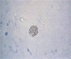
minimal change didesase
most common cause of nephrotic syndrome in children
minimal change disease
Focal segmental glomerulosclerosis (FSGS), membranous nephropathy, secondary causes (SLE, infections, Henoch-Schönlein purpura, hepatitis B, HIV)
causes of nephrotic syndrome in pediatrics
Fluid and salt restriction during edema phase
Diuretics (e.g., furosemide) for symptomatic edema, with caution
Monitor weight, urine output, electrolytes
management of nephrotic syndrome principles
oral corticosteroids
first line treatment of minimal change disease
Hemolytic Uremic Syndrome
a condition that can occur when the small blood vessels in your kiddney become damaged and inflamed
-bloody diarrhea, adominal pain, fever
initial signs of hemolytic uremic syndrome
E. coli O157:H7
etiology of HUS
Microangiopathic hemolytic anemia (Coombs-negative)
Thrombocytopenia
Acute kidney injury (AKI
triad of hemolytic uremic syndrome
hemolytic uremic syndrome
most common cause of AKI in young children
-fluid management
-HTN management
-anemia tx with PRBC
-no abx or anti-motility agents!
mainstay management of typical HUS
Atypical Hemolytic Uremic Syndrome
HUS without the diarrheal prodrome
- can occur due to inherited disorder of complement regulation
- Triggered by pregnancy or infection
Neisseria gonorrhoeae
Often asymptomatic in females
Purulent discharge, dysuria, cervical motion tenderness
Pharyngitis, conjunctivitis possible
Trichomonas vaginalis
Frothy, yellow-green discharge
Vulvar irritation, strawberry cervix
metronidazole
management of trichomonas
Primary: painless chancre
Secondary: rash (palms/soles), mucous patches, lymphadenopathy
Tertiary: neurologic, cardiac, gummas
describe the presentation of syphilis
Uretopelvic Junction Obstruction
blockage somewhere between the renal pelvis and the ureter
most common cause of hydronephrosis in neonates
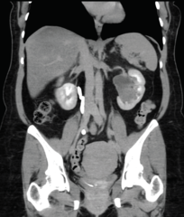
ureterocele
Cystic dilation of the distal ureter within the bladder wall (often associated with duplicated collecting systems)
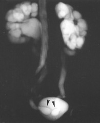
febrile UTIs in infants/children
most common presenting symptom of ureterocele
UPJ obstruction
mosst common cause of antenatal hyrdonephrosis
posterior urethral valves
irregular thin membranes of tissue located within the male posterior urethra that do not allow urine to exit the urethra
cobra head sign
Dilatation of the distal ureter which may be seen in patients with ureteroceles.
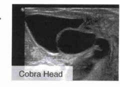
vesicoureteral reflux
backflow of urine from the bladder into the ureters
-usually due to congenital defect of vesicoureteral valve
1. dilation of pelvis
2. papillae flatten
3. decreased parenchyma width
4. atrophy of cortex
5. umbrella with rim of atrophy/fibrosis
sequential changes associated with hydronephrosis
hypospadias
incomplete urethral development leading to malpositioning of the urethral meatus anywhere along the ventral surface
E. coli
80% of UTIs are caused by
fever, FTT, feeding problms, hyperbilirubinemia
signs of UTI in neonate
in
indication for VCUG
testicular torsion
twisting of the spermatic cord causing decreased blood flow to the testis
bell clapper deformity
the condition in which the patient lacks the normal posterior fixation of the testis and epididymis to the scrotal wall
-allows twisting of spermatic cordd
testicular torsion
absent cremasteric reflex indicates
phimosis
narrowing of the opening of the prepuce over the glans penis
-a normal variant if congenital
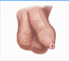
poor hygiene or chronic irritation---> fibrotic ring of tissue close to the opening of the prepuce
acquiredd phimosis is often result of
phimosis
inability to retract the foreskin fully back over glans
-never a urologic emergency, unless ther is an obstructed urinary stream
-manage with manual stretching, steroid creams, circumcision
treatment of phimosis
Paraphimosis
entrapment of retracted foreskin behindd the corona of glans peniss
urologic emergency!
manual reduction attempt immediately
-dorsal slit or circumcision
treatment of paraphimosis
prematurity, family hx
inguinal hernias in pediatric pts are more common in
inguinal hernia
bowel sounds iin the scrotum are strongly suggestive of
silk glove sign
indirect hernia sac in pediatric patient
the sac feels like a finger of a silk glove when rolled under the examiners finger
cryptorchidism
failure of testes to descend ffrom intra-abdominal into scrotum
Do not circumcise (foreskin may be needed for repair)
Surgical repair usually between 6-18 months
management of hyposppadias
tender, firm mass, vomiting
signs of incarcerated hernia
Refer to urology if not descended by 6 months
Orchidopexy ideally by 12 months to reduce infertility/malignancy risk
management of cryptorchidism
Fusion of labia minora; may cause urinary dribbling, recurrent vulvovaginitis
presentation of labial adhesions
Asymptomatic: observe (often resolves spontaneously)
Symptomatic: topical estrogen cream ± gentle separation
management of labial adhesions
Neonates: mucocolpos (vaginal bulge) from maternal estrogen
Adolescents: primary amenorrhea, cyclic pelvic pain, bulging bluish hymen
presentation of imperforate hymen