Clinical Skills III Week 7 (Long Bone Fractures)
 Call Kai
 Learn
Learn Practice Test
 Spaced Repetition
 Match
Match Flashcards
 Knowt Play
1/64

There's no tags or description
Looks like no tags are added yet.
Name | Mastery | Learn | Test | Matching | Spaced | Call with Kai | Chat |
|---|
No analytics yet
Send a link to your students to track their progress
65 Terms
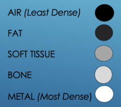
densities on film: least dense to most dense
AIR (Least Dense, black)
FAT
SOFT TISSUE
BONE
METAL (Most Dense, White)
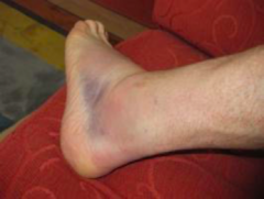
signs and symptoms of long bone fractures
- Edema
- Erythema
- Ecchymosis
- Instability/Crepitus/Deformity
- Pain/Tenderness
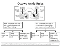
when do you image long bone fractures
- Ottawa Ankle/Foot/Knee Rules
- Possible Retained Foreign Body (consider radiopaque vs radiolucent)
- MOI
- Repeated visits for similar complaint
ottawa foot and ankle rules
ottawa knee rules
1. Age > 55
2. Isolated patella tenderness
3. Tenderness at the head of the fibula
4. Inability to flex knee to 90º
5. Inability to bear weight (4 steps) immediately and upon ER evaluation

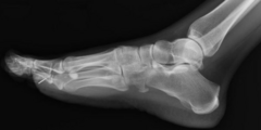
foreign bodies
- Inform pts that not all FBs will be visible on plain film (opaque vs lucent)
- If retained FBs are not removed --> concern for infection
What other modalities could be used to visualize retained foreign bodies?
CT
MRI
U/S
what to image
1. Affected joint
2. Joint Above & Below (ex: Maisonneuve Fracture)
3. Bilateral Bones/Joints (ex: peds & geriatrics for comparison)
anytime there is an ankle injury, you should r/o?
maisonneuve fracture (order tib/fib series and ankle)
Maisonneuve Fracture: spiral fracture of the proximal third of the fibula associated with an avulsion fracture of the medial malleolus secondary to a disruption/rupture of the deltoid ligament
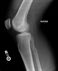
ankle anatomy review
what is a syndesmotic joint?
Syndesmotic Joint: 2 bones and 4 ligaments (fibrous joint linked by network of ligaments)
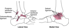
XR labeled of ankle

Bimalleolar Fracture: Fracture of the medial and lateral malleoli (surgery is needed bc unstable) —stirrup + posterior splint typically used

orthopedic physical exam
1. Status of joint above & below affected area
2. Status of the affected joint (ie: erythema, edema, ecchymosis, deformity)
3. Neurovascular status (distal to affected area)
• Strength → bilateral
• Tendon Injury → remember ortho tests (ie: Thompson Test)
• Sensation & Pulses → bilateral (consider comorbidities)
• Additional Injuries → is this an open fracture?
4. ROM (passive & active) → consider pain as a reason for ↓ ROM
simple fracture
no disruption of ligaments or surrounding skin
comminuted fracture
bone is broken into several pieces
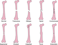
open fracture
external wound (ie: laceration) leads to the fx or bone fragments protrude through the skin
oblique fracture
fx follows a diagonal line

spiral fracture
fx follows a helical line
avulsion fracture
bone fragment tears away from the main bone b/c of trauma usually to a ligament (common in ankle injuries)

greenstick fracture
fracture where one side of the bone is bent & the other side is broken (common in peds)

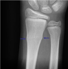
torus/buckle fracture
affected bone compresses causing a bulge in the cortex w/o a break (common in peds)
pathological fracture
fracture of a diseased bone secondary to a force that would not have broken a healthy bone
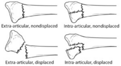
intra-articular fracture
fx within or involving a joint
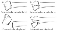
extra-articular fracture
fx that does not affect a joint
displaced fracture
normal anatomic alignment has been disrupted
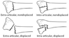
non-displaced fracture
normal anatomic alignment is maintained
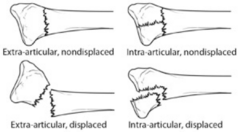
summary for what info to get from fracture?
1. Open or closed
2. Number of fragments (ie: simple vs. comminuted)
3. Intra or extra-articular
4. Pattern of the fracture line (ie: transverse, oblique, spiral)
5. Location (diaphysis, metaphysis, proximal, distal, etc)
6. Area (ie: what bone?)
7. Displacement (defined in terms of the abnormal position of the distal fracture fragment in relation to the proximal bone)
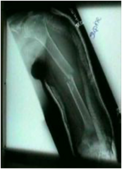
describe the fracture
Closed, extra-articular, comminuted, transverse, mid-shaft, humeral fracture with mild medial displacement
monteggia fracture
fracture of the ulnar shaft with anterior dislocation of the radial head (FOOSH injury—kids and young adults most often)

colles fracture
an extra-articular fracture of the distal radius; specifically, a fracture of the distal radial metaphysis with dorsal or posterior displacement
often seen in pts with osteoporosis (FOOSH with a pronated forearm)

fat pad/sail sign
noted in elbow/humeral plain films; caused by displacement of the fat pad around the elbow joint
highlights an elbow joint effusion & intra-articular fx
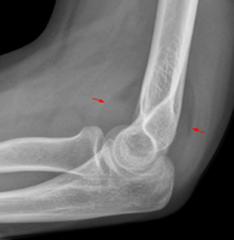
fat pad/sail sign: what fat pad is normal and what is pathologic (anterior vs posterior)
anterior fat pad can be normal
but posterior fat pad on a lateral elbow film is ALWAYS abnormal
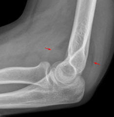
fat pad/sail sign - what does it indicate in adults vs peds?
Adults: radial head fx
Peds: supracondylar humeral fx

if there is no fx on plain film but (+) sail sign, what is the problem and how can you treat it?
occult fx
splint with long arm posterior
sail sign on an elbow X-ray is a triangular shape made by a pushed-up fat pad
salter harris fractures
Classification of Epiphyseal (growth) Plate fractures (5 Types)
All involve or extend through the epiphyseal plate → only in peds as complete fusion yet to occur
SALTR

type I salter harris fracture
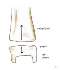
A Transverse Fx along the Physis:
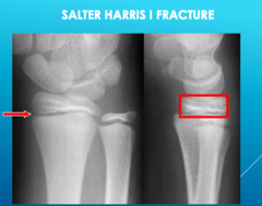
- Difficult to detect on plain films; look for pathological widening of the Physis
- May result in disrupted bone growth (rare)
type I salter harris fracture dx and tx
Often a clinical dx → tenderness, swelling, ecchymosis & joint effusion at epiphyseal plate
Tx: many will require cast immobilization (x3 wks), some require surgery for proper alignment (outpatient Ortho follow-up)
salter harris I fracture (hard to identify on xray)
type II salter harris fx
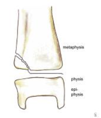
A Fx through the Metaphysis & Physis (spares Epiphysis):
- Most common type
- May cause minimal shortening; however, long-term impairment is rare
- Creates a "Thurston Holland Corner Sign"
type II salter harris fracture tx
cast immobilization w/ outpatient ortho follow-up
Salter Harris II Fracture

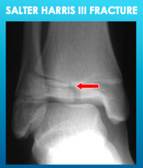
type III salter harris fx
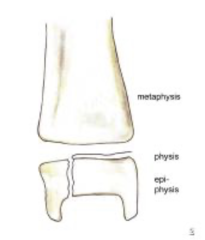
A fracture through the Physis & Epiphysis (spares Metaphysis):
- Fracture line passes through the hypertrophic layer of the Physis & extends to split the Epiphysis → damages the Physis' reproductive layer
- More common in older children
- Chronic disability is common b/c the physis is damaged
type III salter harris fracture tx
internal fixation to ensure proper alignment (Ortho consult in ED)
rarely results in deformity → prognosis is favorable
Salter Harris III Fracture
type IV salter harris fracture
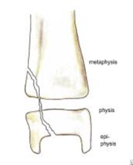
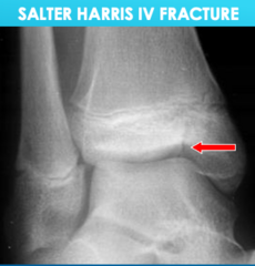
A fracture involving all 3 elements of the bone → the Physis, Epiphysis & Metaphysis:
- Similar to Type III (also an intra-articular fracture) → can result in chronic disability
- Also creates the "Thurston Holland Corner Sign". Small corner of bone in metaphysis. Type II and IV.
Interferes with the growing layer of cartilage cells → often causes premature fusion of the involved bone → leads to deformity (essentially stops bone growth)
type IV salter harris Fx Tx
internal fixation (Ortho consult in ED)
Salter Harris IV Fracture
type V salter harris fracture
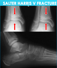
A compression/crush injury of the Epiphyseal Plate (no fracture!):
Initial diagnosis is difficult from plain film radiography
often made retrospectively with premature closing of the Physis

type V salter harris fracture dx and tx
- Clinical history is crucial (Axial Load injury)
- Tx: cast Immobilization (possible surgery) & requires Ortho consult in ED
Poor functional prognosis & almost always results in growth disturbance
Salter Harris V Fracture
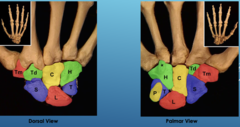
hand bone anatomy review
S - Scaphoid
L - Lunate
T - Triquetrum
P - Pisiform
T - Trapezium
T - Trapezoid
C - Capitate
H - Hamate
*pay careful attention to the pisiform & triquetrum
boxer's fracture
- Fx of the 4th &/or 5th metacarpal (most often the neck)
- Results from a direct blow with a closed fist (striking a hard surface—ie: a concrete wall)
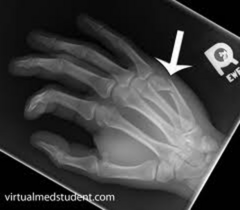
boxer's fracture dx and tx
- Pain & swelling over fx area & depression of the affected knuckle (have pt make a fist)
- Ensure intact neurovascular status
Tx:
- May require reduction in ED (consult Ortho if significant displacement)
- Patient requires Ulnar Guttar splint & Ortho follow-up
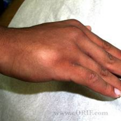
scaphoid/navicular fracture MOI
FOOSH with hyper-extended wrist: compresses the bone between the Radius & 2nd row of carpal bones
s/sx of scaphoid fracture
pain/edema/ecchymosis at Anatomical Snuff Box
OR
pain with Axial Loading of Thumb
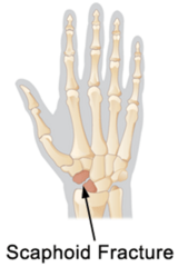
scaphoid navicular fracture dx and tx
Difficult to detect on plain film even with a Scaphoid View (inform pts of this) → if Snuff Box tenderness → Thumb Spica & Ortho follow-up (best appreciated with MRI)
**Important not to miss this fracture due to the limited blood supply to this area from the Radial Artery; if left untreated → avascular necrosis
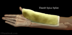
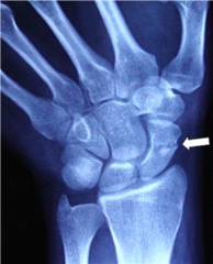
scaphoid navicular fracture
series vs simple films
Important to obtain several views of the affected joint
Most commonly:
- PA
- lateral & oblique (consider Navicular or Scaphoid view for hand/wrist)
It is common to appreciate abnormalities on some views & not others (especially w/ wrist & carpal fractures) because bones overlap
Without multiple angles, it is very easy to miss a fracture
triquetrium fracture
- 2nd most common carpal fx
- Due to forced hyperflexion or avulsion
- Pain at dorsum of wrist

triquetrium fracture dx
Need lateral view → Pisiform overlies Triquetrum on frontal view
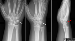
Jones Fracture
fracture of the base of the 5th Metatarsal
Describe the Fracture....
Nightstick Fx: Mid-diaphysis (mid-shaft) ulnar fracture
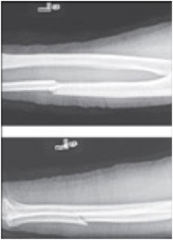
Describe the Fracture....
1. Non-displaced inter-articular fracture of the proximal phalanx
2. Non-displaced fracture of the distal phalanx
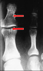
Describe the Fracture....
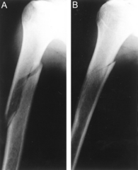
Transverse Midshaft Femoral fracture with associated medial displacement
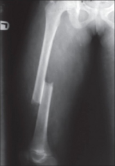
Describe the Fracture....
Jones Fracture: fracture of the base of the 5th Metatarsal
Management: posterior splint, non-wt bearing & refer to ortho
Highly vascular area—can result in non-union
