July 2026 Dose of VetPrep
 Call Kai
 Learn
Learn Practice Test
 Spaced Repetition
 Match
Match Flashcards
 Knowt Play
1/19

There's no tags or description
Looks like no tags are added yet.
Name | Mastery | Learn | Test | Matching | Spaced | Call with Kai | Chat |
|---|
No analytics yet
Send a link to your students to track their progress
20 Terms
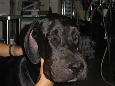
The dog in the photograph was vaccinated earlier in the day. What type of allergic reaction is occurring?
Type II
Type III
Type IV
Type I
Type I
Type I hypersensitivity is IgE-mediated and results in immune mediated hypersensitivity. Type II hypersensitivity is an antibody-dependent reaction and occurs due to IgG or IgM made against normal self antigens or some foreign antigen that resembles some molecule on the surface of host cells. Type III hypersensitivity is an immune complex-mediated reaction. This is caused when soluble antigen-antibody complexes form in large amounts and overwhelm the body instead of being normally removed by macrophages in the spleen and liver. Type IV is considered a delayed hypersensitivity and is cell-mediated. T8-lymphocytes will be sensitized to an antigen and differentiate into cytotoxic T-lymphocytes. T helper 1 type T4-lymphocytes become sensitized to an antigen and produce cytokines.
What is the potential udder fate of goats infected with caprine arthritis encephalomyelitis virus?
The udder is not affected
Hardbag
Bluebag
Gangrene
Hardbag
CAEV is a retrovirus and when it affects the udder it will cause fibrosis and result in a firm udder with agalactia. Treatment is ineffective and the goat should be culled. The disease is usually subclinical but can cause arthritis in adults and encephalitis in kids.
Your client is pregnant and is worried about acquiring toxoplasmosis from her cat. What do you advise?
Have a housemate empty the litter box daily as a simple precaution to prevent infection as it takes 1-3 days for passed oocysts in the stool to sporulate into an infective form.
Submit toxoplasmosis titers from the cat. A IgG titer of 1:64 or greater suggests recent or active infection that could pose a danger.
Toxoplasmosis titer should be performed on the owner by a human physician. A positive titer indicates antibodies to the organism that will prevent infection in the first trimester. A negative titer indicates she should remove her cats from her environment.
Submit toxoplasmosis titers from the cat. A positive cat infected with toxoplasmosis can shed multiple times in its lifetime and pose a zoonotic risk.
Have a housemate empty the litter box daily as a simple precaution to prevent infection as it takes 1-3 days for passed oocysts in the stool to sporulate into an infective form.
Toxoplasmosis gondii is a protozoal organism. The cat is the definitive host; the entire life cycle of the organism can be completed within this host. Most cats become infected when they consume an exposed rodent with bradyzoites encysted in their tissues. Only recently infected cats generally shed oocysts in their stool, and cats typically only shed these oocysts for 1-2 weeks. Most cats will only have one shedding episode in their lifetime.
A IgM (not IgG) titer of 1:64 or greater suggests recent or active infection and that cat is at risk of shedding oocysts in their stools. Oocysts are not infective until they sporulate. This process takes > 24 hours, so emptying the litter box daily is advised, preferably by someone who is not pregnant.
If an owner has owned cats for a long while, it is possible that they may have previously been exposed and therefore have mounted an immune response to the organism. If so, it may be advisable to test for Toxoplasma antibody titers in the owner. A sufficient antibody titer will mean the client is protected from infection during the first trimester.
A 4-year old male Manx cat presents to you because the owners found an empty, opened pill vial in the bathroom and the cat vomited. On physical exam, you note ptyalism and facial edema. The cat's mucous membranes are pale and slightly icteric. You perform a blood smear and detect Heinz bodies in erythrocytes. The cat's packed cell volume (PCV) is 26% (30-45%). The owners provide you a list of the medications in the medicine cabinet which are acetaminophen (Tylenol), finasteride (Propecia), enalapril (Vasotec), and omeprazole (Prilosec). What treatments should you institute for this cat?
Emesis and methylene blue
Acetylcysteine and S-adenosylmethionine
Activated charcoal and whole blood transfusion
Prednisone and amoxicillin
Acetylcysteine and S-adenosylmethionine
Acetaminophen toxicity in cats usually occurs when owners administer the drug, unaware of its significant potential toxicity in cats. In this case, the cat's clinical signs are most consistent with acetaminophen toxicity based on the Heinz body anemia that is present. Cats can die from oxidative damage and methemoglobinemia within 1-2 days of ingestion. It may also be associated with hepatotoxicity in cats, although this is seen more frequently in dogs.
Recall that cats are particularly sensitive to acetaminophen because they have decreased glucuronyl transferase activity which conjugates acetaminophen to glucuronic acid for excretion. As a result, 50-60 mg (a single tablet) may be fatal for a 4-5 kg cat.
Treatment should consist of toxin removal if possible by inducing emesis in some cases. As the cat in this case is already vomiting, this may not be necessary. Activated charcoal is controversial and should only be given if ingestion occurred within hours and should be administered very carefully in cats due to the risk of aspiration.
The specific antidote is acetylcysteine which binds to some of the reactive metabolites of acetaminophen and increases the availability and synthesis of glutathione. Other treatments may include S-Adenosylmethionine (SAMe) which has hepatoprotective and antioxidant properties. Cimetidine can be given to inhibit the p450 oxidase in the liver and limit formation of toxic metabolites. Ascorbic acid can also be used as an adjunct treatment to bind toxic metabolites. In cats with signs of hypoxemia from severe hemolytic anemia (PCV <20%), a transfusion and further supportive care may be warranted.
You are asked to examine some feeder pigs that have stopped eating yesterday. The group is lying down and seems lethargic. They have fevers of 105-106F (40.6 -41.1 C), firm dry feces, and the skin has rhomboid-shaped red blotches scattered on it. What treatment should be recommended?
Streptomycin
Gentamicin
Penicillin
Chloramphenicol
Metronidazole
Penicillin
Erysipelas is susceptible to penicillins, as well as tetracyclines (usually), lincomycin and tylosin. Chloramphenicol and nitroimidazoles (including metronidazole) are not approved for food animal use.
A horse presents to you for chronic, recurrent laminitis and skin disease. You notice on your exam that the horse has a particularly thick, long, wavy, and matted coat. The owner mentioned that this developed many months ago. What is a likely diagnosis?
Cushing's disease (Pituitary Pars Intermedia Dysfunction)
Diabetes insipidus
Pheochromocytoma
Hypothyroidism
Cushing's disease (Pituitary Pars Intermedia Dysfunction)
The correct answer is Cushing's disease. The coat condition described is what horses with glucocorticoid excess develop; it is referred to as hirsutism. They will also be predisposed to infections including laminitis and skin diseases such as Dermatophilus. They are also frequently polyuric, polydipsic, and polyphagic.
A 10-day old commercial dairy calf has diarrhea that is white in color (see photo). The calf is dehydrated, hypovolemic, weak and unable to stand. T=100F (37.8 C), HR=100, and RR=20. No other abnormalities are found on physical examination. Based on these findings, what is the treatment of choice?

Oral fluids containing high levels of both sodium and chloride
IV fluids with added sodium bicarbonate
Oral fluids containing sodium bicarbonate
IV fluids containing only saline
IV fluids containing 50 meq/L of potassium
IV fluids with added sodium bicarbonate
This calf is typical of those suffering from nonspecific calf diarrhea, most often associated with enteropathogenic E coli, rotavirus, or cryptosporidium. The calf develops hypovolemia and metabolic acidosis and requires sodium-containing IV fluids which contain additional alkali such as sodium bicarbonate. One can assess that the calf is severely acidotic given that it is lethargic and unable to stand. While oral fluids may also be useful, at this stage, the calf will require IV fluids.
During your examination of an 18-year old horse, you observe what is shown in the photograph. Based on the location and appearance of this lesion, what is the most likely diagnosis?

Melanoma
Sarcoid
Squamous cell carcinoma
Cuterebra
Habronema
Melanoma
The correct answer is melanoma. Melanoma is one of the most common tumors in horses (about 10% of all neoplasms) and gray horses are at high risk of around 80%. They can occur anywhere but appear most frequently in the perineal region or ventral tail as is evident in this horse.
In horses, they are usually darkly pigmented (as opposed to dogs where amelanotic melanomas occur somewhat commonly). In horses, most are slowly growing but can be locally invasive. Many treatments are out there, but there is no standard of care treatment. Depending on location and extent, consider surgical removal, benign neglect, chemotherapy (systemic or intralesional) and immunotherapy.
A 2-year old female DSH cat presents for weight loss, anorexia, dyspnea, and lethargy. She was previously treated with antibiotics but is still febrile on physical exam. You detect pleural effusion and notice that the abdomen is distended. On CBC there is a non-regenerative anemia, neutrophilia, and lymphopenia. On chemistry there is hyperproteinemia and a slight elevation in liver enzymes. What is your primary differential?
Feline leukemia virus
Feline immunodeficiency virus
Feline infectious peritonitis
Feline calicivirus
Feline infectious peritonitis
The correct answer is FIP. This poor cat is infected with the dreaded wet form of feline infectious peritonitis.
If you think about the pathogenesis of the virus, then these clinical signs make a lot of sense. With FIP, the classical lesion is pyogranulomatous vasculitis due to antigen-antibody complexes depositing in the venular endothelium, which results in pleural and peritoneal effusion. The CBC findings are common for FIP but not too specific. In greater than 50% of cats with the wet form of FIP, there will be hyperproteinemia. In greater than 70% of cats with the dry form of FIP there is hyperproteinemia.
A client calls and says her cat was chewing on her lily plant two days ago and is now acting very sick. You tell her to bring the cat in immediately so that you can perform which of the following?
Abdominal radiographs to diagnose intestinal obstruction
BUN and creatinine levels
ALT, AST, GGT, and total bilirubin levels
Induce emesis and administer activated charcoal
BUN and creatinine levels
Lily plant toxicosis is extremely serious and can cause rapid and fatal acute renal failure in cats. If ingestion is suspected, decontamination and aggressive fluid therapy, and monitoring of renal values are recommended immediately.
In this case, since 2 days have passed, inducing emesis and administering activated charcoal would not be helpful. What you can do is check renal values (BUN and creatinine) and treat for acute renal failure.
Ruling out an intestinal obstruction in a cat with this history is reasonable but not the best of the choices given.
Rubiosis iridis as seen in this cat is a sign of what process in the eye?

Iris atrophy
Anterior uveitis
Glaucoma
Lens luxation
Chorioretinitis
Anterior uveitis
The correct answer is anterior uveitis. Rubiosis iridis along with other signs such as aqueous flare, hyphema, hypopyon, keratic precipitates, and decreased intraocular pressure are all suggestive of anterior uveitis which can be caused by a number of infectious, immune-mediated, traumatic, and idiopathic causes.
A male goat presents for vocalizing and straining to urinate. He has been kicking at his abdomen. You detect crystals adherent to the hairs around the prepuce (see image). What condition should you suspect and try to rule out first?

Lower intestinal obstruction
Acute severe pyelonephritis
Urinary tract obstruction
Upper intestinal obstruction
Urinary tract obstruction
The correct answer is urinary tract obstruction due to calculi. This should be suspected in all male and castrated male goats (and sheep) with non-specific signs of disease or discomfort because it is so common. Common clinical signs associated with urinary tract obstruction are vocalization and dribbling of urine. Heaving or forceful abdominal contractions may be seen. Hematuria, dysuria, prolonged urination, and apparent abdominal pain are also common signs. The most important step of evaluation is exteriorization of the penis and examination of the urethral process because this is the most common site of blockage. In severe cases the entire urethra may be filled with calculi.
Which of these is the most common cause of seizures in the adult ferret?
Hypocalcemia
Idiopathic epilepsy
Intracranial neoplasia
Hypoglycemia
Hepatic encephalopathy
Hypoglycemia
Hypoglycemia secondary to insulinoma is the most common cause of seizures in the adult ferret. Keep in mind, a prolonged seizure can actually cause hypoglycemia, so the finding of low blood glucose in a seizuring ferret does not necessarily confirm a diagnosis of insulinoma. Idiopathic epilepsy has not been reported in ferrets. Hypocalcemia and hepatic encephalopathy can cause seizures, but are not as commonly reported as hypoglycemia.
A 4-year old female spayed mixed Chihuahua presented to the emergency service at approximately 5am this morning after presumptively being attacked by a coyote. The patient has a flail chest and it is questionable if there is direct communication between the thoracic cavity and the environment (it was difficult to examine the dog due to her fractious nature). Exploration of the wound was performed, and once anesthetized, it was apparent she had a pneumothorax. The patient must be ventilated, as there is no vacuum present in the chest for lung expansion to occur. What pressure should the anesthetist not exceed if manually bagging the patient during anesthesia?
20cm H2O
12cm H2O
24cm H2O
8cm H2O
20cm H2O
Pressures above 20cm H20 may result in barotrauma. In an otherwise healthy patient it is not recommended to exceed this pressure. In patients with chronic atelectasis, anesthetists will be much more apprehensive about over ventilating or ventilating the lungs too quickly, as acute expansion can trigger re-expansion pulmonary edema, which may then lead to acute respiratory distress syndrome and death.
A 4-year old male neutered Pit Bull mix comes in to your clinic because his owner thinks he might have gotten into rodenticide while she was out of the house 2 hours ago but is not sure. You send her to bring back the box of rodenticide and induce emesis by administering subconjunctival apomorphine. Within 5 minutes, the dog vomits the material shown in the image below. You rinse out the conjunctiva and administer oral activated charcoal. The owner returns with a box of rodenticide that says brodifacoum. What should you recommend?

Treat the dog with vitamin K1 for 6 weeks
Hospitalize the dog for 24 hours to monitor and treat potential neurologic signs
Check serum calcium levels today and once weekly for 6 weeks
No additional treatment or monitoring is needed
Treat the dog with vitamin E and selenium
Treat the dog with vitamin K1 for 6 weeks
The bright green vomit confirms the owner's suspicion of rodenticide ingestion because many rat poisons contain a bright green dye. Dogs that ingest these products may have bright green vomit or stool. Brodifacoum is a vitamin K antagonist commonly used in rodenticides. Ingestion of this compound causes hemorrhaging after several days due to a lack of production of new clotting factors. Treatment for this condition requires vitamin K1 administration for 4-6 weeks.
A 6-month old feedlot steer, which entered the feedlot 4 weeks ago, has lost weight and is now showing an enlarged left flank as shown in the image below. On exam, you find the left side of the abdomen to be gas-filled under moderate pressure, and the rumen to be otherwise poorly filled and with poor motility. Based on percussion and auscultation, the animal also appears to have chronic bronchopneumonia. What is the most likely cause of the rumen malfunction?

Frothy bloat
Left displaced abomasum
Type 3 vagal indigestion
Cecal dilation
Free gas bloat, failure to eructate
Free gas bloat, failure to eructate
This is sometimes called Type 1 vagal indigestion, or free gas bloat. It is frequently associated with swollen mediastinal lymph nodes caused by pneumonia. The signals to or from dorsal rumen receptors, which detect gas pressure and open the cardia, are compromised such that eructation does not occur normally and free gas bloat occurs. This in turn causes the calf to feel full and it eats poorly and loses weight. One needs to treat the pneumonia and perhaps create a temporary rumen fistula to allow the escape of gas until eructation returns to normal.
A horse is suspected of having the muscle disease rhabdomyolysis, and you are seeking a laboratory test to help confirm the diagnosis. Of the following, which test would likely be the most helpful?
Sorbitol dehydrogenase (SDH)
Ionized phosphate
Gamma glutamyl transferase (GGT)
Anion gap
Creatine kinase (CK)
Creatine kinase (CK)
CK, also called, CPK, is found in muscle cells and is elevated in the serum when muscle damage occurs.
A3-year old, indoor-only, male castrated Maine Coon cat is presented to you for acute-onset of hindlimb pain and paresis. Physical examination reveals a grade III left parasternal heart murmur and minimal movement in the hind legs. Femoral pulses are bilaterally absent, and the hindlimb toes are cold to the touch. The cat is tachypenic, but lung sounds are normal. What is the most likely diagnosis?
Lymphoma of the spinal cord
Chlorpyrifos toxicity
Thrombus at the aortic bifurcation (saddle thrombus)
T3-L3 intervertebral disc disease
Tetanus
Thrombus at the aortic bifurcation (saddle thrombus)
Maine Coon cats are predisposed to development of hypertrophic cardiomyopathy at a young age. Consequently, left atrial enlargement predisposes to atrial thrombus formation, and these clots frequently lodge in the arterial supply to the hindlimbs. The trifurcation is where the aorta divides into the two external iliac arteries and the common origin of the internal iliac arteries. Classic findings due to a clot at the aortic trifurcation include posterior paresis/paralysis, hindlimb pain, cyanotic nailbeds, absent femoral pulses, and a firm leg musculature. Other signs of cardiac disease/failure (murmur or pulmonary edema) are often but not always evident at presentation.
Neither cord lesions, tetanus, nor toxicities should cause the vascular compromise evident on this cat's physical exam.
A 4 year old mare has bilateral swelling and drainage of the mandibular lymph nodes. Rectal temperature is 101.8F (38.8 C). Which of the following is the most appropriate plan for this horse?
Administer corticosteroids
Inform the state veterinarian
Administer penicillin
Culture the discharge for bacteria
Culture the discharge for bacteria
The correct answer to this question is to culture the discharge for bacteria. The most likely diagnosis for this horse is equine strangles. Strangles most commonly affects younger horses (<5 years of age), but can cause disease in any age horse. The etiologic agent of this disease is Streptococcus equi subsp. equi. Currently, the diagnostic test of choice to confirm equine strangles is bacterial culture, but recently there is a trend towards PCR testing on guttural pouch samples. While awaiting test results, the horse should be separated from any other horses, as strangles is highly contagious to other horses. Antibiotic therapy is controversial and thought to lengthen the course of disease rather than shorten it when given at this stage; also, it may possibly interfere with the natural immunity acquired from natural infection. Don't forget to report positive cases of S. equi to the state veterinarian! As of March 2020, S.equi is a monitorable disease in the United States, meaning individual veterinary practitioners are not mandated to report Strangles to the USDA/APHIS. State veterinarians and laboratories include S. equi cases as part of their monthly disease reports to APHIS.
When performing a subtotal colectomy on a feline patient, what blood vessel limits the amount of colon that you are able to remove?
Left colic artery
Ileocolic artery
Caudal mesenteric artery
Pudendal artery
Ileocolic artery
The site for colonic resection is limited by tension on the ileocolic artery when trying to suture your new end of colon to the rectum. Sometimes the tension is too great and instead of a colocolic anastomosis, an ileocolic anastomosis must be performed. Essentially you are trying to connect a section of ascending colon to the rectum. Now that can be pretty far! Performing an ileocolic anastomosis is not ideal because you eliminate the ileocecal valve, and that may predispose the animal to bacterial overgrowth. The caudal mesenteric artery gives branches to the rectum and descending colon. The left colic artery also feeds the descending colon. The pudendal artery supplies the external genitalia. The ileocolic artery provides blood supply to the ascending and transverse colon.