exam 3 - cardiac/shock
 Call Kai
 Learn
Learn Practice Test
 Spaced Repetition
 Match
Match Flashcards
 Knowt Play
1/59

There's no tags or description
Looks like no tags are added yet.
Name | Mastery | Learn | Test | Matching | Spaced | Call with Kai | Chat |
|---|
No analytics yet
Send a link to your students to track their progress
60 Terms
VAD (ventricular assist device)
mechanical pump that helps a weak heart pump blood to the body
Most common is the LVAD (left ventricular assistive device), BiVAD (supports both ventricles), and total artificial heart (TAH) which replaces both ventricles.
Used temporarily for days to weeks to give providers time to decide whether the pt should have a heart transplant, long-term VAD or other treatment.
Used for cardiogenic shock, acute myocarditis, acute reversible HF, or difficulty coming off cardiopulmonary bypass after heart surgery.
Long term VADs may be used when a pt is waiting for a donor heart
Destination therapy is permanent treatment for a patient that has end-stage HF, not a transplant candidate, or heart recovery is not suspected.
VAD - nursing assessments and complications
Nursing assessment: continuous-flow LVAD pts usually have no palpable pulse and no normal systolic/diastolic BP is expected. Should hear a continuous humming sound. Do not use an automatic BP cuff, use a doppler or manual.
Complications: infection, thrombus, thromboembolism, hemorrhage, hemolysis, ventricular arrhythmias, R HF, mechanical failure, multisystem organ failure
IABP (intra-aortic balloon pump)
a temporary mechanical circulatory support device that helps the heart pump more effectively by improving oxygen supply to the heart while decreasing its workload.
Main goals: Increase oxygen delivery to the heart, decrease the heart's workload, and improve cardiac output (CO).
Balloon inflates during diastole and deflates just before systole
Commonly used for cardiogenic shock, severe left ventricular failure, acute MI, patients before or after cardiac surgery
Benefits: inflation- forces blood into the coronary arteries and distally; deflation- afterload is reduced; stroke vol. increases, yet oxygen demands decrease
Complications: bleeding, limb ischemia, infection, and vascular (vessel) injury.
a fib
Cause: Underlying heart disease, such as rheumatic heart disease, CAD, Cardiomyopathy, HF, Pericarditis
Often acutely caused by: Thyrotoxicosis, Alcohol intoxication, Caffeine use, Electrolyte disturbance, Cardiac surgery, Hypertension
Tx:
Treatment Goals: Decrease ventricular response, Prevent embolic stroke
Drugs for rate control: digoxin, B-adrenergic blockers, calcium channel blockers
Long-term anticoagulation:
For some patients, conversion to sinus rhythm may be considered
Antidysrhythmic drugs used for conversion: Amiodarone
Cardioversion may be used to convert atrial fibrillation to normal sinus rhythm
Other treatment: Radiofrequency catheter ablation, Maze procedure, Modifications to the Maze procedure, Use of cold (cryoablation), Use of heat (high-intensity ultrasound)
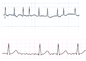
atrial flutter
Cause: CAD, Hypertension, Mitral valve disorders, Pulmonary embolus, Chronic lung disease, Cardiomyopathy, Hyperthyroidism, Alcohol and stimulants (cocaine, diet pills, caffeine, etc.) can contribute
Tx: Primary goal is to slow ventricular response by increasing AV block
Drugs to slow HR: Calcium channel blockers, -adrenergic blockers
Cardioversion may be used to convert the atrial flutter to sinus rhythm emergently and electively
Primary goal is to slow ventricular response by increasing AV block
Anti-dysrhythmic drugs to convert atrial flutter to sinus rhythm or to maintain sinus rhythm (e.g., amiodarone)
Radiofrequency catheter ablation can be curative therapy for atrial flutter
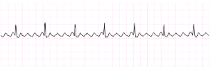
v-fib
Cause: Acute MI, CAD, cardiomyopathy, VF may occur during cardiac pacing or cardiac catheterization, VF may occur with coronary reperfusion after fibrinolytic therapy, Electrical shock, Hyperkalemia, Hypoxia, Acidosis, Drug toxicity
Tx: Immediate initiation of CPR and advanced cardiac life support (ACLS) measures with the use of defibrillation and drug therapy
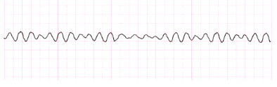
v-tach
Cause: MI, CAD, Electrolyte imbalances, Cardiomyopathy, Mitral valve prolapse, Long QT syndrome, Digitalis toxicity, Central nervous system disorders
Tx: Precipitating causes must be identified and treated (e.g., hypoxia), B-Adrenergic blockers, lidocaine, amiodarone, IV magnesium, lidocaine, Drugs that prolong the QT interval should be discontinued, If the rhythm is not converted, cardioversion may be needed, VT without a pulse is a life-threatening situation, Cardiopulmonary resuscitation (CPR) and rapid defibrillation, Epinephrine, amiodarone, or lidocaine if defibrillation is unsuccessful
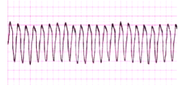
asystole
Cause: Advanced cardiac disease, Severe cardiac conduction system disturbance, End-stage HF
Tx: CPR with initiation of ACLS measures (e.g., intubation, and IV therapy with epinephrine)
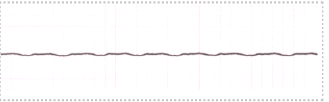
sinus bradycardia
Causes: Occurs in response to, Carotid sinus massage, Hypothermia, Increased Intraocular pressure, increased vagal tone, Administration of B-adrenergic, blockers, calcium channel Blockers, occurs in disease states, Hypothyroidism, Increased intracranial pressure, Inferior wall MI
Tx: Atropine, dopamine, or epinephrine Pacemaker may be required
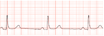
sinus tachycardia
Causes: Associated with physiologic stressors, Hyperthyroidism, Exercise, Pain, Hypovolemia, Myocardial ischemia, Heart failure (HF), Fever
Associated with medications: Epinephrine, dopamine, caffeine, hydralazine, pseudoephedrine
Tx: Determined by underlying cause, B-Adrenergic blockers to reduce, HR and myocardial oxygen consumption, Antipyretics to treat fever, Analgesics to treat pain
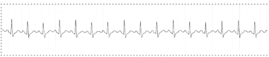
second degree heart block type 1 (Mobitz type 1 or Wenckebach)
Causes: Acute inferior MI or digitalis toxicity.
Tx: Not indicated as there are no symptoms
P wave gets further and further away from the QRS and then the QRS will drop
longer, longer, longer, drop …. then you have Wenckebach

second degree heart block type 2 (Mobitz type 2)
Causes: anterior wall MI, CAD, cardiomyopathy
Tx: artificial pacing
In Mobitz II, the atria are working normally (P waves are present), but some electrical impulses suddenly fail to reach the ventricles, so the ventricles skip beats (no QRS)
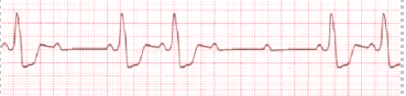
third degree heart block
Causes: Severe heart disease: CAD, MI, myocarditis, cardiomyopathy
Drugs: Digoxin, B-adrenergic blockers, calcium channel blockers
Tx: If symptomatic, transcutaneous pacemaker until a temporary transvenous pacemaker can be inserted
Drugs (e.g., atropine, dopamine, epinephrine): Temporary measure to increase HR and support BP until temporary pacing is initiated. Permanent pacemaker as soon as possible
vasopressors
increase blood pressure by causing vasoconstriction
used for severe hypotension and shock
norepinephrine
epinephrine
dopamine
phenylephrine
vasopressin
inotropes
increase the force of cardiac contraction to improve cardiac output
dobutamine
isoproterenol
IV medications that lower blood pressure (vasodilators)
Nitroprusside
Nitroglycerin
Nicardipine
Clevidipine
Hydralazine
Nesiritide (Natrecor)
ETT complications
aspiration if cuff pressure is too low and secretions leak around the cuff
tracheal injury may occur from excessive cuff pressure, prolonged intubation and can lead to bleeding, ischemia and tissue necrosis.
Accidental extubation (sudden resp. Distress, loss of breath sounds, low oxygen saturation, pt unable to ventilate).
VAP (ventilator associated pneumonia) that can develop 48 hours or more after intubation
ETT care
confirm tube placement, assess bilateral breath sounds, monitor oxygen saturation, assess chest expansion, monitor vent settings, observe respiratory effort, monitor ABGs, secure the tube, document tube depth, prevent accidental extubation, suction only when indicated, monitor cuff pressure (20-25), prevent injection (elevate HOB, oral care, hand hygiene, suctioning secretion's, daily assessment for extubation ready), provide communication board or writing device, use restraints, assess pain and comfort frequently, explain all procedures even if pt is unconscious
interventions for intubated patients that will decrease VAP
elevate HOB to 30 – 45 degrees, perform frequent oral care, suction secretions as ordered, use subglottic ET tubes when available, maintain cuff pressure, hand hygiene, assessing readiness for extubation daily. HOB 30-45 degrees unless contraindicated, No routine changes of ventilator circuit tubing, Spontaneous breathing trials/Spontaneous awakening, trials, Early mobility, ETT with subglottic suctioning, Oral care with chlorhexidine, Ventilator bundle
emergent actions when someone is on a ventilator
Assess the pt first, look for the cause (displacement, obstruction, pneumothorax, equipment failure?), if the vent alarms high pressure something is blocking airflow, if the vent alarms low pressure, air is leaking out. Begin manual ventilation if the pt is deteriorating. Suction if needed, call for help, have emergency equipment at the bedside (Ambu bag, oxygen source, suction set up, suction catheters)
common problems of ICU patients
Nutrition, Anxiety, Pain, Impaired communication, Sensory-Perceptual Problems, Sleep, Family/Caregiver issues, Neurological issues
Neurological issues: Altered LOC—medication induced, severity of illness, sleep deprivation, comorbidities. AEIOUTIPS:
• A—alcohol—withdrawal, intoxication
• E –electrolytes, endocrine, epilepsy (Triple E)—thyroid, sodium, seizure or new meds
• I— insulin—low causes slow
• O— opiates—overdose, too much
• U— uremia—kidney failure
• T— trauma, temperature—low is slow, high can cause seizures
• I— infection—hypovolemia, sepsis
• P – poison
• S—shock, stroke, subarachnoid hemorrhage, space occupying lesion
Delerium: prevent with sleep (cluster care)
airway obstruction
A person may clutch the beck between thumb and fingers (universal sign), weak, ineffective cough; high pitched noises on inspiration (stridor), increased respiratory distress, inability to speak, breathe or cough, collapse, flaring nostrils. Satisfactory management of ventilation may prevent hypoxia and hypercapnia.
Management: head tilt, chin lift (if no cervical spine issue), jaw thrust maneuver (used when cervical spine injury is suspected; opens the airway w/o moving the neck), oropharyngeal airway (a curved plastic airway inserted into the mouth to keep the tounge from blocking the airway; used for unconscious pts, not if the pt is awake and has a gag reflex), sedate and intubate, cricothyroidotomy (emergency airway through the cricothyroid membrane; used when ETT cannot be placed.
hemorrhage
Earliest sign of blood loss is increasing anxiety, as it continues you may see tachycardia, cool, clammy skin, delayed cap refill, decreased BP is a late sign, narrow pulse pressure, decreased urine output.
Management: The goal of emergency management is to control the bleeding, maintain adequate circulating blood volume for tissue oxygenation, and prevent shock. Fluid replacement (two large bore IVs 16-18 gauge), IV fluids (NS or LR), blood products, and massive transfusion protocol, type and screen O- and other blood types, monitor labs (CBC, H&H, coagulation, electrolytes, ABGs). Control external hemorrhage (tourniquet, hold pressure), control of internal bleeding (shows no external signs of bleeding but exhibits tachycardia, falling BP, thirst, apprehension, cool and moist skin, or delayed cap refill, hold pressure). Continue ongoing assessment once bleeding is controlled.
intra-abdominal injuries
Liver: RUQ pain, R shoulder pain, internal bleeding, hypotension, shock.
Spleen: LUQ pain, internal bleeding, L shoulder pain (Kehr sign).
Kidneys or urinary tract: hematuria, flank pain, bruising, reduced urine output.
Pancreas or GI tract: increased lipase, abdominal pain, peritonitis, free air in the abdomen, bowel contents leaking into the abdomen.
Assessment key: bruises, abrasions, penetration, VS, auscultate and palpate (watch for no movement, pain, guarding, etc.), assess other areas of the body also
Management: maintain airway, protect C spine if trauma suspected, control external bleeding, give fluids, keep pt NPO, insert NG tube. Worry about damage to GI tract (surgery needed)
ingested poisons
Some poisons are corrosive, meaning they can burn and destroy tissue in the mouth, throat, esophagus, stomach, and intestines. Airway swelling, difficulty breathing, stridor, drooling, trouble swallowing, burns around the mouth, redness in the mouth or throat, vomiting, chest or abdominal pain, alerted mental status
Management: protect the airway (assess for airway burns and swelling, give oxygen, prepare early intubation if airway swelling worsens, have suction available), support breathing and circulation. Measures to remove the toxin or decrease its absorption: Activated charcoal, Skin cleansing, Eye irrigation, Gastric lavage, Administration of specific antidote, Dialysis, DO NOT induce vomiting, Syrup of ipecac (no longer recommended), Cathartic—increase defecation. Do not automatically perform gastric lavage (perforation, aspiration, worsen burns), do not give activated charcoal automatically (can interfere with examination of the digestive tract)
carbon monoxide poisoning
Early sx: headache (most common early sx), dizziness, fatigue, nausea, palpitations. Moderate sx: confusion, difficulty concentrating, unsteady walking, vision changes, drowsiness. Severe sx: syncope, seizures, coma, respiratory failure, cardiac arrest. Skin changes may occur (cherry-red)
Management: remove pt from the source, remove clothes, 100% oxygen, fresh air. Assess airway, need for intubation, respiratory effort, cont. Oxygen until carboxyhemoglobin is less than 5%
spontaneous breathing trials
a test to determine whether the patient can breathe without significant ventilator assistance.
Benefits: identifies readiness for extubation and reduces ventilator days, lowers risk of pneumonia
spontaneous awaking trials
a daily interruption or reduction of continuous sedation to determine if the patient can safely awaken and follow commands.
Benefits: decrease duration of mechanical ventilation, reduces ICU length of stay, decreases sedation exposure.
spontaneous breathing and awaking trials
Both are evidence-based interventions used in mechanically ventilated patients to determine readiness for ventilator weaning and reduce complications associated with prolonged ventilation.
PEEP
Positive end expiratory volume: keeps the alveoli open and provides more time for gas exchange. Patient will need this when they have problems with keeping alveoli open after exhalation. If they have hypoxemia or low oxygen levels the alveoli will collapse or fill with fluid. Common causes are ARDS, pneumonia, Pulmonary edema(HF), atelectasis. This is used to treat critical cases of ARDS.
ARDS risk factors and sx
Risk factors: sepsis is the most common cause, aspiration of stomach contents, pneumonia, major trauma, burns, shock, pancreatitis, near drowning, smoke or toxic gas inhalation, multiple blood transfusions, fat embolism, air embolism, drug OD, major surgery, cardiopulmonary bypass, DIC, post-cardiac resuscitation
Clinical manifestations: refractory hypoxemia (O2 level stays very low even when supplemental oxygen is given) happens due to shunting (blood flows past collapsed or fluid-filled alveoli that cannot participate in gas exchange) -> alveoli must be opened with PEEP. Severe dyspnea, tachypnea, nasal flaring, intercostal retractations, accessory muscle use, crackles, decreased oxygen saturation, pink, frothy sputum, increased work of breathing, anxiety, agitation, confusion, tachycardia, cyanosis, fatigue, reduced LOC
ARDS medical and nursing management
find the cause and reverse. Mechanical ventilation is required, fluids, vasopressors and inotropes, sedation and analgesia, prone positioning (improves oxygenation) for at least 12 hours per day. Nurses need to improve oxygenation, keep the airway open, help the lungs expand, remove secretions, repositioning
pulmonary HTN sx
main sx is dyspnea that usually progresses from dyspnea with activity, dyspnea with mild activity and dyspnea at rest.
Patients may also have fatigue, weakness, chest pain, dizziness, syncope, hemoptysis, hypoxemia, and exercise intolerance. As R HF develops, they will have peripheral edema, ascites, JVD, hepatomegaly, weight gain, murmur, R sided S-3
pulmonary HTN causes and treatments
Causes:
Idiopathic = cause is unknown.
Secondary causes: COPD, pulmonary fibrosis, pulmonary embolism, left ventricular failure, mitral stenosis, congenital heart disease, HOV, portal HTN, connective tissue disorder, chronic hypoxemia
Treatments: oxygen if hypoxemia, vasodilators, PDE-5 inhibitors (relax pulmonary blood vessels), CCB, diuretics, anticoagulants, digoxin (if R-sided HF develops)
successful fluid resuscitation signs
BP increases, HR decreases, stronger peripheral pulses, improved cap refill, skin becomes warm, pink and dry, improved LOC, less confused or restless, RR decreases, less SOB, urine output is greater than or equal to 0.5mL/kg/hr
anaphylactic shock
Sudden severe allergic reaction that can become life-threatening. Can cause severe airway swelling, bronchospasm, widespread vasodilation, fluid leaking out of blood vessels, hypotension, or cardiovascular collapse.
Characteristics: swelling of the tongue, lips, face, uvula, throat tightness, hoarseness, stridor, wheezing, bronchospasm, SOB, hypoxemia, difficulty swallowing, hives, itching, flushing, redness, angioedema
anaphylactic shock causes, hemodynamic findings and tx
Causes: can be caused by medication, insect, food, contrast dye, abx (especially PCN), NSAIDs, blood products, latex
Hemodynamic findings: hypotension, tachycardia, weak pulses, dizziness, syncope, cardiac arrest or collapse
Treatments: stop the trigger/avoid, call for emergency help, give epinephrine early (0.3-0.5 mg IM in the thigh) and repeat q5-10 minutes PRN, airway support, IV fluids for hypotension, diphenhydramine, nebulizer (albuterol)
cardiogenic shock
happens when the heart cannot pump enough blood forward to meet the body’s oxygen needs.
Characteristics: hypotension, MAP <65, weak peripheral pulses, cool, pale, mottled skin, delayed cap refill, decreased urine output, confusion, anxiety, restlessness, decreased LOC
Hemodynamic findings: systolic BP drops more than 30 mm Hg from baseline for 30 minutes. MAP falls below 65. Tissue perfusion decreases, heart gets less oxygen, heart becomes weaker
cardiogenic shock causes and tx
Causes: most common cause is an MI. Others include cardiomyopathy, severe arrhythmias, valve damage, severe hypoxemia, acidosis, hypoglycemia, hypocalcemia
Treatments: treat the underlying cause; oxygen, pain management, diuretics, beta blockers, inotropic meds, nitrates. IABP and VADs, O2, anticoagulants.
hypovolemic shock
the most common type of shock; occurs when there is not enough fluid or blood in the blood vessels which causes decreased preload, cardiac output and poor tissue perfusion.
Characteristics: early: tachycardia, tachypnea, anxiety, cool, pale skin, delayed cap refill, flat neck veins, decreased urine output, thirst. Late: hypotension, narrow pulse pressure, alerted mental status, oliguria, weak pulses, cold, clammy extremities, severe tissue hypoxia
Hemodynamic findings: everything is decreased
hypovolemic shock causes and treatments
Causes: hemorrhagic (trauma, GI bleed, ruptured AAA, ruptured ectopic pregnancy, hemothorax, fractures, etc). Non-hemorrhagic (fluid loss; vomiting, diarrhea, burns, dehydration, surgery, DKA, DI, etc.)
Treatment: stop the cause, fluids (two large bore IVs; LR or NS→ warm if large amounts are being infused to prevent hypothermia) blood products, albumin, MTP, ongoing assessment, monitoring labs and urine output
hypovolemic shock complications
lactic acidosis, AKI, respiratory failure, decreased cerebral perfusion, DIC, hypothermia during fluid replacement
neurogenic shock
type of disruptive shock caused by a loss of sympathetic nervous system activity. Blood volume is present, but the blood vessels are too relaxed and wide.
Characteristics: hypotension, bradycardia, warm, dry skin
Hemodynamic findings: BP, MAP, HR decreased, CO may decrease
neurogenic shock tx and causes
Causes: spinal cord injury above T6, C-spine injury, high thoracic spinal injury, spinal aesthesia, nervous system damage, severe hypoglycemia
Treatment: spinal immobilization, fluids, vasopressors (norepinephrine to constrict BVs), atropine for symptomatic bradycardia, watch for orthostatic hypotension, prevent DVT
obstructive shock
occurs when a physical blockage prevents blood from flowing normally through the heart or major blood vessels. CO decreases, BP falls, organs get less oxygen, tissue perfusion decreases
Characteristics: tension pneumothorax (sudden chest pain, severe dyspnea, tachypnea, tachycardia, absent or decreased breath sounds on one side, JVD, tracheal deviation, crepitus, hypotension. Cardiac tamponade (hypotension, JVD, muffled or distant heart sounds aka Beck’s triad). PE (sudden SOB, pleuritic chest pain, tachycardia, hypoxia, tachypnea, anxiety)
obstructive shock causes and tx
Causes: tension pneumothorax, cardiac tamponade, PE
Treatments: The priority is to remove the obstruction. Tension pneumothorax (needle decompression followed by chest tube insertion). Cardiac tamponade (pericardiocentesis). PE (anticoagulants, thrombolytics, surgical embolectomy, mechanical ventilation, hemodynamic support)
septic shock
the most common type of distributive shock. Occurs when severe infection causes an uncontrolled autoimmune and inflammatory response throughout the body.
Characteristics: persistent hypotension, poor tissue perfusion, tissue hypoxia, fever
septic shock causes and tx
Cause: infections
Treatments: Fluid replacement, FIND THE SOURCE, ANTIBIOTICS, Drain abscesses--“pus pockets”, 20-40 ml/kg fluids (1-2 Liters minimum), CVP, MAP, Urinary output, CVP > 8, MAP >65, urine output at least 0.5 ml/kg, SvO2 >70, Vasopressors (norepinephrine); epinephrine 2nd line treatment, Low dose corticosteroids—assess for complications of use
stages of shock - compensated (nonprogressive)
the earliest stage of shock. The body is fighting for its life – the sympathetic nervous system turns on and the body will enter fight or flight mode and will release norepinephrine and epinephrine which causes a faster HR and stronger heart contractions. The blood vessels will constrict, RAAS is activated. BP will remain normal, BG increases, RR increases, anxiety, feeling of impending doom, cool or clammy skin, delayed cap refill (vasoconstriction), narrow pulse pressure, mildly decreased urine output
stages of shock - uncompensated (progressive)
happens when the body can no longer keep up to its compensatory responses. In the compensated stage, the body was able to maintain blood pressure by increasing the heart rate and constricting blood vessels. In the uncompensated stage, those efforts begin to fail. The heart starts to fail, the kidneys get less blood, anaerobic metabolism increases which causes a ride in lactic acid, serum lactate and metabolic acidosis, the clotting system becomes abnormal and can lead to DIC, blood flow to the GI tract decreases. Heart and kidneys will show the first signs. Hypotension, oliguria, diaphoresis, failing cardiac pump, Tachycardia, lethargic, metabolic acidosis (myocardia, depression), Resp—shallow, rapid mottled LOC changes slow cap refill
stages of shock - irreversible
this is the last and most severe stage of shock. The body has reached a point where the organs are permanently damaged and begin to shut down. Tissue perfusion is critically low and cells begin to die b/c they are not getting oxygen and nutrients, severe metabolic acidosis will develop, kidney failure will develop, liver failure will develop, the intestines will fail, the lungs fail, the heart fails and the pt will develop MODS.
MODS (multiple organ dysfunction syndrome)
multiple organ dysfunction syndrome occurs when two or more organ systems become severely dysfunctional in a critically ill patient. Must affect two or more organ systems and last 24-48 hours to be MODS. Most common organ dysfunctions seen with MODS are acute kidney failure and ARDS
MODS prevention
Prevention is top priority. Early detection and documentation of initial signs of infection are essential in managing MODS in older patients. Subtle changes in mentation and a gradual rise in temperature are early warning signs. Other patients at risk for MODS are those with chronic illness, malnutrition, immunosuppression, or surgical or traumatic wounds. If preventive measures fail, treatment measures to reverse MODS are aimed at (1) controlling the initiating event, (2) promoting adequate organ perfusion, and (3) providing nutritional support.
sepsis
a life-threatening organ dysfunction caused by a dysregulated response to an infection. Always caused by an infection. Can rapidly progress to septic shock and multiple organ dysfunction if untreated.
SIRS (systemic inflammatory response syndrome)
a widespread inflammatory response to ANY INSULT (infectious or not). Maybe caused by my infection, trauma, burns, pancreatitis, surgery, hemorrhage, etc. it may or may not progress to sepsis.
aortic stenosis
Causes: buildup of calcium deposits on heart valves (most common), congenital malformation
syncope, angina, dyspnea (SAD). Increase HR without improving cardiac output. Chest pain, dizziness, orthopnea, difficulty sleeping, difficulty with small amounts of activity. Assess systolic murmur (loud), S4
mitral valve stenosis
Dyspnea on exertion, orthopnea, paroxysmal nocturnal dyspnea, dyspnea at rest (as it gets worse), Progressive fatigue, Low CO, chest pain, palpitations, a-fib, paroxysmal a-tach, Hemoptysis, hoarseness, respiratory infections, Heart failure symptoms--peripheral edema, ascites, Loud S1 and murmur, heart enlarged, a-fib. Can cause strokes. Can take a long time for sx to develop.
Causes: rheumatic fever or endocarditisà thickens leaflets. Radiation therapy to the chest area could also result in MS
aortic valve regurgitation
Asymptomatic to start. Palpitations, neck vein pulsation, dyspnea, fatigue, angina, orthopnea, pulmonary congestion
Causes: untreated syphilis, calcifications on valve, bicuspid aortic valve, high BP, aging
mitral valve regurgitation
Fatigue, SOB (lay flat or exercise), murmur, palpitations, dyspnea on exertion, S3
Causes: aging, mitral valve stenosis or prolapse, rheumatic fever, HA
management/treatment for valve disorders
Echocardiogram, cardiac Cath helps diagnose all valve disorders
symptom relief: goals → decrease regurgitation, improve cardiac output, reduce pulmonary congestion. Diet changes (limit sodium, alcohol, caffeine), avoid strenuous activity.
Medications:
Beta-blockers, CCB-rate control, reduce palpitations, when having CAD
ACE inhibitors- vasodilation- be careful with AS, as it can cause syncope
ARB, nitrates, hydralazine- reduce afterload
Diuretics, vasodilators, sodium restriction—reduce preload and pulmonary congestion—be careful
Antiarrhythmics and Cardioversion—for a-fib and prevent clot