NPTE Final Frontier Integumentary System
1/59
There's no tags or description
Looks like no tags are added yet.
Name | Mastery | Learn | Test | Matching | Spaced | Call with Kai |
|---|
No analytics yet
Send a link to your students to track their progress
60 Terms
integ system
skin is the largest body organ: 15-20% of BW
primary function of skin
Protection, insulation, holding organs together, sensory, fluid balance, temperature control, absorbing UV radiation, metabolizing vitamin D, and synthesizing epidermal lipids
Epidermis
superficial
keratinocytes, melanocytes, langerhans cells, Basal cells
dermis
Deep
collagen, reticulum, fibroblasts, macrophages, lymphatic glands, blood vessels, nerve fibers
Meissner corpuscles
touch and vibration
Krause end bulbs
Cold
golgi tendon organs
contraction
ruffini endings
hot sensation
pacinian corpuscles
pressure
venous insufficiency
refers to inadequate drainage of venous blood from a body part, usually resulting in edema and/or skin abnormalities and ulcerations
arterial insufficiency
refers to a lack of adequate blood flow to a region of the body
clinical presentation: venous insufficiency
- proximal to the medial malleolus
- irregular, shallow appearance
- flaking, brownish discoloration- hemosiderin staining
- mild to moderate pain
- elevation decreases pain
clinical presentation: arterial insufficiency
- Lower 1/3 of leg, toe and lateral malleolus
- Smooth edges, well defined, tend to be deep
- Thin and shiny, hair loss, yellow nails
- Severe pain
- Elevation increases pain
"painful cramping"
claudication >> struggling to get blood >> arterial
pressure ulcers
located over bony areas and are staged related to the depth of the wound bed
pressure ulcers: stage 1
reddened area that does not go away
pressure ulcers: stage 2
first 2 layers of skin, superficial in nature
pressure ulcers: stage 3
subcutaneous fat may be visible
FAT: 3 letters
pressure ulcers: stage 4
down to the bone and including the bone
BONE: 4 letters
pressure ulcers: unstageable
related to not visualizing the wound base because of necrotic tissue
do pressure ulcers change stages after initial naming?
NO THEY DO NOT
even if they progress/heal they will always be referred to the initial stage name
diabetic ulcers
generally located on the weight-bearing surface of the foot
venous insufficiency ulcers
frequently are proximal to the medial malleolus; they are edematous
arterial ulcers
generally located on the lateral malleolus, distal toes, or areas of trauma
pressure ulcers
result of unrelieved external pressure on an area
Herpes zoster
"SHINGLES"
dermatomes
- herpes zoster (shingles) has initial symptoms of pain and paresthesia localized to the affected dermatome
integumentary
- presents as a rash
- mostly unilateral
- raised to palpation (<2mm height)
- pink w silvery white appearance
- CN 3 and 5
- NO HEAT
precautions: contact and airborne
herpes simplex virus type 1
located above the waist
cold sores
herpes simplex virus type 2
located below the waist
wheals
hives
pustules
pus filled
vesicles
fluid filled, dome
wound healing basics
- too much moisture will delay healing
- too moist, the periwound becomes macerated
- macerated wounds can be white, friable, over hydrated, and sometimes wrinkled skin
- maceration may be caused by uncontrolled wound drainage, perspiration, or incontinence
dressing: very mild exudate
transparent film
dressing: minimal exudate
hydrogel, hydrocolloid
dressing: moderate exudate
foams
dressing: heavy exudate
calcium alginates, hydrofiber
Dressings: Nonadherent
films > hydrogel > hydrocolloids > foam > calcium alginates, hydrofibers
dressing: antimicrobials
films > hydrogel > hydrocolloids > foam > calcium alginates, hydrofibers
debridement: selective
removal of ONLY non viable tissues from a wound
dead tissue < alive tissue
types of selective debridement
sharp: use of scalpel, scissors, forceps
enzymatic: use of topical application
autolytic: body's own mechanism
Debridement: nonselective
removal of both nonviable and viable tissues from a wound, >50% necrotic
types of nonselective debridement
wet to dry: application of moist gauze over area of necrotic tissues to be completely dried and removed
wound irrigation: move necrotic tissue from wound bed using pressurized fluid
hydrotherapy: using a whirlpool w agitation directed toward a wound requiring debridement
Red-Yellow-Black System
- red: pink granulation tissue, protect wound, maintain moist environment
- yellow: moist yellow slough, remove exudate and debris, absorb drainage
- black: black thick eschar firmly adhered, debride necrotic tissue
Povidone -iodine solution
infection- surgical site
zinc oxide cream
dental
nitrofurazone solution
burns
Burns: superficial
epidermis
dry, red skin, w/o open areas
heals in 5 days w/o scarring
burns: superficial partial thickness
epidermis and some dermis
weeping blisters, blanches to pressure with quick capillary refill, extremely painful
heals within 10-14 days
minimal scarring
burns: deep partial thickness
epidermis and dermis
mottled red and white areas, blanches to pressure w/ slow capillary refill, decreased pinprick sensation
can take up to 3 weeks
large wounds can be managed surgically
burns: full thickness
epidermis, dermis, some subcutaneous tissues
dry, rigid, leathery eschar, lack of pain, pressure, temp sensation
requires > 3 weeks; will require surgical closure, may have contractures
burns: subdermal
epidermis, dermis, subcutaneous tissues
charred, dry and exposed deep tissue
requires surgical interventions, amputation and paralysis possible
epidermal
no scar and have intact skin
superficial partial thickness
painful to touch, temp, have brisk capillary refill, produces minimal scarring
deep partial thickness
waxy white, have pain of deep pressure, delayed capillary refill, result in excessive scarring and development of hypertrophic and keloid scars
subdermal
charred, dry and exposed to deep tissue and often require amputation
Rule of 9s adults
head = 9% (front 4.5, back 4.5)
each arm = 9% (front 4.5, back 4.5)
trunk = 18% front, 18% back
each leg = 18% (9 front, 9 back)
genitals = 1
Upper body: 63%
Genitals: 1 %
Lower body: 36%
Rule of 9s children
Head 18% (8.5 front, 8.5 back)
each arm 9% (front 4.5, back 4.5)
chest 18%
back 18%
each leg 14% (6.5 front, 6.5 back)
genitals 1%
upper body: 72:% (71%)
genitals: 1 %
lower body: 28%
scars: normal
flat and similar to skin color
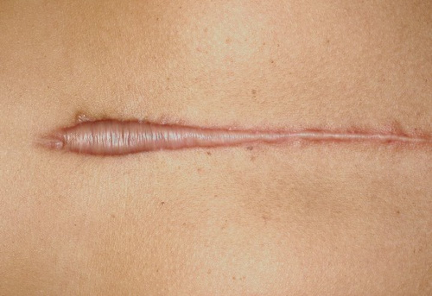
scars: hypertrophic
a healed wound w thick fibrous tissue that remains WITHIN the original wound border
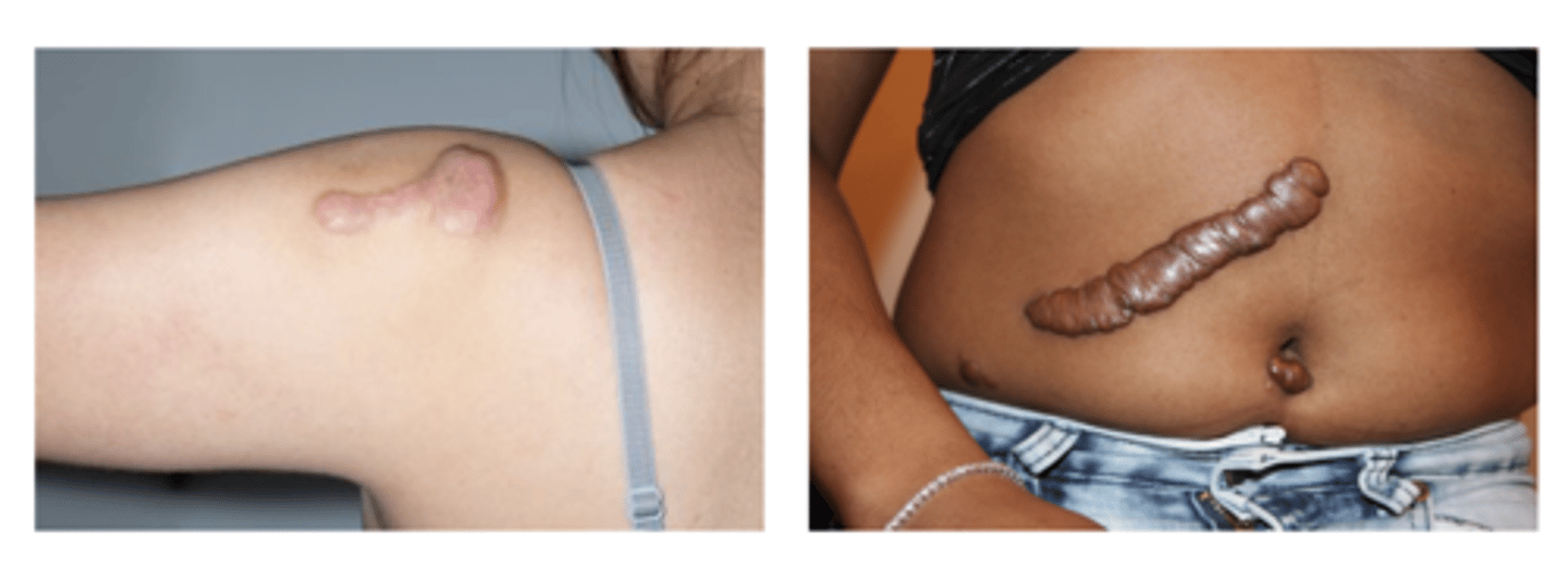
scars: keloid
excessive scar tissue grows OUTSIDE of the original margins of the wound