Neurological examination and brainstem
1/129
There's no tags or description
Looks like no tags are added yet.
Name | Mastery | Learn | Test | Matching | Spaced | Call with Kai |
|---|
No analytics yet
Send a link to your students to track their progress
130 Terms
What are the 4 steps to a neurological exam?
general examination
head and cranial nerves
trunk and spine
limbs
What do we mean by a general examination?
general observation of the animal in a hands-off manner and obtaining information from the owner
taking a detailed history (need video footage from the owner)
observe the animal upon approach to the room: its interaction with the owner and it’s surroundings
what are we assessing in the general examination stage?
mental status
posture
gait
muscle condition
response to surroundings
what should we be able to conclude from the general examination?
probable location and extent of nervous system dysfunction that we can assess with the rest of the exam.
what are the 5 levels of consciousness
normal
confused and disorientated
depressed
stuporous
comatose
If an animal is circling in one direction, where does this indicate the lesion it?
lesion is on the side of the brain that the animal is circling towards
what do we do to test CNI
smell
not easy to test
waft food and see if the animal follows it?
How do we test CNII
consciousness
vision
menace response (also VII)
visual placing
fixating response
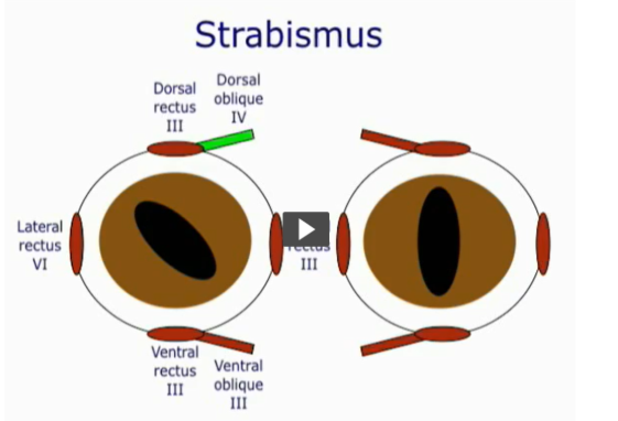
how do we assess CNIII
PLR (II afferent, III efferent)
anisocoria
strabismus
what is anisocoria
different sized pupils in each eye
what are the 2 pathways of PLR
direct = efferent pathway
consensual/indirect = afferent pathway
how do we assess CNIV
strabismus
How do we assess CNV
skin sensation (jaw tone)
palpebral reflex (V, VII)
corneal reflex (V, VII)
How do we assess CNVI
strabismus
How do we assess CNVII
facial muscle symmetry
lacrimal gland function
nasal gland function
How do we assess CNVIII
hearing and nystagmus
nystagmus (III, IV, VI too)
How do we assess CNIX
Swallowing (gag)
How do we assess CNX
laryngeal function (bark)
GI function
How do we assess CNXI
neck position
muscle tone
How do we assess CNXII
tongue position and tone
Fill in the gaps
olfactory
optic
vision
trigeminal
three
sensation
facial
expression
salivation
vestibulocochlear
glossopharyngeal
parasympathetic
vagal
accessory
hypoglossal
intrinsic
What supplies the afferent pathway of the palpebral response
CNV
what nerve supplies the efferent response of the palpebral reflex
CNVII
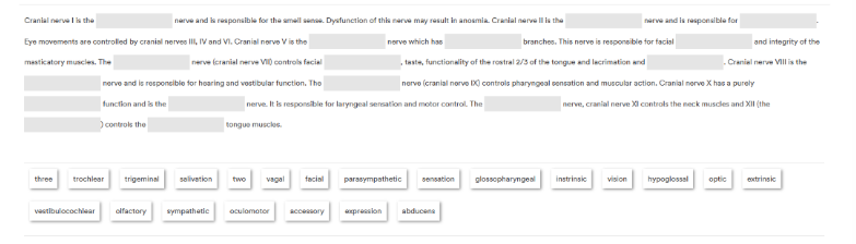
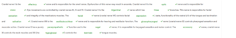
What kind of strabismus do we see if CNIII is damaged?
What muscles are affected?
lateral and ventral
Muscles = dorsal rectus, medial rectus, ventral rectus and oblique
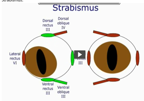
What kind of strabismus do we see if CNVI is damaged?
What muscle(s) is affected?
medial
Muscle = lateral rectus
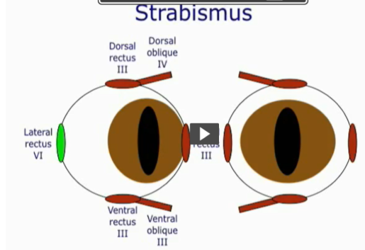
What kind of strabismus do we see if CNIV is damaged? What muscles are damaged?
rotational
Muscle = dorsal oblique (only 1 muscle affected)
What kinds of spontaneous nystagmus are there, what is it concurrent with?
horizontal
vertical
rotational
spontaneous nystagmus is concurrent with a vestibular lesion
Outline the neuronal pathway of the PLR response
optic nerve
optic chiasm
optic radiation
pre-tectal nucleus
Edinger-Westghal nucleus
CNIII
ciliary ganglion
What is the swinging test?
swing the light from eye to eye, resting for a few seconds at each eye.
What are some symptoms of horner’s syndrome in cats?
ptosis
miosis
enophthalmos
What can also indicate Horner’s syndrome in horses
ipsilateral excessive sweating
What can indicate Horner’s syndrome in cows?
Ipsilateral dry nose
Outline the menace response (afferent, efferent supply and what it does)
It causes a blink response to a threatening stimulus of the eye
AFFERENT SUPPLY = CNII
EFFERENT SUPPLY = CNVII
what do we need to prevent when conducting a menace response and why
need to prevent a draught/airflow
stimulates a corneal response
what are the brain regions involved in the menace response?
conscious visual pathway
contralateral visual cortex
contralateral motor cortex
ipsilateral cerebellar hemisphere
Outline the components of the menace response pathway
Visual threat → optic nerve → visual cortex → motor cortex → cerebellum → facial nerve
During the menace response to which sides of the brain do the nervous impulses travel?
impulse crosses over to the contralateral side through the optic chiasm (some decussation) → visual cortex
returns to ipsilateral side when it enters the cerebellum from the motor cortex
what do we test when we have an absent menace response?
palpebral reflex
Absent menace but present palpebral = CNVII problem
Why do we use a fixating response?
able to demonstrate conscious vision of a patient
Outline the pathway of conscious vision
optic nerve → lateral geniculate nucleus → optic radiation → occipital cortex (prosencephalon)
What Cn is associated with the Schirmer Tear Test and how?
CNVII
Parasympathetic function, lacrimal function is tested by doing an STT
What are the aims of assessing the trunk and what stage of the neurological exam is this?
Identifying any focal regions of pain or dysfunction by careful manipulation
Step 3 of the neurological exam
How do we assess the trunk?
panniculus response (from scapula to pelvis)
spinal manipulation
palpation
what is the panniculus response?
Pinch the skin on either side of the spine (one side at a time)
Cutaneous trunci muscle causes muscle contraction
should be able to see this response from the scapula to the pelvis
What degree of manipulation should we get in the spine
get head parallel to spine laterally
get head fully vertical to the ground
What do we do when palpating the spine
wings of the atlas
down the sides of the vertebrae (one side at a time)
press down onto the spinous processes
What is stage 4 of the neurological evaluation and what 4 things do we assess?
Stage 4 = limb evaluation
proprioception
sensation
segmental reflexes
muscle mass and tone
What are 6 proprioceptive tests we conduct?
knuckling
hopping
hemiwalking
tactile placing
paper slide
extensor postural thrust
What is knuckling and what should we see?
a weight bearing/pain free paw is placed with dorsum on the ground
there should be a return to normal positioning
What is hopping and what should we see?
Hold one limb up against the trunk and move the body outside the line of balance
there should be a rapid and accurate sideways repositioning of the limb
what is Hemiwalking and what should we see?
both limbs on one side of the body are held up against the trunk
see more subtle changes on a single limb hopping
What is tactile placing and what should we see?
the animal is brought up to the edge of a table whilst blindfolded
when the limbs touch the edge of the table they should be accurately placed immediately
What is paper sliding and what should we see?
place a limb onto a piece of paper and pull the paper laterally
weight bearing/pain free paw is moved to the outside the body and the limb should rapidly and accurately be returned to a normal position
What is the extensor postural thrust and what should we see?
dog is lifted off the ground and replaced on the table
as the hindlimbs touch the table there should be a reflex extension of hindlimbs which also precipitate the dog to step backwards (makes the dog look like it jumps)
How do we conduct an ECR myotatic reflex
percuss the muscle belly (Extensor carpi radialis)
should see the carpus reflex extension
How do we conduct a myotatic reflex for the biceps?
percuss a finger/thumb which is pressing onto the tendon of insertion of the biceps
reflex contraction generates: flexion of the elbow (not always seen), increased tension at tendon of insertion and movement of skin over muscle belly/visible contraction
How do we conduct a myotatic reflex for the triceps
percuss a finger/thumb onto tendon of insertion
reflex contraction of the triceps generates: reflex extension of the elbow (sometimes) and movement of the skin over muscle belly/visible contraction
How do we conduct a myotatic reflex for the patella
percuss the straight patellar tendon
reflex extension of the stifle
How do we conduct a myotatic reflex for the cranial tibial
percuss muscle belly, see a reflex flexion of the hock
What receptor organ is used to initiate a myotatic reflex
muscle spindle
what peripheral nerve is tested by the biceps reflex
musculocutaneous
Which peripheral nerve is tested by the triceps reflex
radial
which peripheral nerve is tested by the ECR reflex
radial
Which nerve is tested by the patellar reflex
femoral
which peripheral nerve is tested by the cranial tibial reflex
sciatic
How can we use muscle contraction to determine if we’ve got an UMN or LMN lesion?
LMN cause spasms - continual contraction of the muscle
UMN usually inhibit these
Therefore:
if we’ve got a LMN lesion = no spasms/weakness, rapid muscle atrophy, reduced reflexes, low tone
if we’ve got an UMN lesion = spasm, increased/normal reflexes, high tone and slow muscle atrophy
what are indicators of a LMN lesion?
rapid muscle atrophy
reduced reflexes
low tone
What are indicators of an UMN
slow muscle atrophy
increased/normal reflexes
high tone
what is withdrawal/sensation
squeezing toes tests: a) sensation and b) strength of withdrawal
what is a withdrawal reflex
local segmental reflex
what is necessary to confirm the presence of sensation
demonstrable cortical resposne
are sensation and withdrawal independent of each other and why/why not?
yes they are
it’s for the necessity of integrity of the peripheral nerves
what are the 3 tests for the lumbosacral spinal cord
perineal reflex
anal tone
tail tone
what should we be aware of with neurological tests of dogs vs cats?
many cats won’t tolerate the restraint and invasion of personal space needed for a thorough neurological exam
choose 3 tests that will most likely confirm the lesion localisation and severity
Name the parts of the brain stem
midbrain
pons
medulla oblongata
tectum
tegmentum
basilar area
What do we find in the midbrain
Basilar portion: crus cerebri
Tegmentum: red nucleus, nuclei of CNIII-IV
Tectum: superior and inferior colliculi
what do we find in the pons?
Basilar portion: descending tracts, pontine nuclei
Tegmentum: Ascending tracts, nuclei of CNV-VIII
hat do we find in the medulla oblongata
Basilar portion: pyramids, inferior olivary nucleus
Tegmentum: gracile and cuneate tubercles, nuclei of CN IX,X,XII
How is the brain stem vascularised?
Arteries: vertebrobasilar system
Veins: deep plexus that drains into several veins
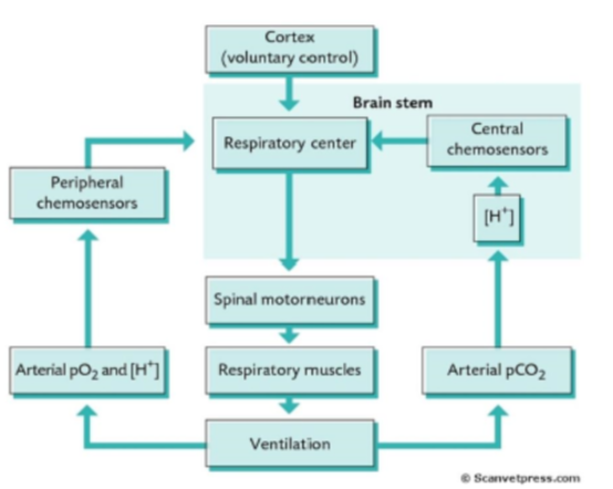
What are the general functions of the brain stem?
breathing regulation
circulation
consciousness
audio-visual reflexes
balance and co-ordination
passageway for ascending and descending tracts
houses cranial nerve nuclei
what part of the brain stem controls the cardiovascular system
medullar oblongata
what part of the brain stem controls the respiratory system
medulla oblongata
what part of the brain stem controls pain
periaqueductal nuclei
rostral ventromedial medulla
what part of the brain stem controls alertness, awareness and consciousness?
midbrain (via CNIII and CNV)
what part of the brain stem controls the communication b/w cerebrum and cerebellum
pons
What do we notice in this diagram about a)ascending pathways and b) descending pathways
a) ascending pathways pass through the brainstem
b) descending pathways include upper motor neurones synapsing on the lower motor neurones in the ventral and intermediate horn of the spinal cord
What are the 4 kinds of haemorrhage
subdural
extradural
subarachnoid
intracerebral
Give some examples of causes of subdural haemorrhage
head trauma
coagulation problems
rupture of the bridging veins
Give some examples of causes of extradural haemorrhage
trauma to temporoparietal region
rupture of middle meningeal artery
Give some examples of causes of subarachnoid haemorrhage
arterial bleed - spontaneous or associated trauma
Give some examples of causes of intracerebral haemorrhage
Also known as haemorrhaging stroke
bleeding from a ruptured blood vessel: intraparenchymal or intraventricular
what are the different ways we can treat brain tumours in dogs
surgery
radiation
symptomatic management
surgery and radiation
surgery and chemotherapy
solely chemotherapy
What are peripheral vestibular disorders
pathology of the inner ear vestibular structures as well as the vestibular portion of CNVIII
it diminishes available sensory information regarding head position and movement
What are central vestibular disorders
They involve:
vestibular nuclear complex and cerebellum
structures of reticular activating system
midbrain
higher centres of cortical function
Affects:
integration and processing of sensory input from vestibular, visual and somatosensory systems
what are examples of the most common CVD?
Most common CVD:
strokes
head trauma
migraine related vestibulopathy
multiple sclerosis
cerebellar degeneration
what do both CVD and PVD affect and what symptoms may they cause?
reduce appropriate neural output for spatial orientation, postural control and eye movement control
May see:
dizziness
imbalance
falls
visual blurring (oscillopsia)
what is Ischaemia
decreased blood supply to tissues
leads to a decrease in oxygen and nutrients to the area → necrosis
what is a stroke
when blood flow is restricted to a part of the brain
what 2 causes of strokes are there
ischemic = blocked vessed
haemorrhagic = bleeding
what can we refer to the midbrain as?
mesencephalon