Epithelial 1 Tissue Notes
1/30
There's no tags or description
Looks like no tags are added yet.
Name | Mastery | Learn | Test | Matching | Spaced | Call with Kai | Chat |
|---|
No analytics yet
Send a link to your students to track their progress
31 Terms
What are cells? What are tissues?
The cell is the basic functional unit of the body
The cells and extracellular components are characterized by distinctive pattern of organization.
This organized arrangement reflects the cooperative effort of cells performing a particular function.
Cells that function in a collective manner is called tissue : Four fundamental tissue types
Tissues are characterized by different structural organization and physiologic properties. Tissues reflect different body organs .
What gives rise to all the tissues and organs?
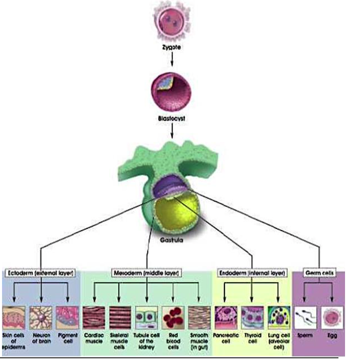
In the early developing embryo during the gastrulation phase, a trilaminar germ disc is being formed.
ectoderm,
mesoderm,
endoderm,
which give rise to all the tissues and organs.
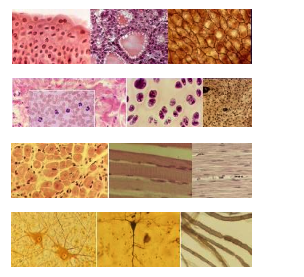
Name the Four Basic Tissue Types
Epithelial Tissue
Body surface, lines body cavity, form glands
Connective Tissue
underlies or supports the other three basic tissues, both structurally and functionally.
Muscle Tissue
is made up of contractile cells and is responsible for movement.
Nerve Tissue
Nerve tissue consists of nerve cells (neurons) and associated supporting cells of several types.
what are the Two different epithelial tissues:
Two different epithelial tissues:
Lining epitheium ( skin, body cavites)
Secretosy epithelium: Exocrine glands
Endocrine glands
All 3 germ layers are involved
Ectoderm: nasal and oral mucosa, epidermis, cornea, mammary and Cutaneous glands
Endoderm: epithelial lining of respiratory, intestinal tracts and glands (ie. pancreas, liver)
Mesoderm: urogenital system, mesothelium

Explain diagram
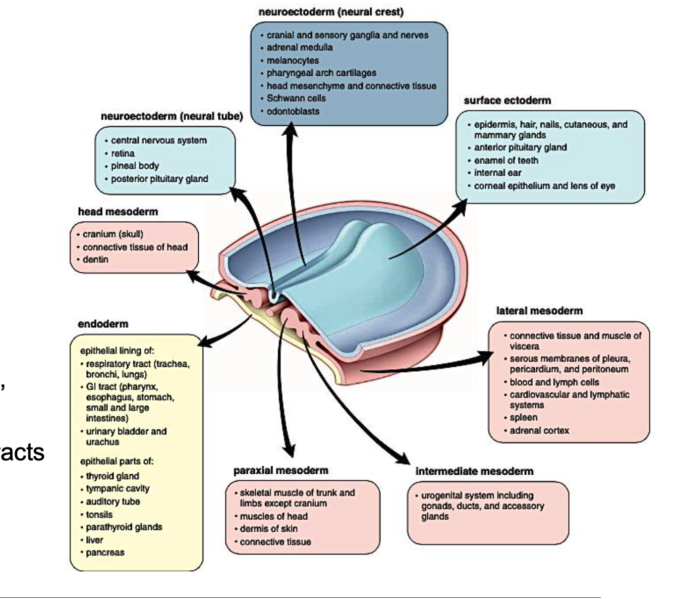
The image provided is a diagram illustrating the derivatives of the three primary germ layers—ectoderm, mesoderm, and endoderm—during embryonic development. These germ layers give rise to all the tissues and organs in the body.
Here's a breakdown of what each section of the diagram explains:
Central Diagram (Embryo): This stylized 3D representation shows an early embryo with the three germ layers distinguishable:
Ectoderm (Blue): The outermost layer.
Mesoderm (Red): The middle layer.
Endoderm (Yellow): The innermost layer.
Neuroectoderm (Neural Crest) - (Light Blue Box, Top Left): This is a specialized part of the ectoderm. Its derivatives include:
Cranial and sensory ganglia and nerves
Adrenal medulla
Melanocytes
Pharyngeal arch cartilages
Head mesenchyme and connective tissue
Schwann cells
Odontoblasts
Neuroectoderm (Neural Tube) - (Light Blue Box, Left): Another specialized part of the ectoderm, formed from the folding of the neural plate. Its derivatives include:
Central nervous system
Retina
Pineal body
Posterior pituitary gland
Surface Ectoderm - (Light Blue Box, Top Right): This refers to the general outer layer of the ectoderm. Its derivatives include:
Epidermis, hair, nails, cutaneous, and mammary glands
Anterior pituitary gland
Enamel of teeth
Internal ear
Corneal epithelium and lens of eye
Head Mesoderm - (Red Box, Left): Derivatives of the mesoderm in the head region:
Cranium (skull)
Connective tissue of head
Dentin
Lateral Mesoderm - (Red Box, Right): Derivatives of the mesoderm that forms the outer body walls and visceral organs:
Connective tissue and muscle of viscera
Serous membranes of pleura, pericardium, and peritoneum
Blood and lymph cells
Cardiovascular and lymphatic systems
Spleen
Adrenal cortex
Paraxial Mesoderm - (Red Box, Bottom): This mesoderm lies alongside the neural tube. Its derivatives include:
Skeletal muscle of trunk and limbs (except cranium)
Muscles of head
Dermis of skin
Connective tissue
Intermediate Mesoderm - (Red Box, Bottom Right): This mesoderm is located between the paraxial and lateral mesoderm. Its derivatives form:
Urogenital system including gonads, ducts, and accessory glands
Endoderm - (Yellow Box, Bottom Left): The innermost germ layer. Its derivatives primarily involve the lining of various internal systems and glands:
Epithelial lining of:
Respiratory tract (trachea, bronchi, lungs)
GI tract (pharynx, esophagus, stomach, small and large intestines)
Urinary bladder and urachus
Epithelial parts of:
Thyroid gland
Tympanic cavity
Auditory tube
Tonsils
Parathyroid glands
Liver
Pancreas
In summary, the image provides a comprehensive overview of how the three primary germ layers differentiate and specialize to form the diverse tissues and organs of the human body.

Epithelial Tissue Function and main features
Absorption
Secretory
Transportation
Mechanical protection,
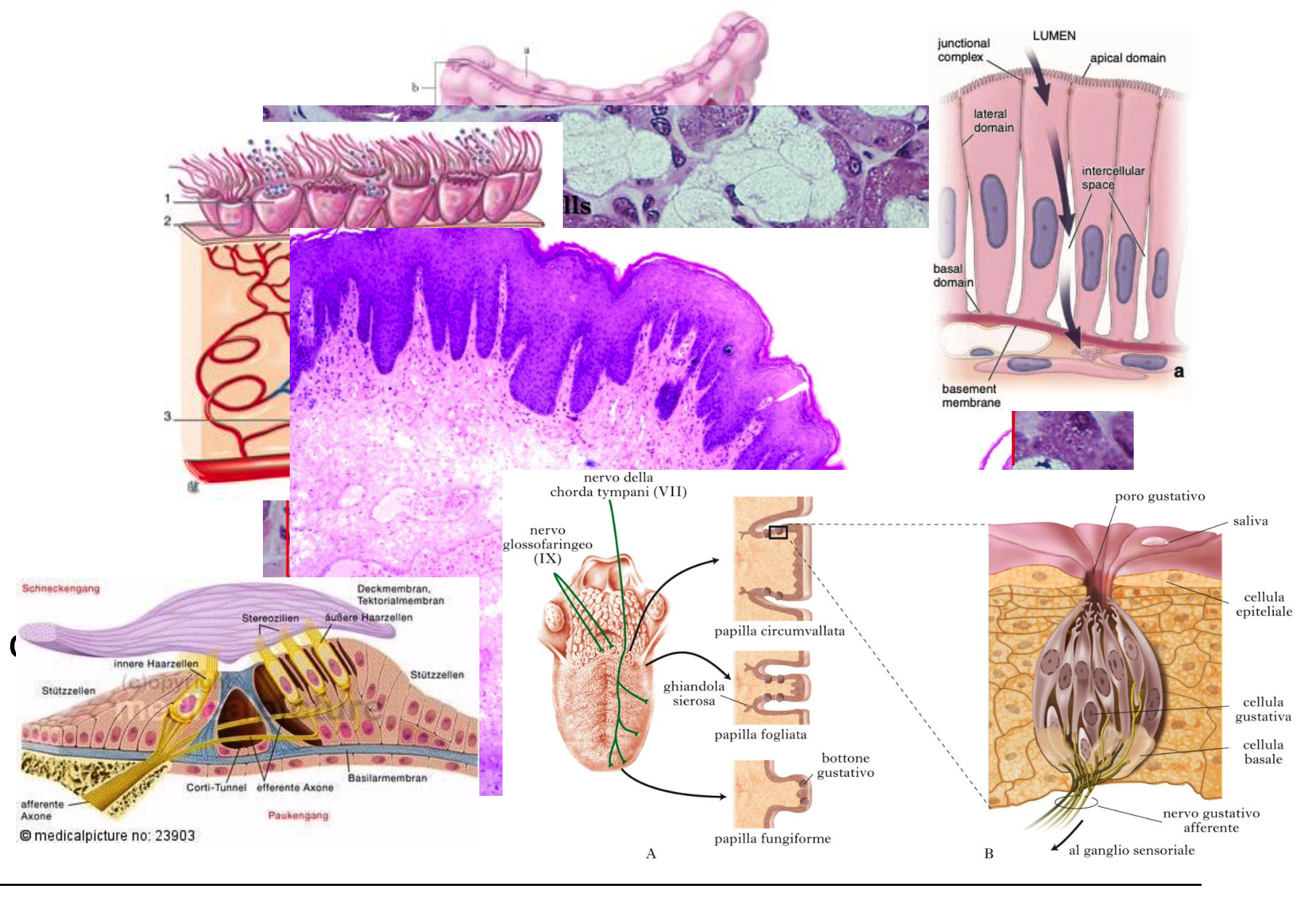
Receptor function
(taste of buds, retina, hair
MAIN FEATURES
Poor extracellular matrix
Numerous cell junctions
Presence of the basal lamina
Nourishment and oxygenation for diffusion
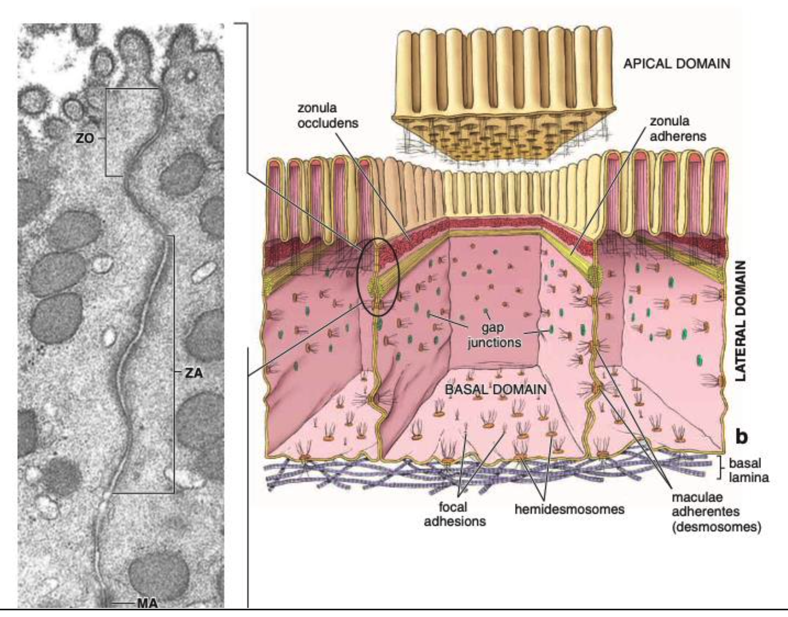
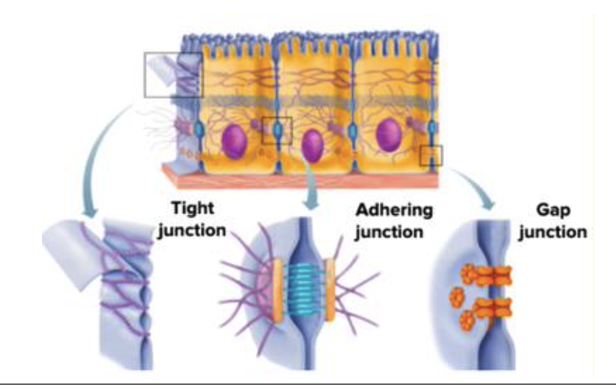
Name 3 CELL JUNCTIONS
Tight junctions, zonula occludens
Adhering Junctions:
Cell-Cellcontact:desmosomes, zonula and macula adhaerens
cell-matrix contact: hemidesmosomes, focal contacts and podosomes
Gap Junctions, chemical synapses
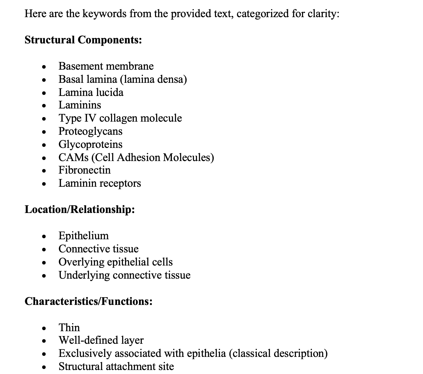
Describe THE BASEMENT MEMBRANE
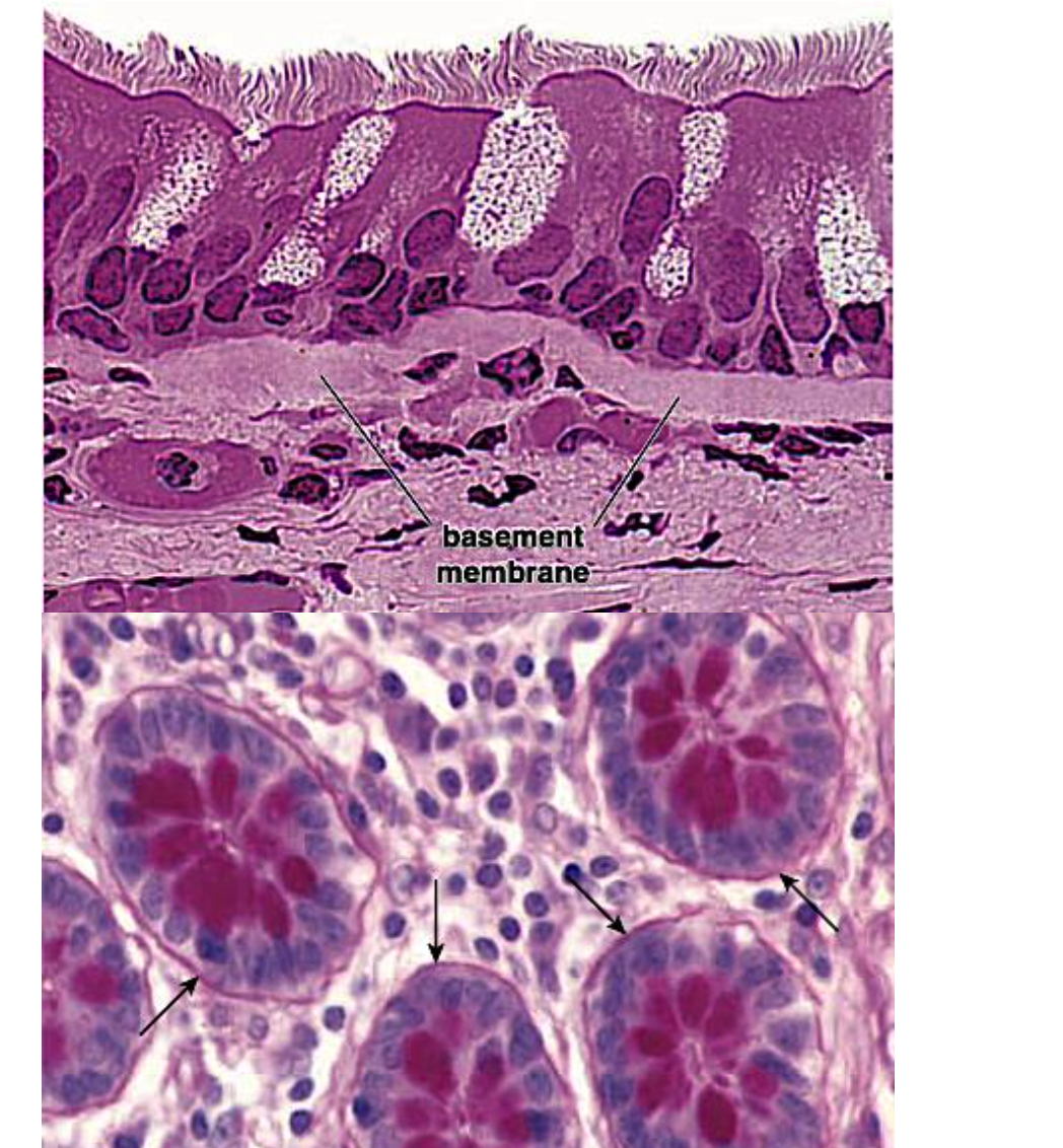
The basement membrane is a thin, well-defined layer between the epithelium and the connective tissue.
The Basement membrane is classically described as exclusively associated with epithelia
The basal lamina is the structural attachment site for overlying epithelial cells and underlying connective tissue. It is also called lamina densa, made of laminins, a type IV collagen molecule, and various associated proteoglycans and glycoproteins
Between the basal lamina and the cell is the lamina lucida, wich contains extracellular portions of CAMs, mainly fibronectin and laminin receptors.
CLASSIFICATION OF EPITHELIUM
Explain LINING EPITHELIUM
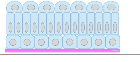
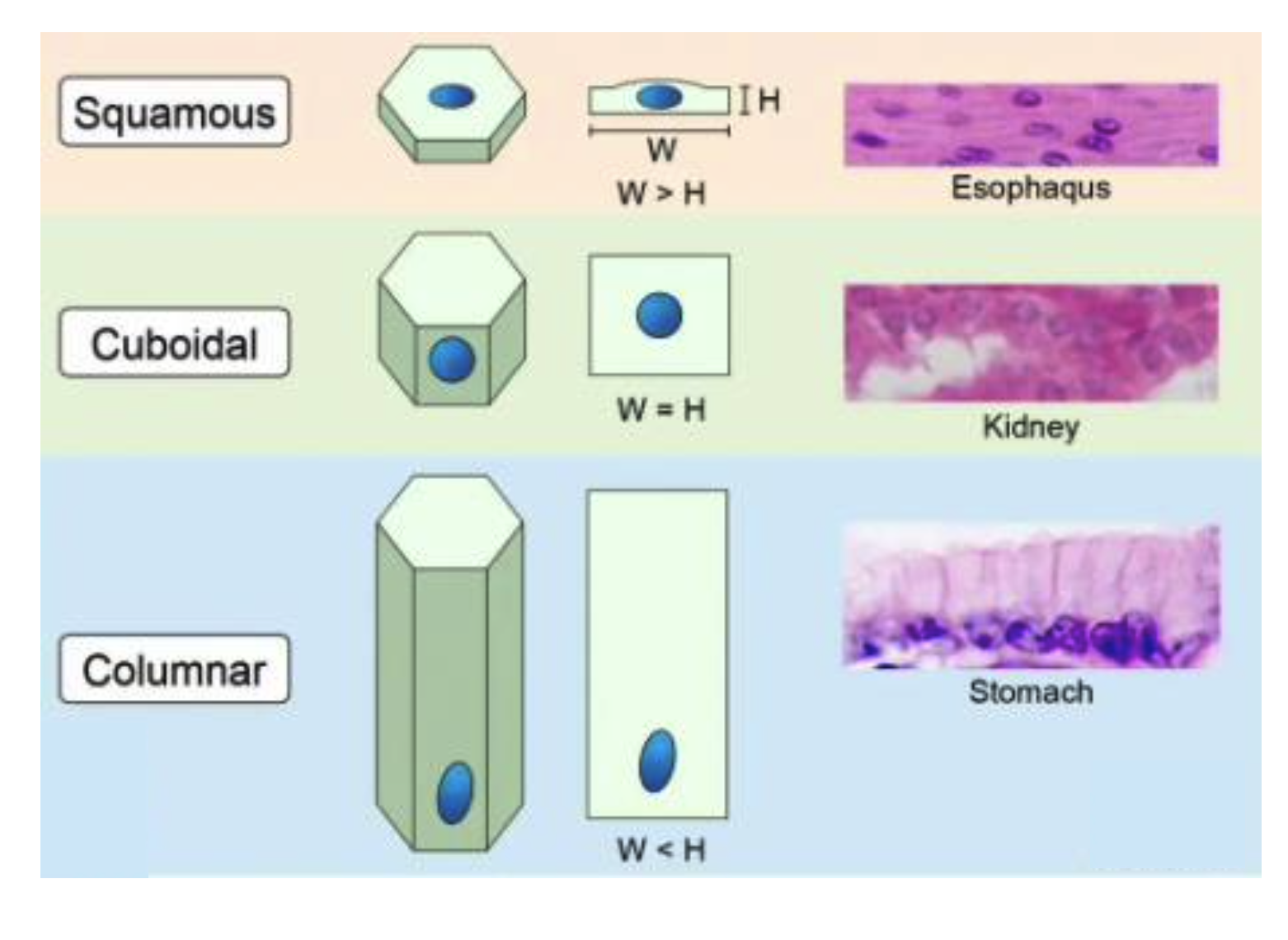
The lining epithelia are classified both on the number of cell layers and the shape of the cells
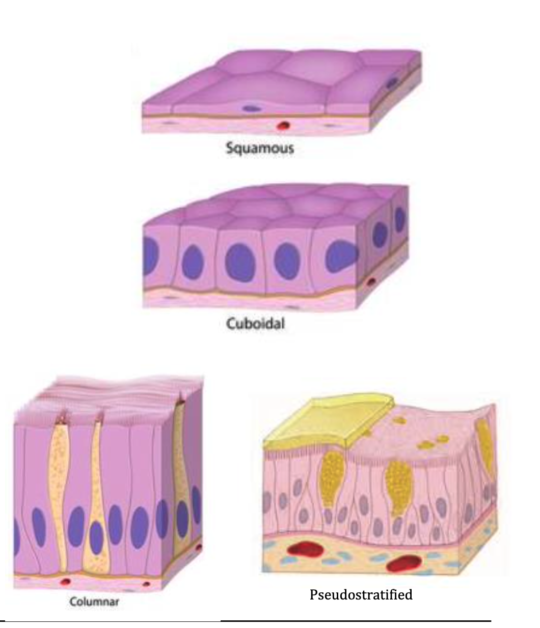
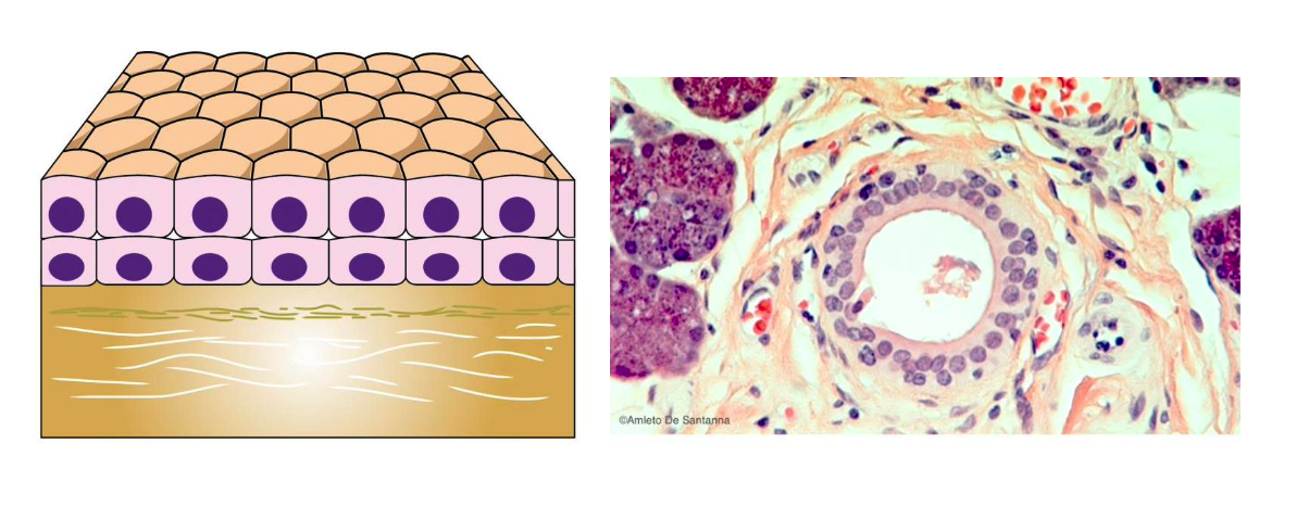
SIMPLE EPITHELIA
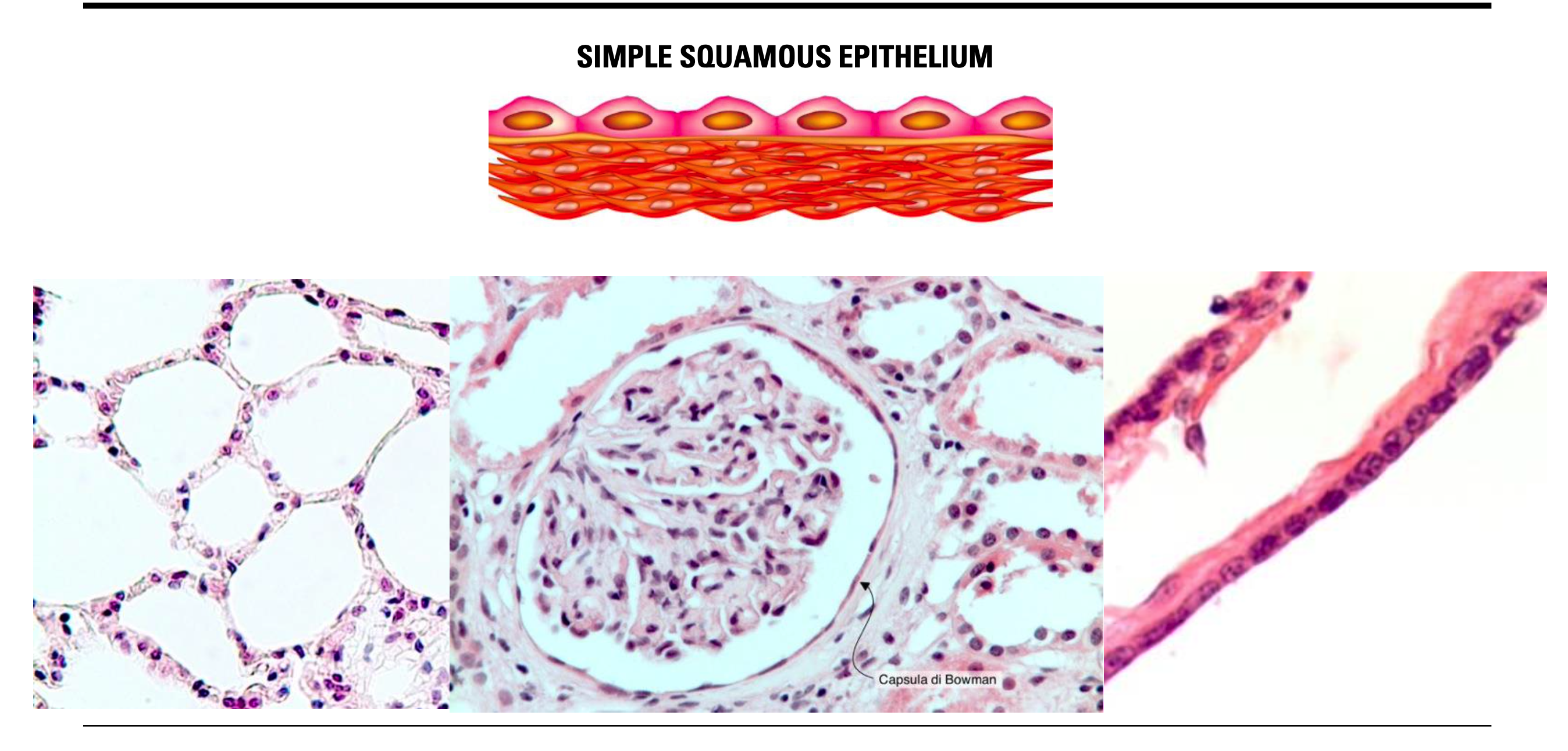
Squamous flat or floor cells (pulmonary alveoli, end of capillaries, Loop of henle)
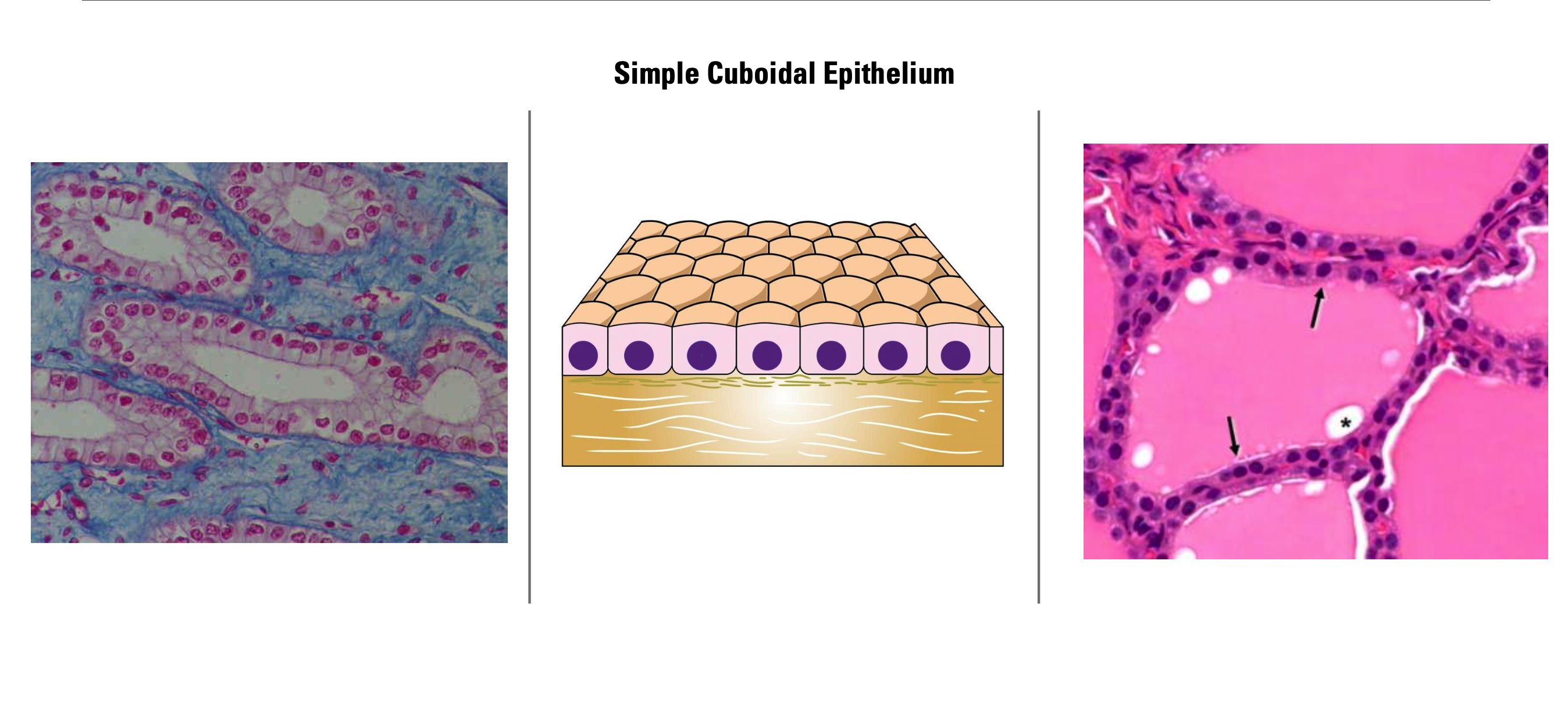
Cuboidal or isoprismatic (lining of the ovaries, collecting tub. of the kidney, ducts)
Columnar or bathyprismatic (digestive tract lining, gallbladder) Ciliated variant (uterine tube, small bronchi, paranasal sinuses)
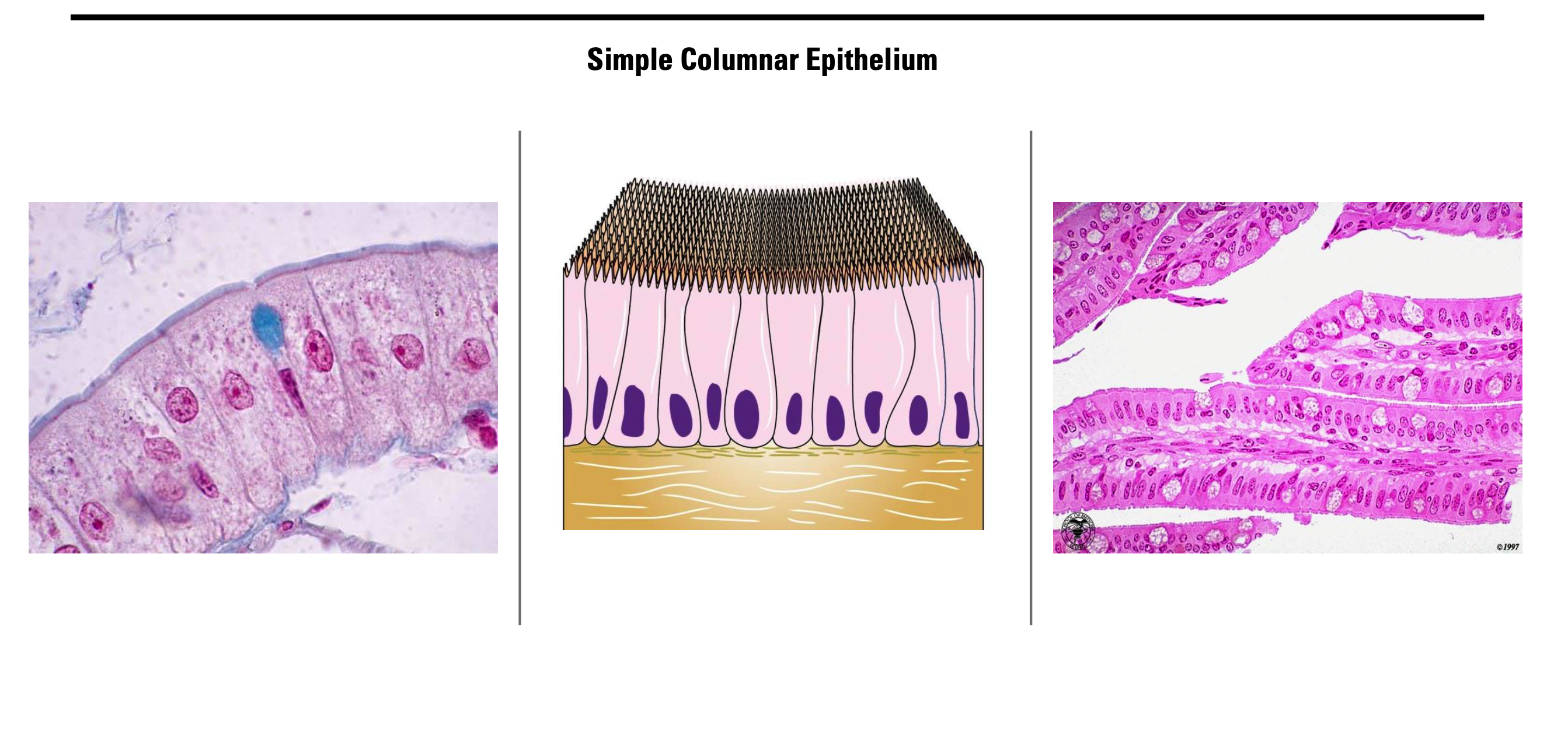
Pseudostratified (nasal cavity) non-ciliated variant (epididymis, male urethra,)
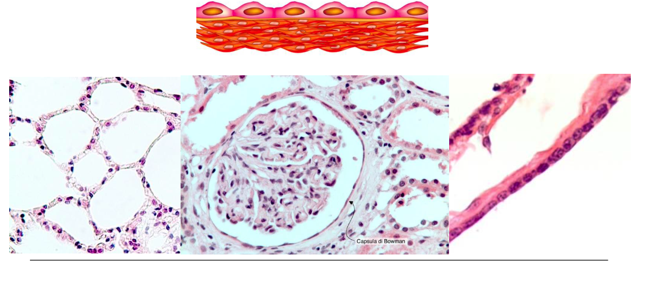
What’s this?
SIMPLE SQUAMOUS EPITHELIUM
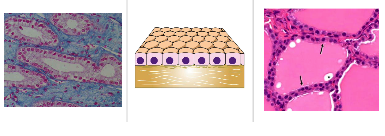
What’s this?
Simple Cuboidal Epithelium
What’s this?
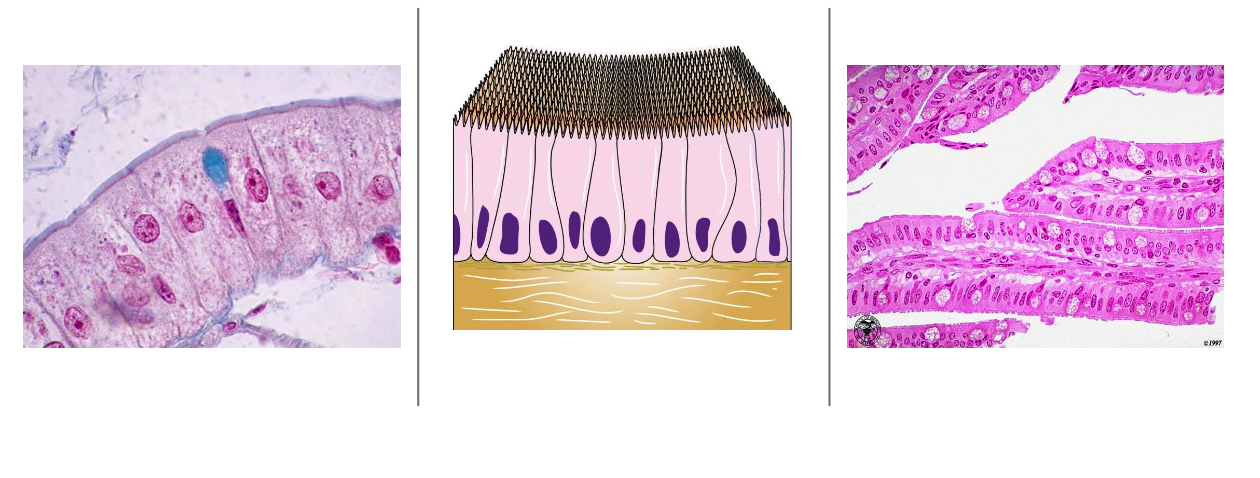
Simple Columnar Epithelium
What’s this?
Pseudostratified Epithelium
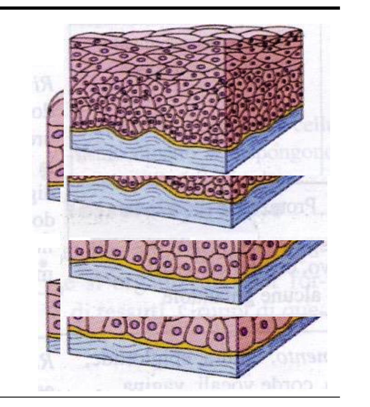
Describe STRATIFIED EPITHELIA
In the stratified epithelia, the shape of the cells usually vary from layer to layer:
Squamous nonkeratinized flat cells with nuclei (lips, cervix)
Keratinized squamous flat cells without nuclei (skin)
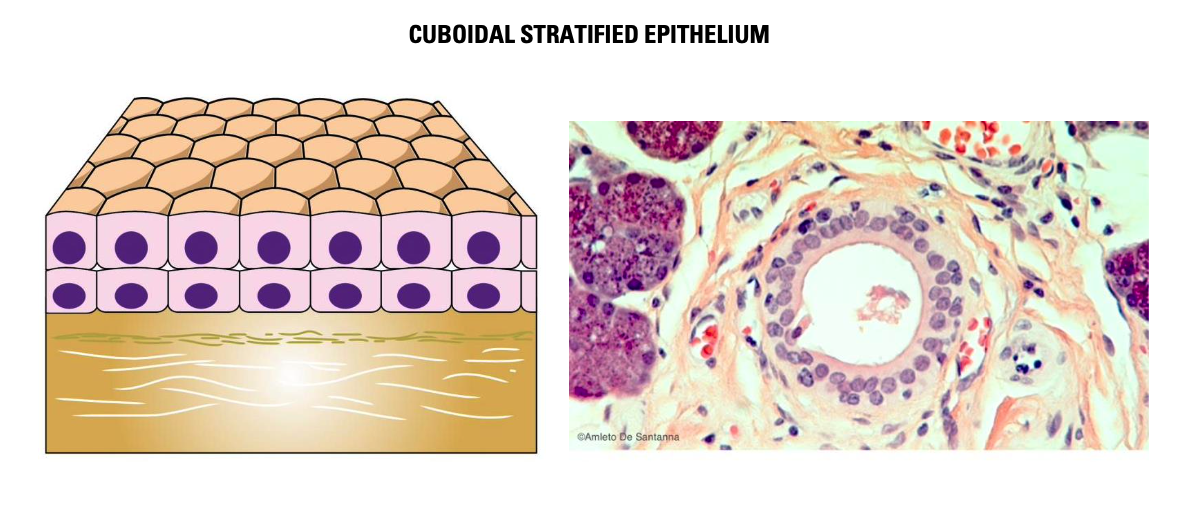
Cuboidal, cubic cells (gland ducts)
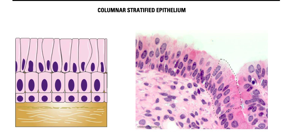
Columnar, columnar cells (conjunctiva, large
ducts)
Transitional dome-shaped cells when the organ is relaxed, flat when the organ is tense (urinary system)
What’s this?
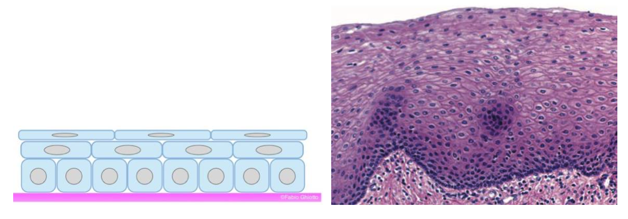
SQUAMOUS NONKERATINIZED EPITHELIUM
Only the top layer is in contact with the lumen. Not all the cells in this epithelium are squamous.
what’s this?
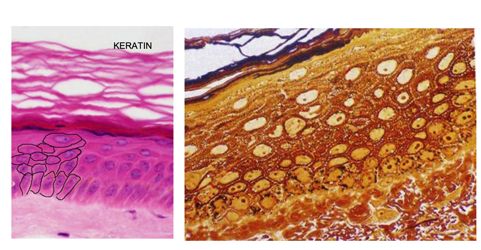
KERATINIZED SQUAMOUS EPITHELIUM
The squamous keratinized epithelium is characterized by the outer cell layes made of cells transformed into squamous lamellae. This layer is made of keratinocytes wich allow the protection of the inner layers.
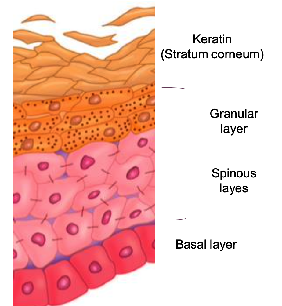
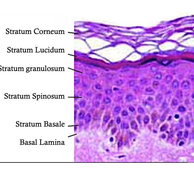
In the EPIDERMIS four distinct layers can be identified. Name them.
the stratum basale, also called the stratum germinativum because of the presence of mitotically active cells, the stem cells of the epidermis;
the stratum spinosum, also called the spinous layer or prickle cell layer because of the characteristic light microscopic appearance of short processes extending from cell to cell;
the stratum granulosum, which contains numerous intensely staining granules;
the stratum corneum, which is composed of keratinized cells.
Describe photo
The stratum basale is represented by a single layer of cells that rests on the basal lamina
The stratum spinosum is at least several cells thick. Keratinocytes in this layer are larger than those of the stratum basale.
The stratum granulosum is the most superficial layer of the nonkeratinized portion of the epidermis.
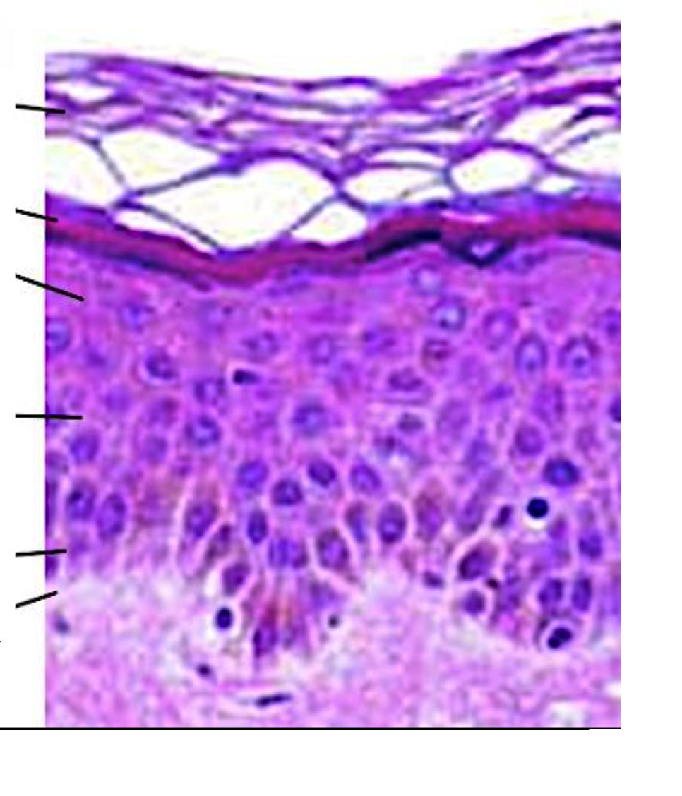
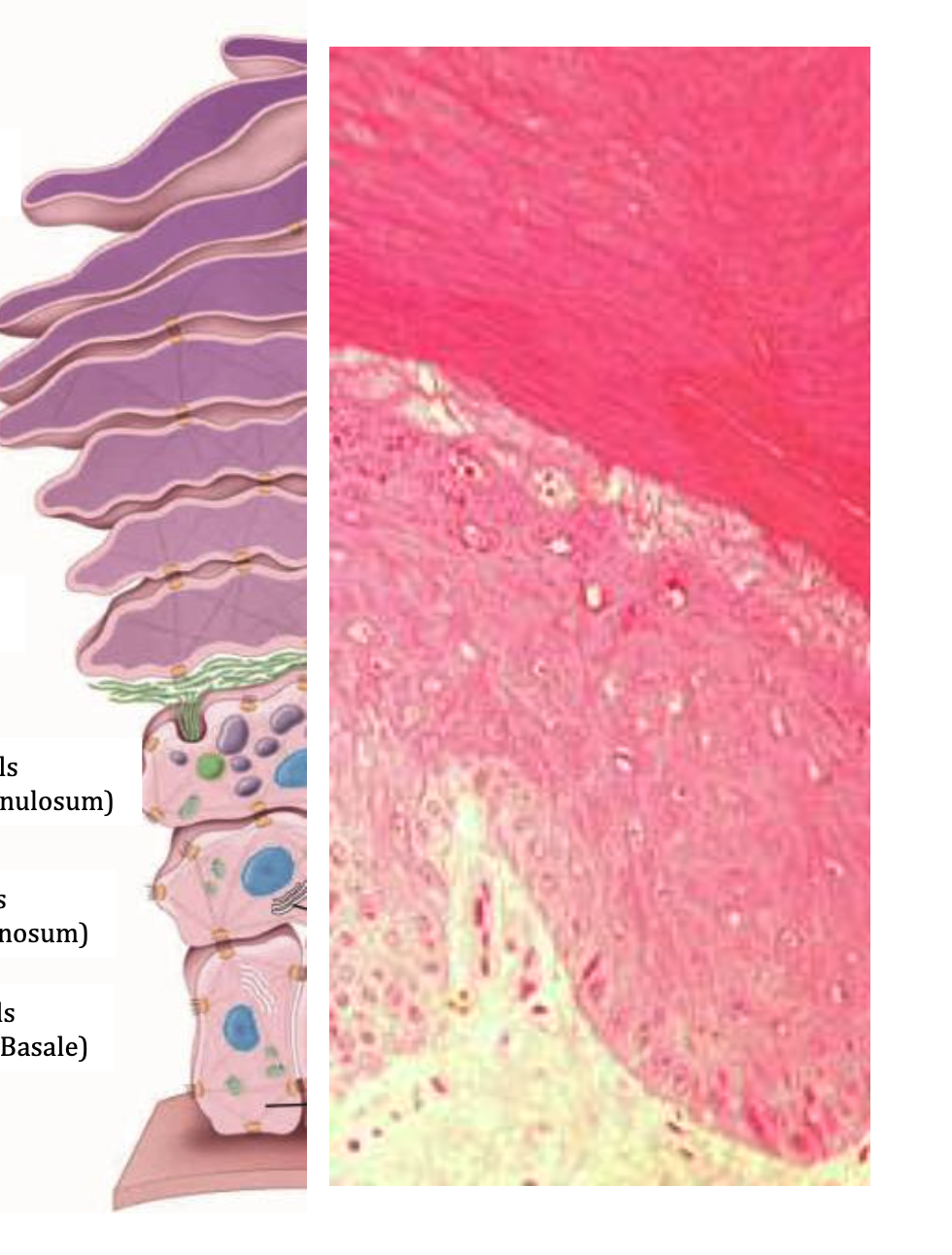
What can you see?
The stratum lucidum, considered a subdivision of the stratum corneum. Not distinguishable throughout the epidermis.
It is present in the epidermis of the palm of the hand and sole of the foot. It has flattened cells with large keratohyalin granules that contain Flaggrin.
Superimposed on the granular layer, it appears as a thin, clear and refractive line. The cells are devoid of granules

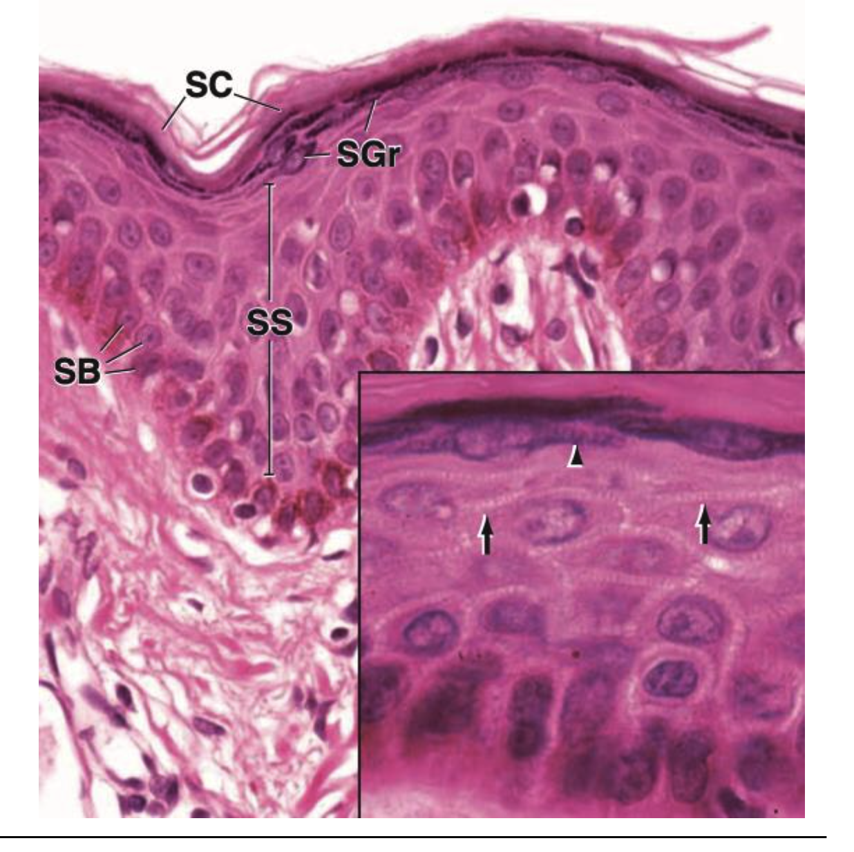
The cell layer that occupies the deepest location is the……?
The cell layer that occupies the deepest location is the stratum basale (SB).
Just above this is a layer several cells in thickness, the stratum spinosum (SS). It consists of cells that have spinous processes on their surface. These processes meet with spinous processes of neighboring cells and, together, appear as intercellular bridges.
The next layer is the stratum granulosum (SGr), whose cells contain keratohyalin granules. On the surface is the stratum corneum (SC) which consists of keratinized anucleated cells. The keratinized cells are flat and generally adhere to other cells above and below without evidence of cell boundaries.
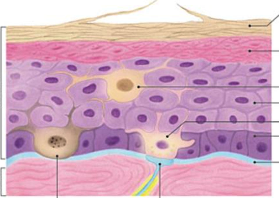
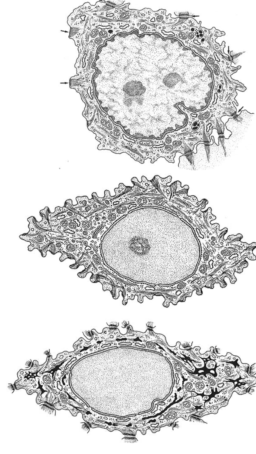
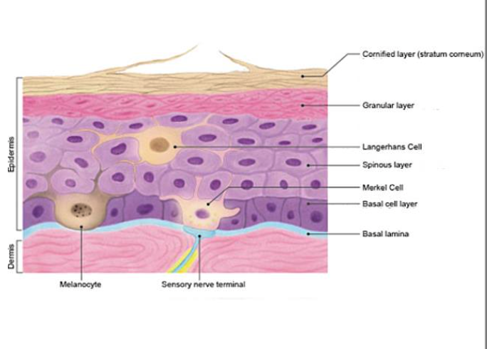
THE CELL OF THE EPIDERMIS
Keratinocytes are highly specialized epithelial cell
The melanocyte has long dendritic processes that contain accumulated melanosomes and extend between the cells of the epidermis, which are also visible on the electron micrograph.
The Langerhans’cell is a dendritic cell often confused with a melanocyte but is actually part of the mononuclear phagocytotic system and functions as an antigen-presenting cell of the immune system in the initiation of cutaneous hypersensitivity reactions (contact allergic dermatitis).
Merkel’s cells are the sensitive mechanoreceptor cells associated with sensory nerve endings.
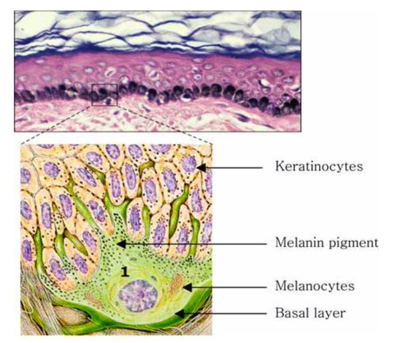
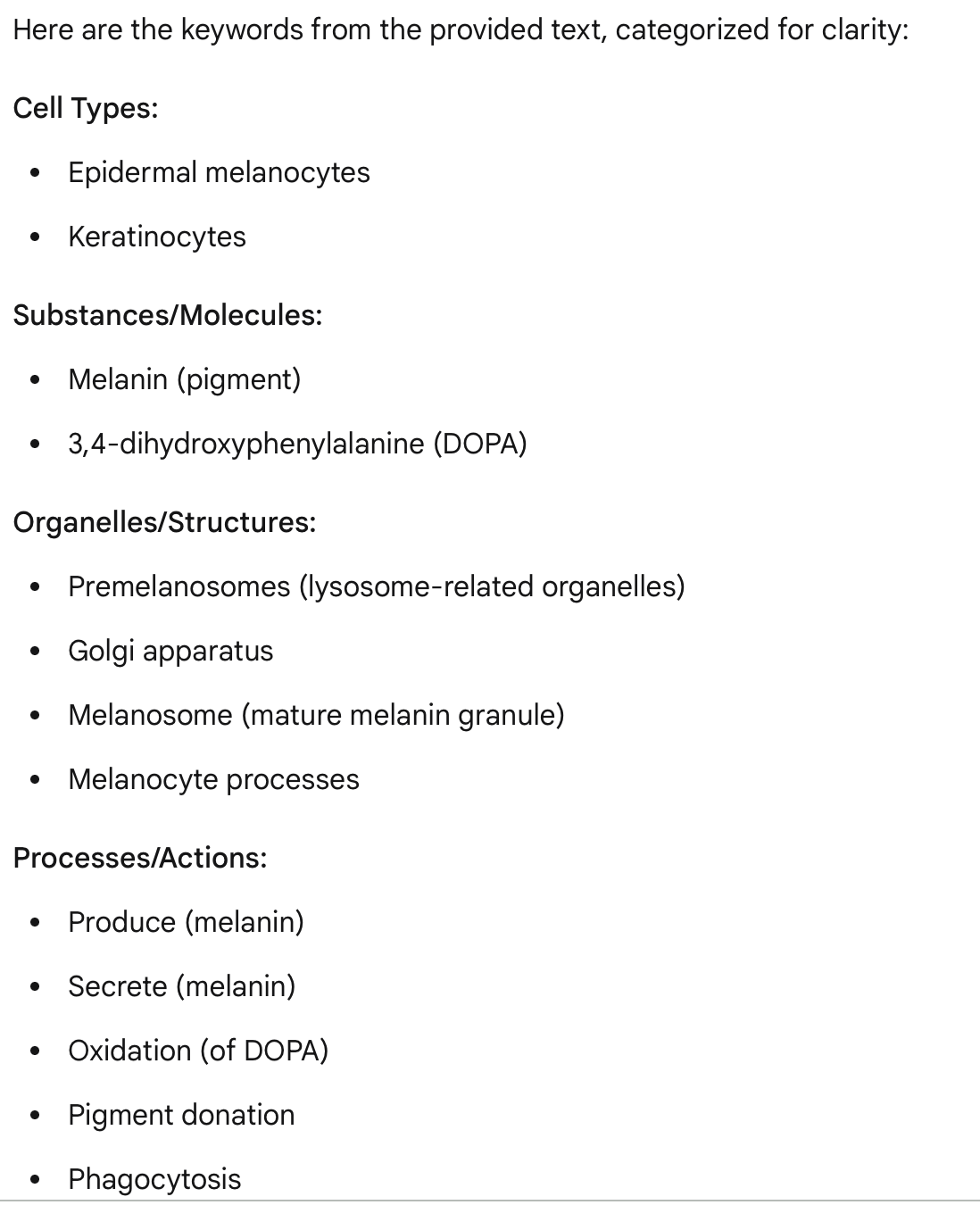
MELANOCYTE what is it?
The epidermal melanocytes produce and secrete the pigment melanin.
Melanin is produced by the oxidation of 3,4- dihydroxyphenylalanine (DOPA) , a reaction that occur in lysosome-related organelles called premelanosomes (from the Golgi apparatus).
As more melanin is produced the premelanosome becomes obscured until the mature melanin granule, the melanosome, is formed. Premelanosomes are concentrated near the Golgi apparatus while mature melanosomes most commonly in and at the ends of the processes. Melanosomes and their melanin contents are transferred to neighboring keratinocytes by pigment donation.
This process involves the phagocytosis of the tips of the melanocyte processes by keratinocytes.
What’s this?
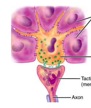
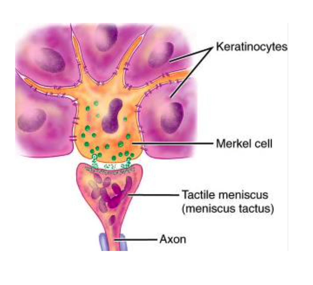
Merkel cell
They are dendritic cells located in the stratum basale. They are most abundant in skin where sensory perception is acute, such as the fingertips. Merkel cells are bound to adjoining keratinocytes by desmosomes and contain intermediate (keratin) filaments in their cytoplasm.
Merkel cells are best characterized by the presence of dense-cored neurosecretory granules that resemble those found in the adrenal medulla.
Merkel cells are closely associated with the expanded terminal bulb of afferent myelinated nerve fibers. The neuron terminal loses its Schwann cell covering and immediately penetrates the basal lamina, where it expands into a plate-like ending called a disc receptor that lies in close apposition to the base of the Merkel cell. The combination of the neuron and epidermal cell, called a Merkel corpuscle, is a sensitive mechanoreceptor.
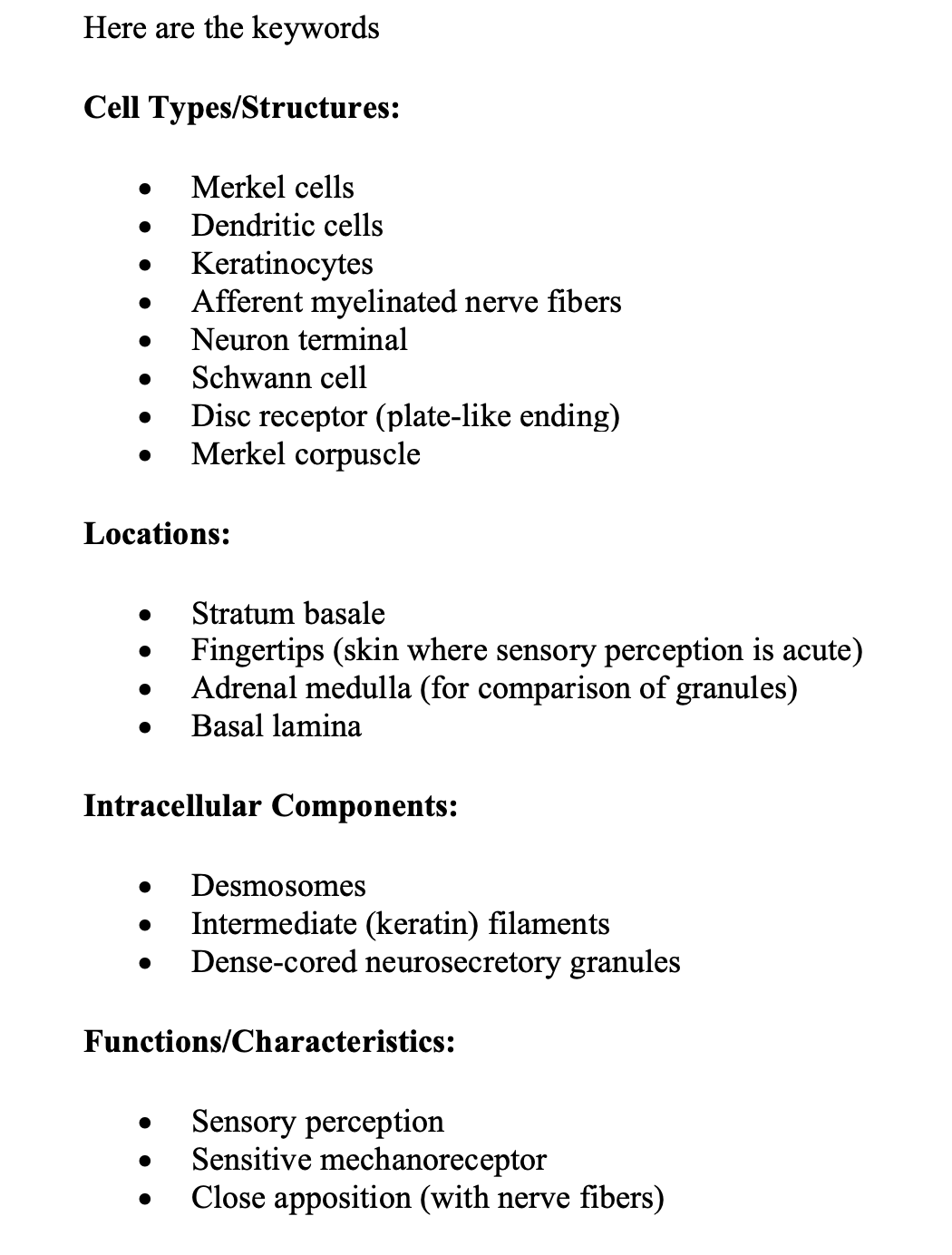
what are the key words you have to remember to describe Merkel cell?
describe image
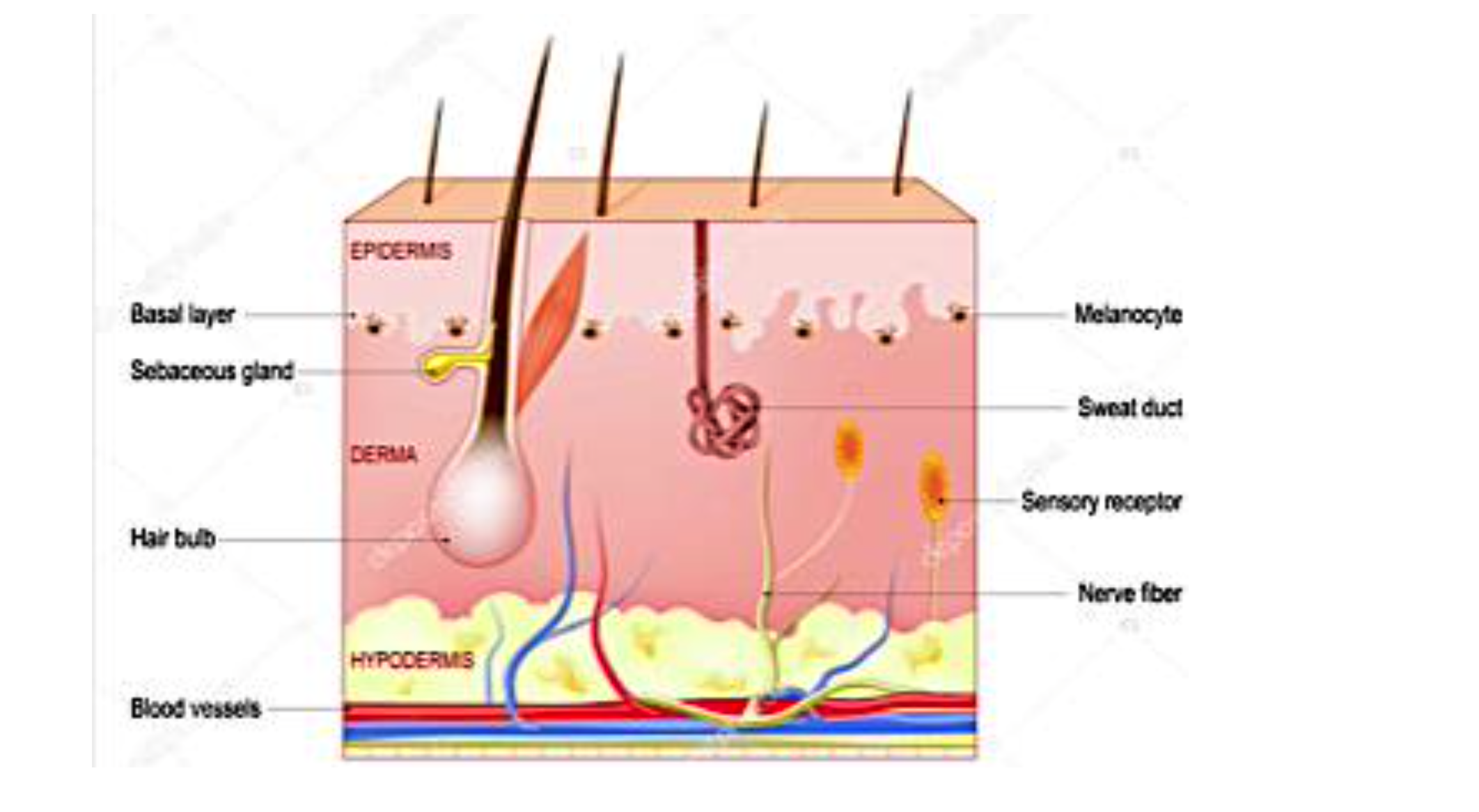
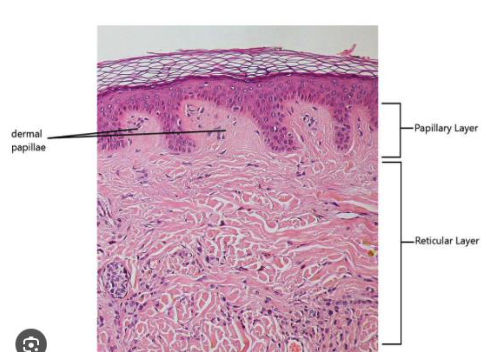
Describe the different layers of the dermis?
The dermis is composed of :
The papillary layer, the more superficial layer, consists of loose connective. The collagen fibers contains predominately type I and type III collagen molecules.
The papillary layer is relatively thin and includes the substance of the dermal papillae and dermal ridges.
It contains blood , nerve processes that either terminate in the dermis or penetrate the basal lamina to enter the epithelial compartment.
The reticular layer lies deep to the papillary layer.
It is characterized by thick, irregular bundles of mostly type I collagen and by coarser elastic fibers.
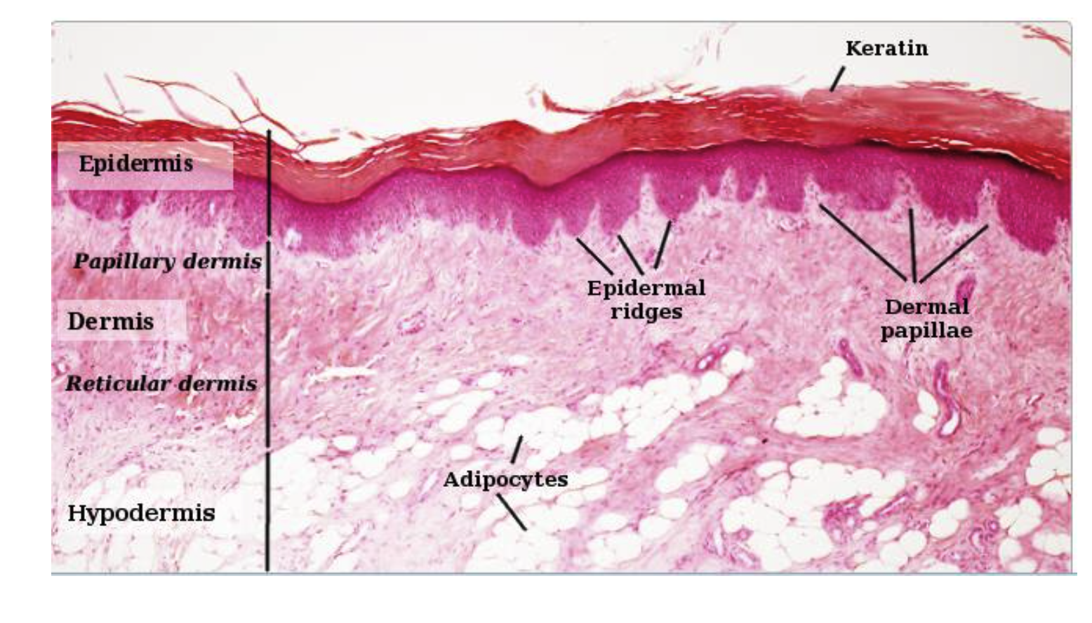
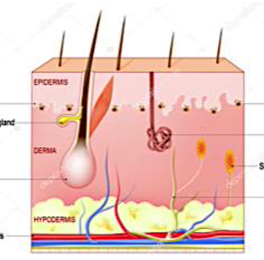
What is the hypodermis? and what is it made up of?
The hypodermis (subcutaneous layer).
Hypodermis is made of:
airolar layer,characterized by
adipose tissue
lamellar layer, in wich collagen is arranged in parallel bundles
What’s this?
CUBOIDAL STRATIFIED EPITHELIUM
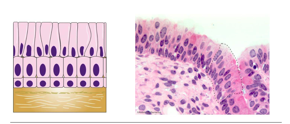
what’s this?
COLUMNAR STRATIFIED EPITHELIUM
what’s this?
THE TRANSITIONAL EPITHELIUM (UROTHELIUM)
is composed of at least three layers: 🇦
1. The superficial layer contains single or multinucleated large polyhedral cells that bulge into the lumen. They are frequently described as dome-shaped or umbrella cells because of their apical surface curvature.
2. The intermediate cell layer contains pear-shaped cells that are connected to each other and the overlying dome-shaped cells by desmosomes. The thickness of this layer varies with the state of the urinary tract expansion.
3. The basal cell layer consists of small cells containing a single nucleusthat rests on the basement membrane. This layer contains stem cells for the urothelium.