[ODCOP1] Ocular Dominance & Interpupillary Distance
 Call Kai
 Learn
Learn Practice Test
 Spaced Repetition
 Match
Match Flashcards
 Knowt Play
1/70

There's no tags or description
Looks like no tags are added yet.
Name | Mastery | Learn | Test | Matching | Spaced | Call with Kai | Chat |
|---|
No analytics yet
Send a link to your students to track their progress
71 Terms
Ocular Dominance
Ocular Dominance (OD)
Refers to the tendency of one eye to be preferentially used or relied upon by the visual system over the other
NOT simply which eye has better visual acuity
Two patients can have equal VA and still have clear ocular dominance
Ocular Dominance
Clinically relevant in:
Spectacle and contact lens prescribing (monovision; multifocal CLs)
Refractive surgery planning (LASIK monovision; LASIK full correction eye selection)
Binocular vision assessment
Sports performance and visual training
Sighting (Motor) Dominance
Sensory Dominance
2 clinically recognized types of ocular dominance
Sighting (Motor) Dominance
The eye used as the preferred “lead eye” when:
Aligning at a target
Aiming at a target
The eye instinctively used when:
Sighting through a small aperture
Pointing at a target
Sighting (Motor) Dominance
Also called motor dominance — it reflects the motor behavior of preferentially aligning the dominant eye with the visual axis to a target
It is the most commonly tested form of ocular dominance in routine optometry
Relatively stable and consistent within individuals — right eye dominance is indicated in approximately 71% of the population
Hole in a Card Test
Porta / Triangle / Ring Test (Miles Test)
Tests for sighting dominance
Hole in a Card Test
Purpose:
Identify the sighting dominant eye
Patient aligns a small aperture with a distant target
Both eyes remain open
Materials:
Card with a small circular hole
Approximately 3–5 cm diameter
Held at arm’s length
Or a commercially prepared target
Hole in a Card Test
Procedure
Ask the patient to hold a card with a central hole in both hands at full arm’s length.
Identify a small target at 4–6 m
Example:
A letter on the VA chart
A door handle
Instruct the patient:
“With both eyes open, look through the hole at the target.”
Observe which eye is naturally aligned with the hole and the target from in front of the patient.
Confirm by alternately occluding each eye
The dominant eye is the one that, when occluded, causes the target to disappear from the aperture.
Perform 3 consecutive trials
Record each result.
Recording
☐ OD dominant
☐ OS dominant
☐ Equal/Undetermined
Porta / Triangle / Ring Test
Purpose:
A second sighting dominance test using the patient's own hands
Should be consistent with the Hole in a Card result
Porta Test / Ring Test
Procedure
Ask the patient to:
Extend one arm
Form a ring using the thumb and index finger of that hand
Ask the patient to:
Look at a distant target through the ring
Keep both eyes open
Alternately occlude each eye
Dominant eye: keeps the target centered in the ring
Non-dominant eye: causes the target to shift out of the ring
Triangle / Miles Test
Procedure (Alternative)
Ask the patient to:
Extend both arms
Overlap the thumbs and index fingers
Form a small triangular aperture
Ask the patient to:
Look at the distant target through the triangle
Keep both eyes open
Ask the patient to:
Slowly bring the triangle toward their face
The aperture naturally drifts toward the dominant eye
Observe:
Which eye the aperture lands in front of
Recording
☐ OD dominant
☐ OS dominant
☐ Equal/Undetermined
Sensory Dominance
Refers to the eye to which the brain gives processing priority when both eyes are receiving information simultaneously
Sensory Dominance
It is the eye whose input the brain favors when the two eyes are in competition.
When the two eyes receive slightly different images:
The dominant eye's image is more strongly represented in conscious perception.
Important Distinction:
(BLANK) dominance and (BLANK) dominance do not always agree
In one study:
41% of participants showed a different dominant eye by (BLANK) testing compared to sighting testing.
Sensory Dominance
(BLANK) is Particularly Important for:
Monovision contact lens and spectacle fitting
Multifocal CL optical zone selection
Refractive surgery monovision planning
Binocular vision suppression assessment
+1.50 D Blur Test (Resistance to Blur Test)
Test for Sensory Dominance
+1.50 D Blur Test
Principle
The dominant eye resists blur more strongly
It is harder for the brain to suppress clear vision from the sensory dominant eye.
Placing a (BLANK) blur over the dominant eye:
Causes more noticeable visual disturbance than the same lens over the non-dominant eye.
The sensory test, also called “resistance to blur,” allows for the determination of the “strength” of ocular dominance.
+1.50 D Blur Test
Procedure
Ensure the patient is wearing their best distance correction
Or is uncorrected if emmetropic
Ask the patient to view a line of letters on the distance VA chart
Approximately 2 lines above their best VA
With both eyes open
Place a +1.50 D lens over the RIGHT eye only
Ask:
“Does the chart look blurred or different now?”
“On a scale of 1 to 5, how bothersome is this blur?”
Record response.
Remove the lens and place +1.50 D over the LEFT eye only
Repeat the same question.
Ask:
“Which was more noticeable — the blur over the right or the left?”
The eye over which the blur is more noticeable and bothersome =
Sensory dominant eye
Repeat 2–3 times for reliability.
Recording
☐ OD sensory dominant
☐ OS sensory dominant
☐ Equal
Clinical Significance of Ocular Dominance
Monovision correction
Multifocal CL fitting
Refractive surgery
Sports
Documentation
Interpupillary Distance
The distance, in millimetres (mm), between the centres of the two pupils
Phoropter / Trial Frame
Spectacle Lenses
2 Primary Clinical Reasons for Measuring PD
Incorrect PD measurement
optical centres misaligned → unwanted prismatic effect → symptoms including asthenopia, headaches, diplopia, and reduced VA — especially significant in high-powered prescriptions
Distance (Far) PD
Near PD
Intermediate PD
Monocular PD (half-PD)
Binocular PD
Types of Interpupillary Distance
Distance (Far) PD
Definition: Separation between visual axes of both eyes when fixating a distant object (at optical infinity)
When Used: Measured for distance spectacle prescription; routine for most patients
Near PD
Definition: Separation between visual axes at the plane of the spectacle lenses when fixating at the near working distance
When Used: Separation between visual axes at the plane of the spectacle lenses when fixating at the near working distance
Intermediate PD
Definition: Separation between visual axes at an arm's-length working distance (approximately 60–80 cm)
When Used: Measured for computer or intermediate spectacles; progressive lens fitting
Monocular PD (half-PD)
Definition: Distance from either the right OR left visual axis to the center of the bridge of the nose
When Used: Essential for progressive addition lens dispensing; asymmetric faces; any case where precise monocular centration is required
Binocular PD
Definition: Total distance PD measured across both eyes simultaneously
When Used: Routine screening; standard spectacle orders
Women
Distance PD most commonly 55–65 mm (Average Values)
Men
Distance PD most commonly 60–70 mm (Average Values)
Young children
PDs as low as 45 mm (Average Values)
Near PD at 40 cm
Near PD = Distance PD − 3–4 mm
The eyes converge inward for near tasks.
Therefore:
Distance PD is usually 3–4 mm greater than the near PD at 40 cm
Reason:
Both eyes rotate nasally (converge)
This brings the visual axes closer together when fixating on a near target.
63–65 mm
Average adult binocular distance PD
Interpupillary Distance
Clinical Importance
The PD is one of the most clinically critical measurements in spectacle dispensing
It determines exactly where the optical centre of each lens is positioned relative to the patient's visual axis
Correct Optical Centre Alignment
When the optical centre of a lens is correctly aligned with the visual axis:
Light passes through the lens without deviation
The patient experiences the full corrective effect of the prescription
There is no unwanted prismatic disturbance
Effect of Prescription Power
The higher the prescription power, the more clinically critical the PD measurement becomes
Low prescription:
A small PD error causes negligible prismatic deviation
High prescription:
The same PD error induces clinically significant prism
Prentice's Rule
governs the relationship between PD error and induced prism:
Δ = F × c
Where:
Δ = prism in prism dioptres
F = lens power in dioptres
c = decentration in centimetres
PD Too WIDE
(Optical centres are further apart than the patient's visual axes)
The patient is looking through the nasal portion of each lens
This induces base-out (BO) prism in each eye
An opposing prismatic force that fights the eyes' natural convergence
Symptoms:
Eyestrain and fatigue
Difficulty maintaining single vision at near
Headaches
Blurred or unstable vision
Diplopia in severe cases
Discomfort wearing the glasses for extended periods
Patient reporting:
“I keep wanting to take them off”
PD Too NARROW
(Optical centres are closer together than the patient's visual axes)
The patient is looking through the temporal portion of each lens
This induces base-in (BI) prism in each eye
A convergent prismatic force
Symptoms:
Blurred or double vision
Reduced depth perception
Spatial distortion
Headaches
Nausea in severe cases
Patient feeling:
“Pushed forward”
Perceiving the world as different in depth
Single vision distance
Single vision near (reading)
Bifocal / Multifocal
Progressive Addition Lenses (PALs)
High minus or plus lenses
Lens Type
Single vision distance
PD Error Consequence: Base-out or base-in prism depending on direction of error; generally tolerated in low Rx but symptomatic in high Rx
Why It Is Especially Critical: Higher power = more prism per mm of error (Prentice's Rule)
Single vision near (reading)
PD Error Consequence: Same as above PLUS misalignment at the specific convergence demand of near tasks — patient cannot sustain comfortable reading
Why It Is Especially Critical: Near tasks require sustained convergence — even small amounts of opposing base-out prism cause rapid fatigue
Bifocal / Multifocal
PD Error Consequence: If distance PD is wrong, both zones are misaligned; if near PD (seg inset) is wrong, reading zone is misaligned even if distance zone is correct
Why It Is Especially Critical: Must measure AND correctly incorporate BOTH distance and near PD
Progressive Addition Lenses (PALs)
PD Error Consequence: Monocular PD error causes the patient's visual axis to miss the progressive corridor entirely — they cannot find the clear intermediate or near zones
Why It Is Especially Critical: (BLANK) are the most PD-sensitive lens type — monocular PD must be precise to within ±0.5 mm ideally
High minus or plus lenses
PD Error Consequence: Significant induced prism from even small PD errors; also prismatic jump at the lens edge
Why It Is Especially Critical: Any prescription ≥ ±4.00 D warrants pupillometer measurement for maximum accuracy
Progressive Addition Lenses (PALs)
Provides a gradual change in lens power from distance → intermediate → near without visible segment lines.
Progressive corridor
The narrow, usable zone through which the lens power progressively changes from distance toward near.
Monocular PD
What measurement is used to position the progressive corridor during PAL manufacturing?
PD Is Measured Correctly
Light passes through the optical centre of each lens
Results in:
No unwanted prism
Full corrective effect of the prescription
Comfortable, stable vision
PD Is Measured Incorrectly
Consequences in order of severity:
Mild error in low Rx
Usually no noticeable symptoms
Patient adapts
Moderate error in medium Rx
Asthenopia
Headaches
Fatigue
Particularly at near
Significant error in high Rx
Diplopia
Spatial distortion
Nausea
Inability to wear the glasses
Any error in PAL
Loss of clear corridor
Swimming/rocking sensation
Progressive lens intolerance
Can occur even with a correct prescription
Anatomical PD Measurement (PD Ruler)
What It Measures
The distance between the anatomical centres of the pupils
The geometric centre of each pupil
Clinical Use
The most widely used method in routine clinical optometry worldwide
Requires only a millimetre ruler
No specialist equipment
Repeatability of anatomical binocular PD measurements is similar to that of a pupillometer
Anatomical PD Measurement (PD Ruler)
Advantages: quick; inexpensive; no special equipment; usable by any trained clinician; reliable for binocular PD in most patients
Disadvantages: slightly less accurate for monocular PDs than a pupillometer; susceptible to parallax error (particularly when the examiner's PD differs significantly from the patient's); requires the examiner to be binocular
Between the center of 2 pupils
Right temporal pupil margin to left nasal pupil margin
Right temporal limbus to left nasal limbus
Anatomical PD Procedure
Monocular PD Measurement
Why (BLANK) Matters
Many patients have asymmetric faces
The nose bridge is not perfectly centered
This makes the right and left monocular PDs unequal
Progressive addition lens dispensing requires monocular PDs
If binocular PD is used for both lenses in an asymmetric face:
The optical centres will be misaligned
Monocular PD Measurement
Procedure (Elliott, 2007)
Use the centre of the pupil as the reference point
Not the margin
Align the zero mark with the centre of the patient's RIGHT pupil
Read the distance to the centre of the nose bridge
This is the right monocular PD (OD PD)
To find the LEFT monocular PD:
Subtract the OD monocular PD from the total binocular distance PD
Monocular PD Measurement
Formula
OS monocular PD = Total distance PD − OD monocular PD
Most Common Errors
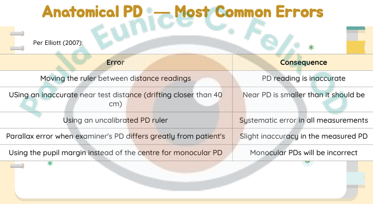
Monocular PD Measurement
Catoptric / Physiologic PD (monocular)
Photographic PD Measurement
ANATOMICAL PD METHODS
Catoptric / Physiologic PD (Monocular)
What It Measures
Measures the distance between the corneal light reflexes
Rather than the anatomical pupil centres
Measures the visual axes (physiologic PD)
Rather than the lines of sight (anatomical PD)
Pupillometer
The (BLANK) uses a light source directed at the cornea
The corneal reflex of each eye is aligned with reference markers in the instrument
Catoptric Method
Comes from the Greek katoptron, meaning mirror
Refers to the use of corneal reflections as the measurement reference
Physiologic PD
Distance between the two principal corneal reflexes
This locates the visual axes of the eyes
Anatomical PD
Distance between the anatomical pupil centres
This locates the lines of sight (optical axes)
Pupillometer
How it Works
A light source illuminates both corneas simultaneously.
The examiner aligns reference markers with the corneal reflexes of each eye using a single viewing aperture.
Monocular measurement
No parallax error
Digital pupillometers give a direct readout of:
Binocular PD
Monocular PDs
Advantages
More accurate for monocular PD measurements than the anatomical method (Holland & Siderov, 1999).
Particularly beneficial for progressive addition lens dispensing
No parallax error
Examiner uses one eye only.
Quick and simple
Can be performed by a clinical assistant.
Considered the gold standard instrument for routine IPD measurement (Chu, 2024).
Limitations / Important Considerations
The PD measured with a corneal reflection pupillometer is typically 0.5–1.0 mm smaller than the anatomical PD.
Because it measures the physiological PD (visual axes), not the anatomical PD (optical axes).
Inaccuracies can occur if the pupillometer sits higher or lower on the bridge than the intended spectacle frame.
Photographic PD Measurement
What It Measures
The inter-pupillary distance (IPD) estimated from a calibrated photograph of the patient's face.
How It Works
Take a calibrated photograph of the patient's face
A known reference scale is placed in the same plane as the patient's pupils during the photograph.
Examples:
Ruler
Reference card of known width
Frame dimensions
Measure the pupil-to-pupil distance
The center-to-center pupil distance is measured from the photograph.
Calculate the scaling factor
The known reference scale is used to convert the measured pixel or millimetre distance on the photograph into the patient's true PD in millimetres.
Photographic PD
PD estimated from a calibrated facial photograph using a known reference scale.
Anatomical
Instrument: PD ruler (mm ruler)
Measures: Pupil centres; lines of sight
Clinical Use: Routine binocular PD for most patients
Key Limitation: Parallax error; less accurate for monocular PD
Catoptric / Physiologic
Instrument: Pupillometer (corneal reflex)
Measures: Corneal light reflexes; visual axes
Clinical Use: Monocular PD; progressive lenses; high Rx
Key Limitation: PD ~0.5–1.0 mm smaller than anatomical; instrument positioning Positioning of the corneal reflex (light)
Photographic
Instrument: Camera / smartphone app
Measures: Pupil centres from image
Clinical Use: Online ordering; remote measurement
Key Limitation: Lens distortion; reference scale accuracy; not reliable for high Rx
Gerstmann's Three-Quarters Rule
What It Is
A clinical calculation rule for determining the near segment inset per eye and therefore the near PD when:
Direct near PD measurement is not possible, or
The working distance is non-standard.
The Rule
For every diopter of dioptric demand, the optical centre of each reading lens or the geometrical centre of each bifocal addition should be inset 0.75 mm (three-quarters of a mm) from the distance optical centre.
Segment Inset per Eye
(Gerstmann's Three-Quarters Rule)

Dioptric Demand
(Gerstmann's Three-Quarters Rule)

Near PD
(Gerstmann's Three-Quarters Rule)

Recording PD
PD: distance PD / ne ar PD (in mm) (binocular)