Complex Concepts in Musculoskeletal Function and Alterations
 Call Kai
 Learn
Learn Practice Test
 Spaced Repetition
 Match
Match Flashcards
 Knowt Play
1/85

There's no tags or description
Looks like no tags are added yet.
Name | Mastery | Learn | Test | Matching | Spaced | Call with Kai | Chat |
|---|
No analytics yet
Send a link to your students to track their progress
86 Terms
what is the musculoskeletal systems
protects vital organs
provides body support and structure
allows movement
holds bones together at joints
produces heat (through contractions)
helps blood return to the heart
stores important minerals (calcium, phospohorus, magnesium, fluoride in bone)
acts as a reservoir for immature blood cells
produces bblood cells in bone
Bone structure and tissue - shape + construction & 2 primary tissue types
shape and construction
determined by its specific function and the physicial forces exerted on it
two primary tissue types
cancellous (trabecular) or cortical (compact) bone tissues
4 types of bone cells
osteoprogenitor cells: stem cells that replace needed bone cells
osteoblasts: cells responsible for building new bone tissue
osteocytes: mature cells that maintain bone tissue
osteoclasts: cells that break down old bone tissue
Hormones involved with bones: Active Vitamin D (Calcitriol)
Vitamin D incr. calcium in the blood by helping the small intestine absorb more calcium from food for the body
Also helps calcium and phosphate get deposited into the bone matrix to which maintains bone density and strength
Hormones involved with bones: Parathyroid (PTH)
Is released when blood calcium is low : it does this by
Pulling calcium out the bones
helping the kidneys keep calcium
incr. calcium absorption in the intestines
lowering phosphate levels (by secretion of kidneys)
Hormones involved with bones: Calcitonin
Lowers blood calcium by:
Inhibiting osteoclasts, reducing bone breakdown and calcium release from bone
Helps the kidneys to reabsorb more calcium in the urine
Moves calcium into bone
What are joints
point btwn 2 or more bones
Primary function: permits mobility, allows fluid skeletal movement, and provides structural support
Key components
Ligaments: fibrous connective tissue bands that hold bone to bone
Bursae: fluid filled sacs that facilitate movement and reduce friction
Joints: synarthoses (solid)
Immovable structural connections securely joined by solid cartilage or fibrous connective tissue (e.g. skull sutures)
joints: diarthrodial (synovial)
highly movable joints encapsulated with a fluid filled synovial cavity and protexted by smooth articular cartilage
how muscles attach?
tendons and aponeurosis
tendons and their use?
a strong band of connective tissue that attaches muscle to bone
when a muscle contrcats, the tendon pulls on the bone to create movement at a joint
aponeurosis and its uses?
wide flat sheet of connective tissue
i works similarly to a tendon but is not cord like
connects muscle to bone or muscle to muscle
eample: abdominal bones
isometric tone
constant length
The length of the muscles remains constant but the force generate is incr.
Eample: pushing against an immovable wall
isotonic tone
shortening muscle
characterized by shortening of the muscle with no incr. in tension within the muscle
Eample: fleing the forearm
Normal activities of muscle tone and contraction
most muscle movements are a combination of both contraction
patient problems of musxle tone and contraction
when issues occur, tone presents as
Flaccid
Spastic
Atonic
age related changes in musculoskeletal systeem: bones and hormones
numerous metabolic changes, including reduction of estrogen, testosterone and growth hormone, and related decreased physicial activity, contribute to osteoporosis
age related changes in musculoskeletal systeem: cartilage
cartilage degenerates in weight bearing areas and heals poorly
age related changes in musculoskeletal systeem: ligaments
become weak, and this contributes to the development of osteoarthritis (degenerative joint disease)
age related changes in musculoskeletal systeem: muscles
deacr. in muscle mass and ROM can cause functoinal impairment in older individuals
Good newurovascular assessment
newurovascular status is important for the nurse to perform frequent neurovascular assessments of pts with musculoskeletal disorders. it is usually called CSM (circulation, sensation, movement)
Pain
pallor
poikilothermia
pulses (includes capilarry refill)
Diagnosis studies
ray - initial structual imaing
CT scan - detailed bone and joint imaging
Labs - Ca, Vit. D, Phosphorus, Hormones, CBC, ESR, CK, Myoglobin
MRI
Anthrography
Arthroscopy
DEXA Scan
What is bursitis
Inflammation of bursa, a small fluid filled sac that acts as a cushion to reduce friction between bones, tendons, and muscles near joints
what is tendonitis
is the irritation or inflammation of a tendon, the thick fibrous cords that attach muscle to bone often caused by repetitive strain or ouveruse
clinical manifestations of bursitis and tendonitis
localized pain: pain that intensifies with movement or diect pressure on the join
swelling and redness: visible localized swelling, warmth, and erythema around the affected region
stiffness: reduced rangge of motion often descriibed as a “locked” or severely stiff joint
treatment and care of bursitis and tendonitis
RICE: Rest, Ice (20 min sessions_, compressions, and elevations
Medical therapy: NSAIDS for pain, corticosteroid inections for advamced swelling
what is carpal tunnel syndrome?
an entrapment neuropathy where the median nerve at the wrist is compressed.
causes include: thickened fleor tendon sheath, skeletal encroachment, edema, or a soft tissue mass
what is median nerve function?
responsible for forearm movement and providing feeling to the fingers and hands
risk factors of carpal tunnel
repetitive movement
pregnancy
obesity
hypothyroidism
age and trauma
signs and symptoms of carpal tunnel
numbness/tingling
wrist and hanf pain
loss of control
burning sensation
diagnostic tools for carpal tunnel
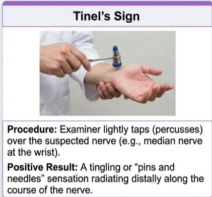
tinels ssign
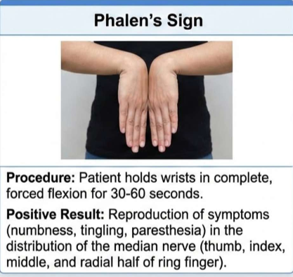
phalen’s test
ray
electromyography
ultrasound
MRI
treatment plan of carpal tunnel
wrist splints: worn at night for neutral positioning
anti inflammatories
steiroid injections
activity changes
alternatives
release surgery
phalens sign
tinels sign
Always assess first: injury to the musculoskeletal system requies elevated of:
damaged area assessment of the fracture site
underlying structures: muscles, blood vessels, and nerves
secondary organs: check for trauma beneath the bone
Always assess first: injury to the musculoskeletal system requies elevated of: treatment and education
treatment: provide support until healingn is complete
education: essential for optimal patient outcomes
common soft tissue injuries: contusion
injurt from blunt force (blow, kick, fall) causing bleeidnf into soft tissues (ecchymosis/brusing)
common soft tissue injuries: strain
a pulled muscle affecting a musculotendinous due to overuse or overstretching
common soft tissue injuries: sprain
a ligamentous injury often caused by traua or a wrenching/twisting motion
managemtn and treatment of common soft tissue injuries
history: what were you doing
Phsycial: compare and contrast areas of edema, ecchymosis, tenderness, and abnormal motion
diagnostics: use neurovascular assessment RAY, ultrasound, or CT/MRI later
may require brace or physical therapy
RICE
treatment for musculoskeletal
some experts advocating for movement based approaches like “Police”
protet, optimal loading, ice, compression, elevation
some say excessice use of ice suppresses inflammatory response which is necessary for repair
some say complete rest after injury can delay healing and that controlled mild movement might promote recovery
risk factors of fractures
6.3M+ incidents
All ages young and older
Fragility fractues
cushings syndorme
osteoporosis and neoplasms
anorexia and postmenopausal status
type of fractures structural integrity
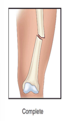
complete: broken all the way through
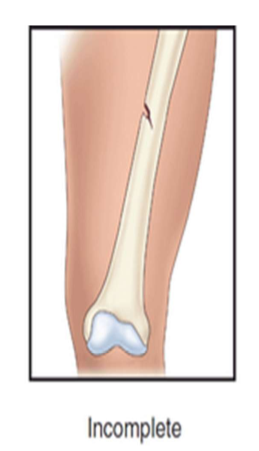
incomplete: partially broken
example: greenstick fracture
type of fractures structural integrity: clinical classifications
oblique: diagonal break (45-60 degrees)
comminuted: multiple fragments
closed: skin remains intact
open: skin broken (infection risk)
complete fracture
incomplete fracture
closed fracture simple nondisplaced

closed simple displaced fracture

what are signs and ysmptoms of a fracture - clinical presentation
pain, loss of function, swelling, deformity, shortening (hip), crepitus (hear it)
what are signs and ysmptoms of a fracture - systemici impact
adjacent structures are often affected, resulting in
soft tissue edema and hemorrhage
ptoential joitn dislocations
ruptured tendons and severed nerves
damaged blood vessels
immediate care of fractures
stabiize patient: ensure stability and rule out other vascular injuries
immobilize secure the affected body part immediately
good neurovascular checks (before and after)
wound care
debridement or cover: if open cover to prevent infection
medical protocoles for fractures
pain management
may ice or elevate, depend on the MD
If it is open:
cover
antibiotics
look at other stuff (trauma)
treatment options of fractures
closed reduction: setting a bone without surgery; requires a cast or splint
ORIF: open reduction with internal fiation using hardware
traction: may be required with ORIF or closed reduction
open fractures lead to infections
fracture management: closed reduction
set (reduce) a broken bone without surgery
nurse role: assist rhe HCP during the procedure
patient needs: sedation is required during the process
Post-procedure: cast application and neurovascular “good checks”
additional needs: may require temporary traction
fracture management: open reduction (ORIF)
internal fiation via surgical intervention
utilizes hardware: plates and screws to secure bone
major goals of fracture management
treatment knowledge
understand annd follow the treatment regimen
pain relief
monitor continuously; address persistent pain
physical mobility
improve ROM and use SCDs effectively
self care mastery
achieve maimum possible level of self care
tissue healing
heal trauma lacerations and abrasions
neurovascular function
maintain adequate system function
complication prevention
ensure complete absence of slinical complications
compartment syndrome
pain out of proportion to injury
most common: lower leg and forearm
6 Ps: pain, pressure, paresthesia, pallor, paralysis, pulselessness (late)
risk can lead to tissue necrosis
t: prompt recognition, remove cast, or fasciotomy
fat embolism syndrome
mechanism: fat globules released from bone marrow
high risk: long bone fractures (24-48hr post injury)
S/S: AMS, petecial rash, hypotension
Respiratory distress: tachyppnea, tachycardia, crackles
T: IV hydration, support respiratory status (O2/intubation)
other complications of fractures
Healing issues
delayed union, malunion, and nonunion
Vascular risks
Venous thromboemboli (DVT/PE)
Coagulation disorders
Disseminated INtravascular Coagulation
Device sensitivity
Reaction to internal/eternal devices
Pelvic fractures and intability: stable fractures
do not break the pelvc ring: just need rest and pain medication
Pelvic fractures and intability: unstable fractures
may result in rotational instability
Pelvic fractures and intability: severe cases
life threatening complications and systemic problems
pelvic fractures clinical manifestations
phsyical signs (Ecchymosis and local edema)
localized tendernedd
symphysis pubis, iliac spines, sacrum, or coccy)
sensory changes
Numbness, tingling of pubis, genitals, anf thighs
Mobility impact
inability to bear weight, pain with movement
diagnostic and treatment of pelvic fracture
Good neuro assessment
No pulse of lower etremities means blood pool closing up flow from arteries and veins
because of the fracture may not be able to void
look at urine
think of what organs and other vascular that is around pelvic for potential complications, it is a very vascular area
stabilize pelvic w pelvic binder
prevent or minimize compications such as bleeding and shock
once pt. is stable then think about fiing the pelvic
use of FAST (focused abdominal ST - ultrasound over pelvic cavity)
CT/CTA scan
retrograde cystourethrogram
importance of hip fracture ? in elederly
risk of falls
disease processes
meds
area rugs, loose carpeting, inadequate lighting, pets
S/S of hip fracture
acute pain (groin pain. hip or knee)
loss of function
leg is shortened, abducted and rotated
swelling, discoloration
crepitu
management of hip fracture
good history and physical (good neurovascular checks)
RAY
may need CT
MRI
initiral or urgent
immobilize (may need traction)
splinting
ice and elevation
pain management
orthopedic consult
if trauma, surgery in 24-48 hrs
pre nursing care of hip fracture
informed consent
blood transfusion
cardiac clearance
PT/OT/Rehab
Education: abduction pillow, no fleing hips, no crossing elgs
post nusing care hip fracture
good neurovascular assessment
pain management
wound check and care (drains)
documentation
complications
Day 1: OOB, DVT precaution, PT at bedside
Discharge: rehab? diet? education
Always follow facilitys protocol
Hip replacement rules
use adductor pillow
no more than 90 degree fleion of the hip
do not cross legs
watch for dislocation signs
acute paain
popping sensation
knee replacement rules
use a knee immobilize
elevate the leg
complications of joint surgery
imeediate complications post-op
hypotension
bleeding
hypovolemima
infeciton
infection
dislocation or subluation
what is traction
application of pulling force to a part of the body
Purposes:
Reduce muscle spasms
Reduce, align, and immobilize fractures
Reduce deformity
2 types of sekeletal and skin (bucks traction)
skeletal traction
use of pins, tongs, screws and wires that are surgically secured to the bone (need to do pin care)
frequently the pts body weight and positioning in bed supply the counterforce
traction is never interrupted
weights are not removed
ropes must be unobstructed and weights must hang freely
skin traction (bucks)
use of a boot or harness with a weight
good positioning, maintain alignment, use trapeze, prevent complications
complications:
nerce damage, infection, vascular injury, prolonged immobilization, skin breakdown
cast assessments
assessment prior to placement (neurovascular)
management of wounds first
s/s to report of cast
persistent pain or swelling
changes insensation, movement, skin color or temperature
signs of infection or pressure areas
abnormalities distal to cast
complications of a cast
compartment syndrome (need to remove cast and or fasciotomy)
disuse
pressure ulcers (may need a window)
pt education for cast
keep clean and cry
postioning: elevation of etremity, use of slings (Weight across shoulder, not neck)
should be able to insert a finger btwn cast and skin
elevate limb higher than heart for first 24-48 hrs
do not scratch or stick anything under cast
no pwder or deoderant under cast
petal/cusion rough edges-cau use tape
report mustiness or unpleasant odor, painful “hot” spot under the cast - can be infection
amputations
surigcal removal of a limb or part of a limb
etiologies may be due to disease processes "(such as progressive peripheral vascular disease, infection or malignant tumor) or traumatic injury
elective amputation is usually due to disorders that lead to ischemia or cell death
amputations is used to relieve, improve functiom, or improve QOL
elective amputations
surgical procedure (try everything first, diagnostic/t)
monitor pt, s/s of bleeding (1st 24 hr)
manage pain (phantom pain)
alternative pain relief
good neurovascular checks
have them use a trapeze
stump care
they are a surgical patient
long term goals of amputation
interprofessional team approach
commicate a positive attitude to facilitate acceptance and participation in rehabilittion
prostheses fitting anf use
PT
vocational or occupational training
encourage active participation in care
focus on safety and mobility
psychological and emotional spport
complications of amputations
hemorrhage (traumatic)
infection
contractures
phantom limb pain
pharmacotherapy for musculoskeletal pain: NSAIDS
ibuprofen, naproxen, celecoxib
stops prostaglandin production at site of injurt to systemically lower bothlocalized swlling and acute physical pain
commonly used as a first line treatment ofor acute inflammation and arthritis flare ups
pharmacotherapy for musculoskeletal pain: corticosteroids
prednisone, methylprednisolone
fast, powerful, anti-inflam. hormones that act globally to suppress overwhelming immune responses and reduce severe joint swelling
typically reserved for short-term management of acute, severe inflam, episodes
pharmacotherapy for musculoskeletal pain: analgesics
acetaminophen
acts primarily on CNS to block pain signaling and regulate body temp. offering effective relief without targeting localized inflam.
suitable for mild to moderate pain managment when anti-inflam. action is not required