extraoral radiography
 Call Kai
 Learn
Learn Practice Test
 Spaced Repetition
 Match
Match Flashcards
 Knowt Play
1/48

Earn XP
Description and Tags
lecture given 3/12/2026
Name | Mastery | Learn | Test | Matching | Spaced | Call with Kai | Chat |
|---|
No analytics yet
Send a link to your students to track their progress
49 Terms
what does a lateral cephalometric projection display?
lateral profile and skull
what are some indications for a lateral cephalometric projection?
ortho, orthognathic surgery, oral surgery
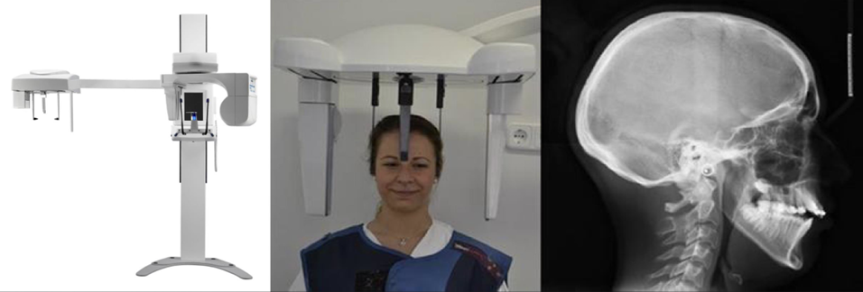
what is this?
lateral cephalometric projection
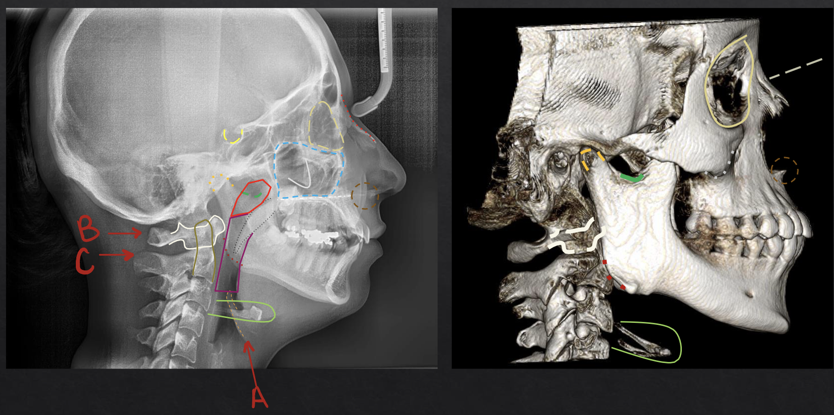
a? (orange-ish outline)
epiglottis
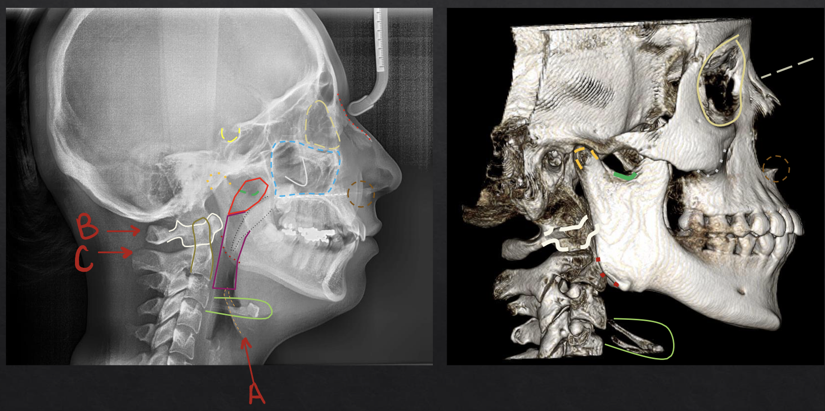
b?
c1 atlas
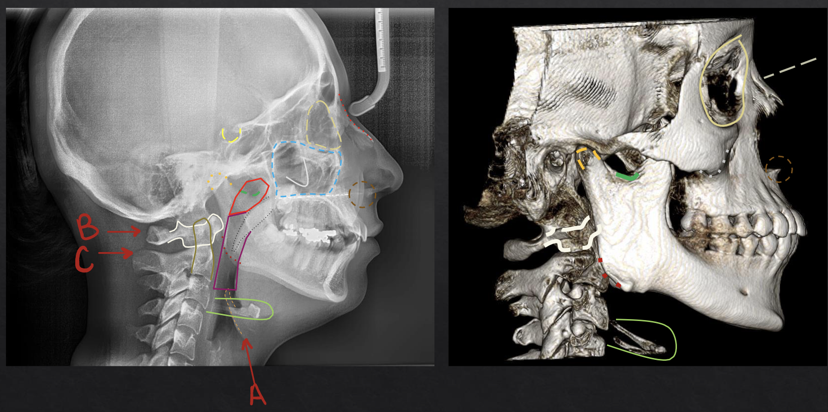
c?
c2 axis
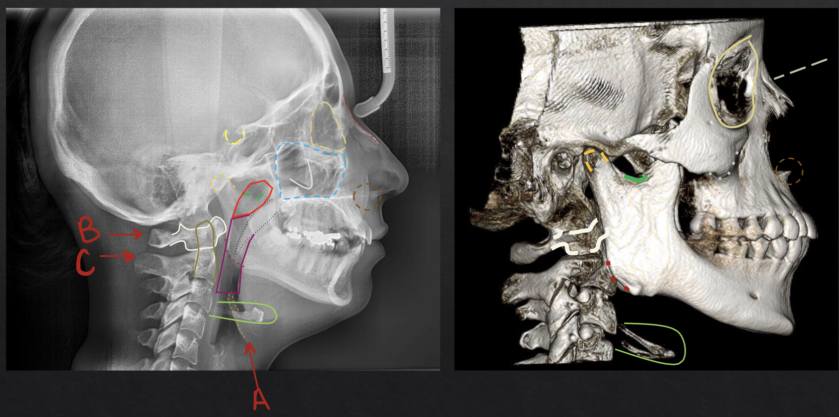
what structure is outlined in yellow? (not full circle, only half circle)
sella turcica
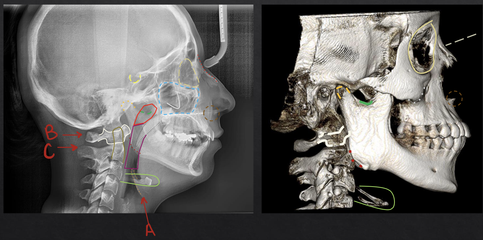
what structure is outlined in purple?
oropharynx
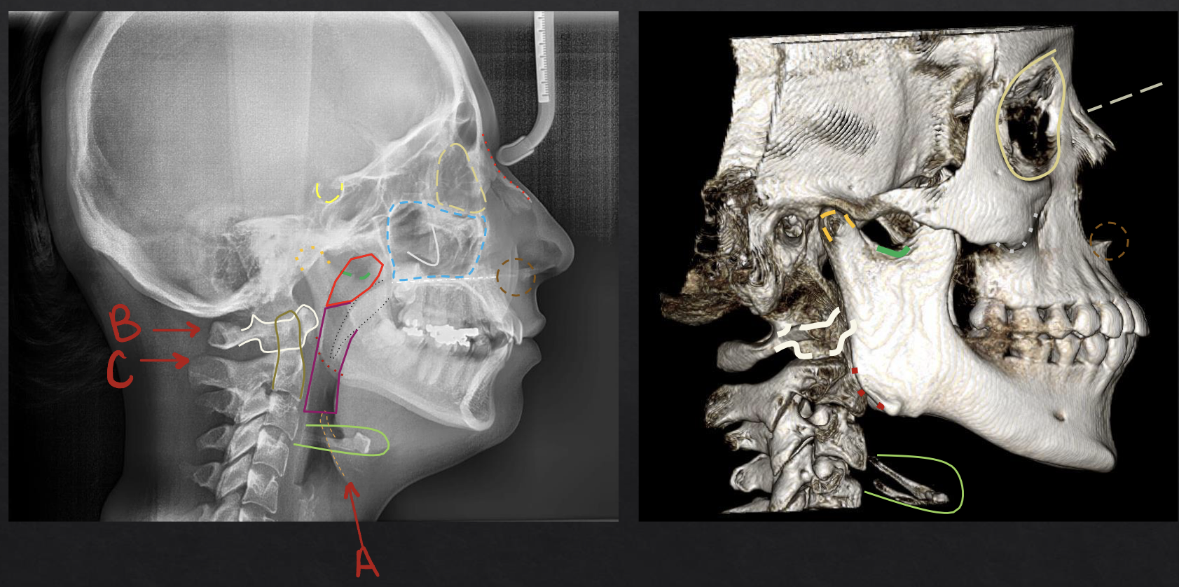
what structure is circled in green?
hyoid bone
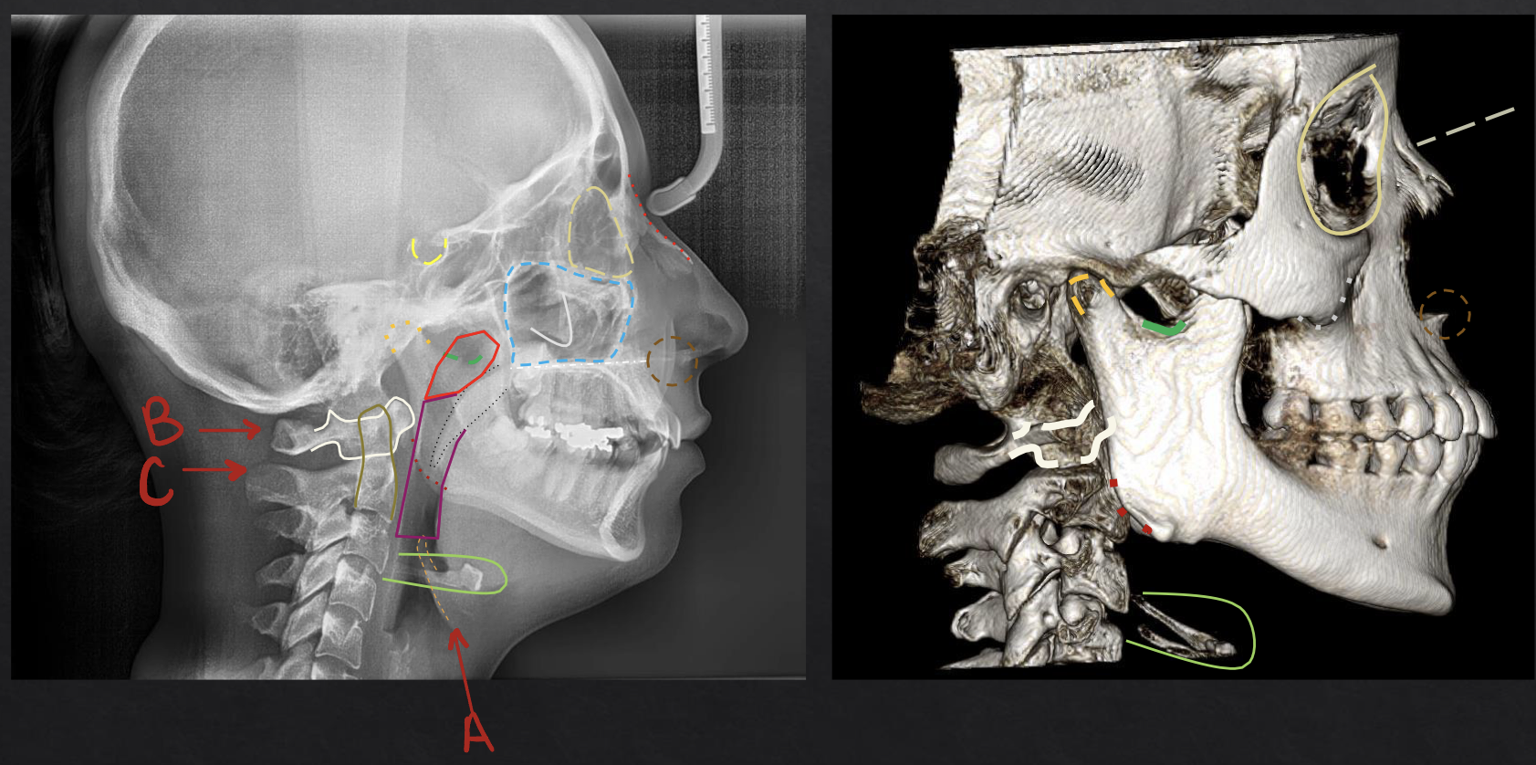
what structure is circled in blue?
maxillary sinus
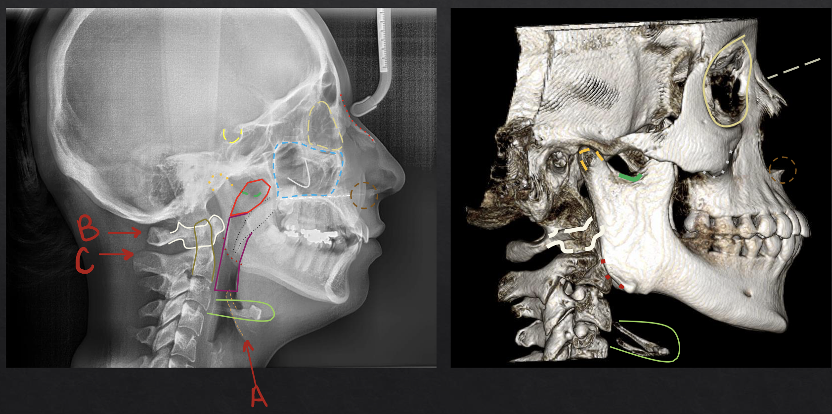
what structure is circled in brown?
anterior nasal spine
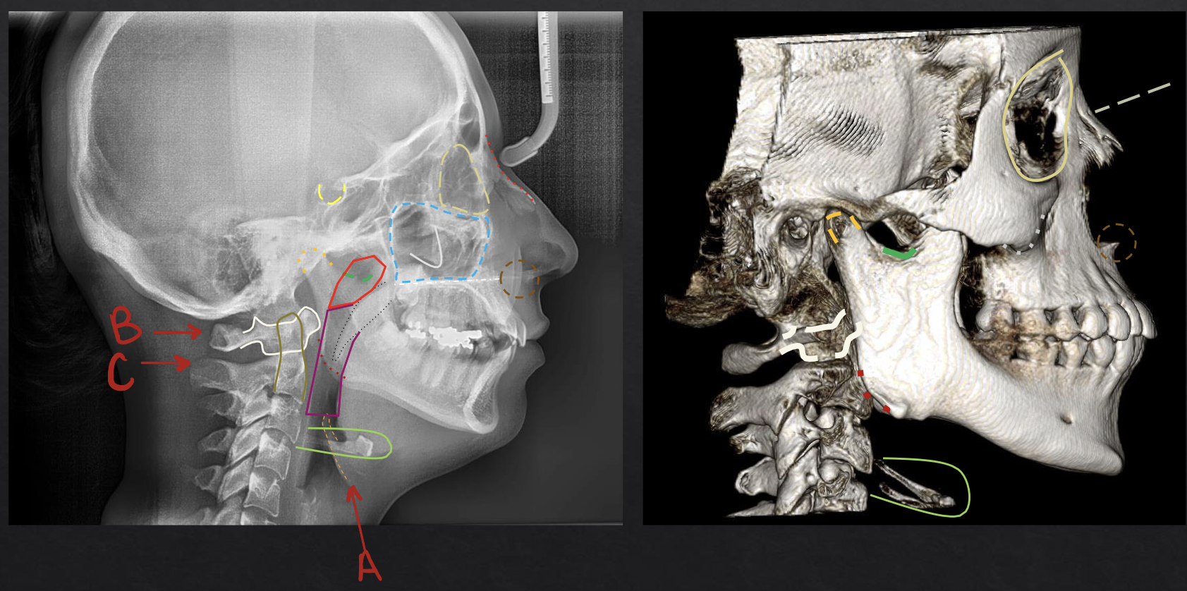
what structure is lined in red? (in the front of the face)
nasal bone
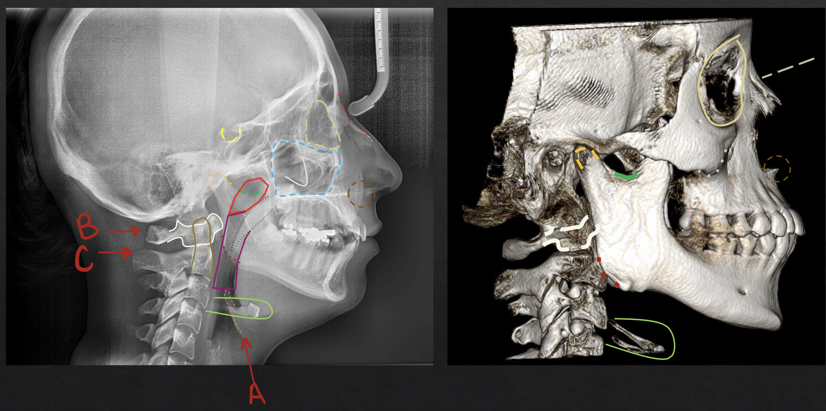
what structure is circled in yellow? (full circle)
orbit
what sits in the sella turcica?
pituitary gland
what are the indications for posteroanterior (PA) cephalometric
ortho, oral surgery, trauma, facial asymmetry malocclusion
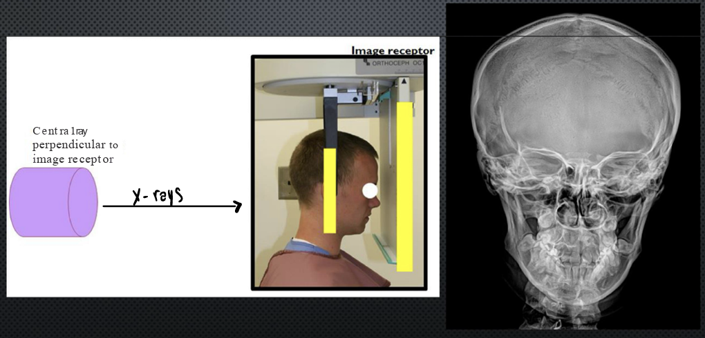
what is this?
posteroanterior (PA) cephalometric
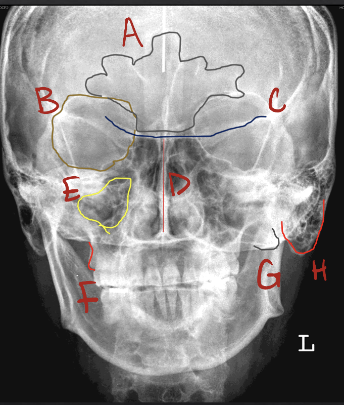
a?
frontal sinus
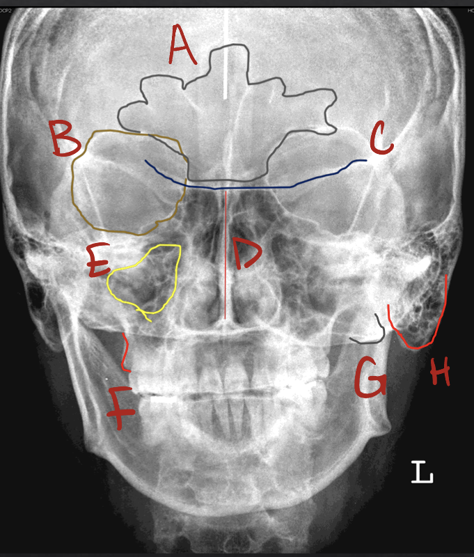
b?
orbit
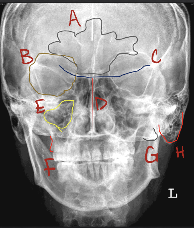
c?
lesser wing of the sphenoid
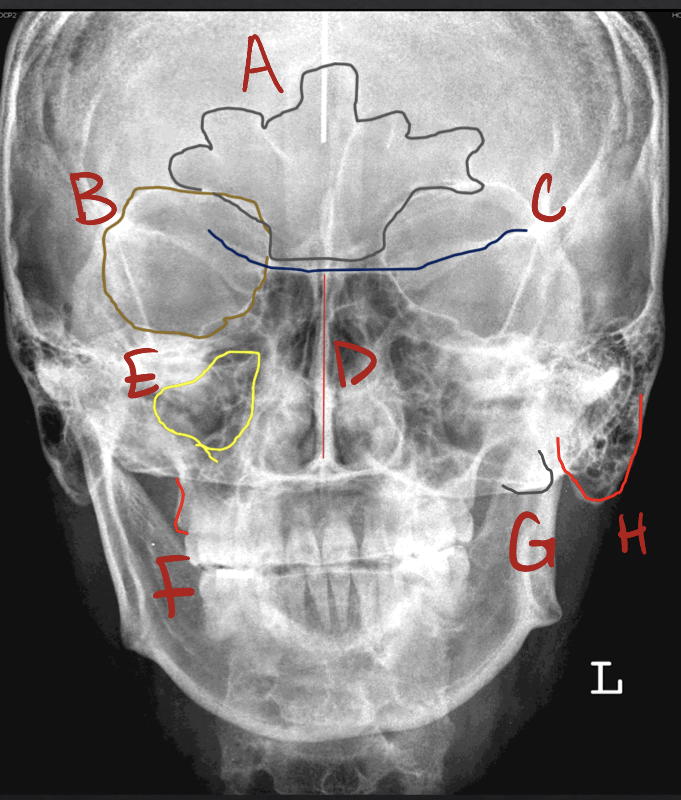
d?
nasal septum
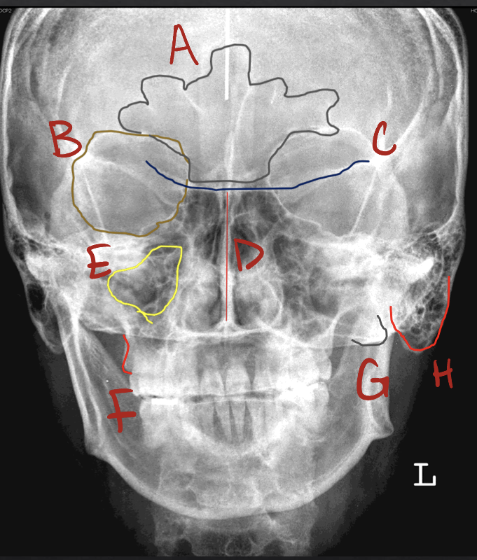
e?
maxillary sinus
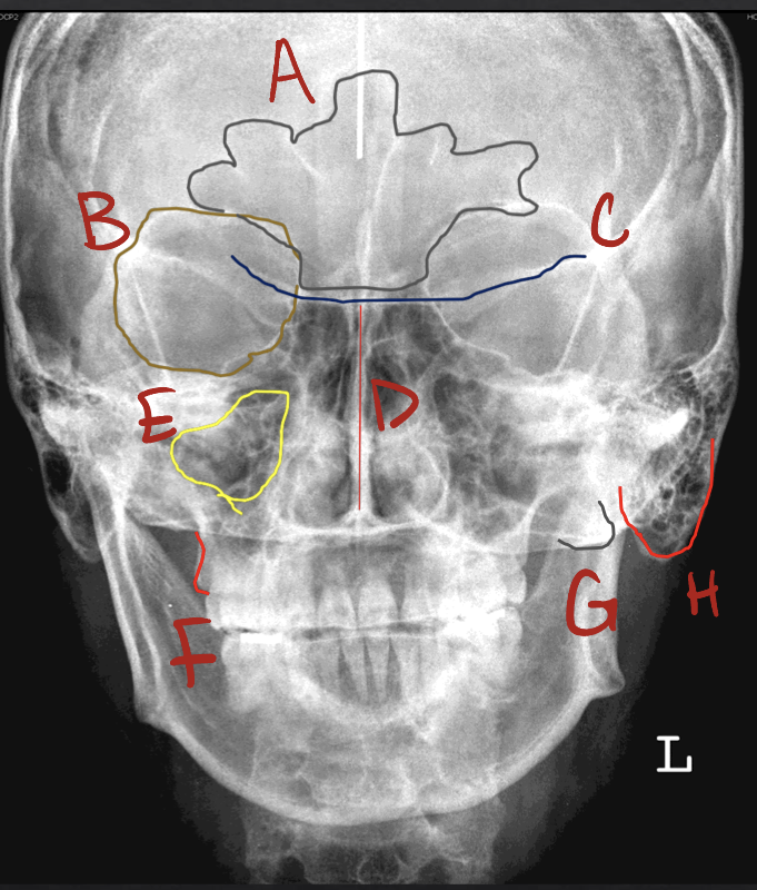
f?
posterior border / tuberosity of the maxilla
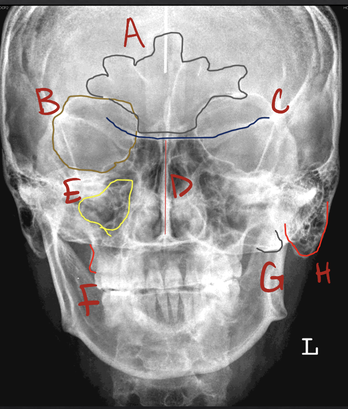
g?
zygomatic process of the maxilla
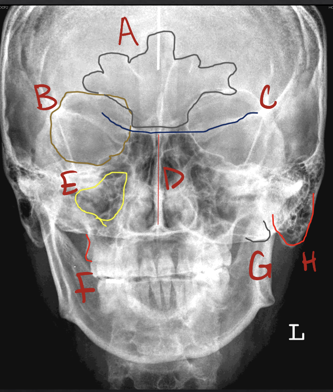
h?
mastoid process
waters projection
lifted at an angle
what are the indications for waters projection?
image sinuses after trauma (can visualize fluid accumulation)
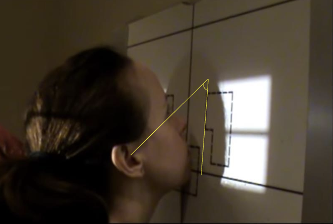
what is this?
waters projection
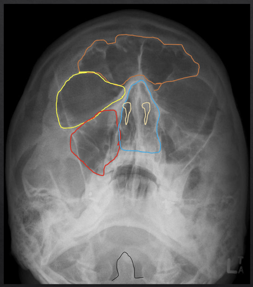
what structure is circled in orange?
frontal sinus
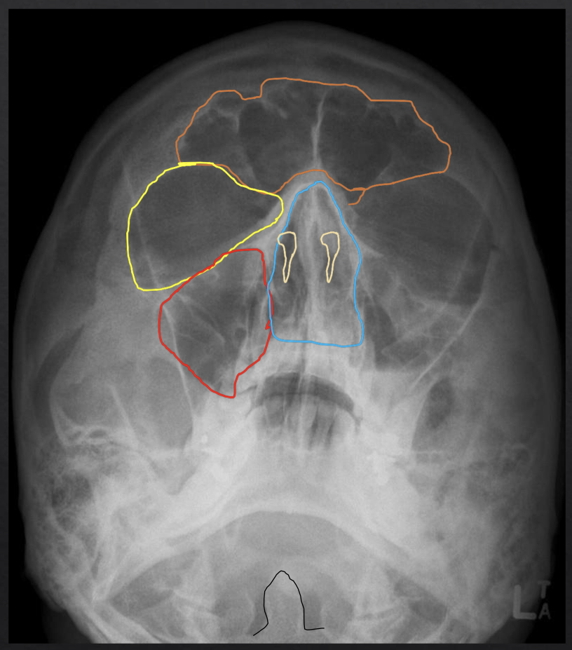
what structure is circled in yellow (big one, not 2 little ones)?
orbit
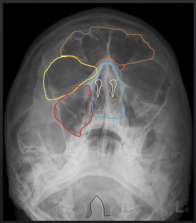
what structure is circled in red?
maxillary sinus
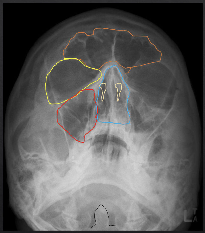
what structure is circled in two lil yellow tear drops?
nasal aperature
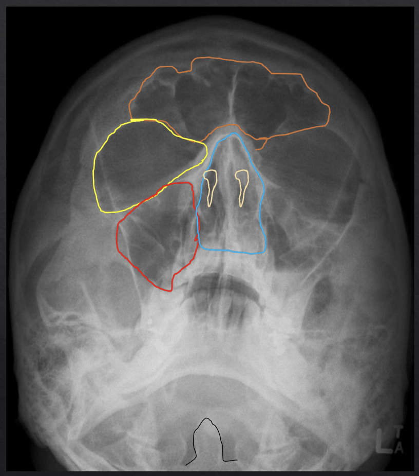
what structure is circled in blue?
nasal cavity
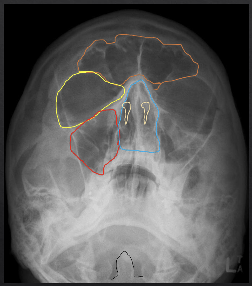
what structure is outlined in black?
odontoid process of C2
t/f in a panoramic image, the x-ray source and receptor are moving
true!
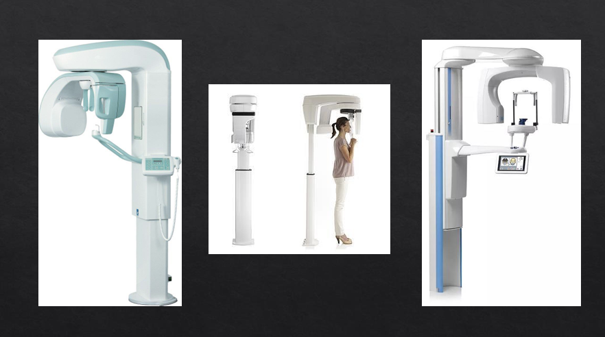
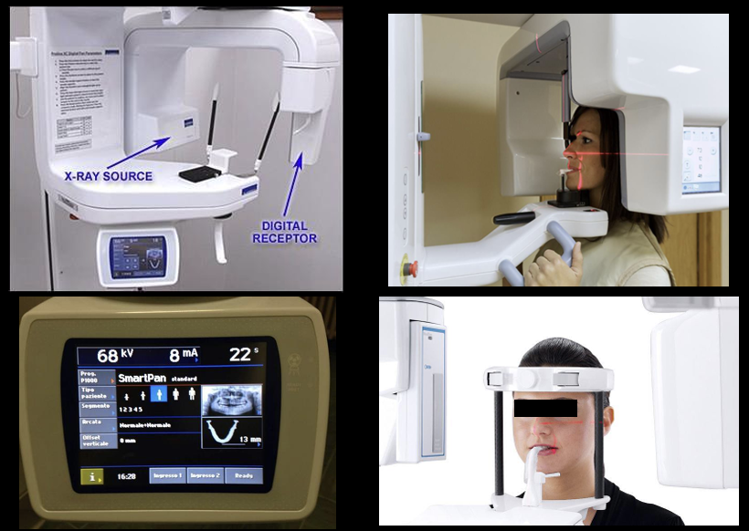
what is this?
panoramic
*what are the indications for pano?
overall evaluation of dentition, examine for intraosseous pathology like cysts / tumor / infections, gross evaluation of TMJ, evaluation of position of impacted teeth, evaluation of eruption of permanent dentition, denomaxillofacial trauma, developmental disturbances of maxillofacial skeleton
what should you tell your pt to do when taking a pano?
put the tip of your tongue on your hard palate- this closes palatoglossal space (which will be superimposed on maxillary teeth)
*what are the advantages of a pano compared with a full-mouth examination?
board coverage of facial bones and teeth, low radiation dose, easy of panoramic radiographic technique, can be used in pts with trismus or in pts who cannot tolerate intraoral radiography, quick and convenient radiographic technique, useful visual aid in pt education and case presentation
*what are the disadvantages of pano?
lower resolution images that do not provide the fine details provided by intraoral radiograph, magnification across image is unequal making linear measurements unreliable, image is superimposition of real/double/ghost images and requires careful visualization to decipher anatomic and pathologic details, requires accurate pts positioning to avoid positioning errors and artifacts, difficult to image both jaws when pt has severe maxillomandibular discrepancy
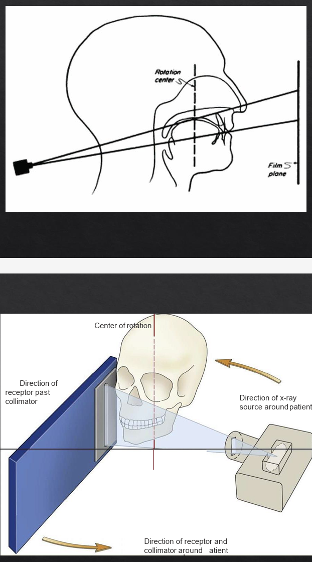
what is this?
angulation of pano
focal trough
a zone of sharpness generated by a moving source and receptor
the closer an anatomic structure is positioned to the center of the trough…
the more clearly it is imaged on the resulting radiograph
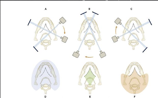
*purple
real images- structures between the moving center of rotation and the receptor form real images
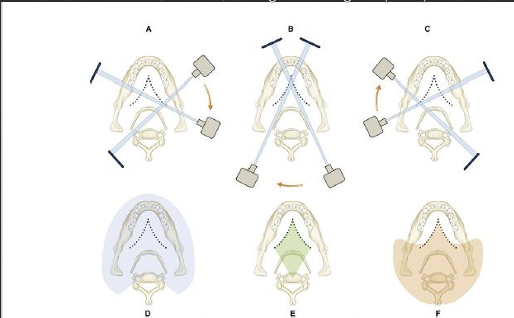
*green
double- structures lying between the moving centers of rotation and the receptor that are imaged twice
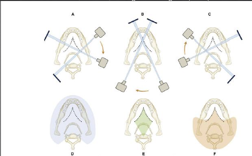
*orange
ghost- structures located between the x-ray source and moving center of rotation cast ghost images
where do ghost images appear?
on the opposite side of, and superior to the original image
also more blurry
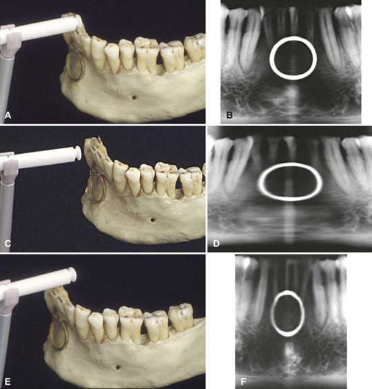
what does the first row show?
pt is biting correctly
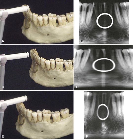
what does the second row show?
the bite plate is too anterior / the patient is too posterior
the mandible is posterior to the focal trough and images will be magnified
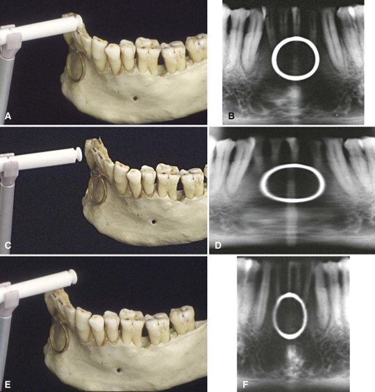
what does the third row show?
bite plate is too posterior / pt is too anterior
the mandible is anterior to the focal trough and images will be minified