MICR 270 Module 3
1/53
There's no tags or description
Looks like no tags are added yet.
Name | Mastery | Learn | Test | Matching | Spaced |
|---|
No study sessions yet.
54 Terms
Overall immune response
From the entrance of the pathogen in the body to its elimination
Includes innate and adaptive immune response
Phases of adaptive immune response
Antigen recognition (antigen presentation)
PAMPS are seen by antigen-presenting cells (APCs), such as dendritic cells and macrophages
APCs present antigens to naïve T-cells via their surface MHC proteins
Lymphocyte activation
series of cellular interactions/communication which lead to T-cell and B-cell differentiation and clonal expansion
B-cells → Plasmocytes
T-cells → Helper T-cells or cytotoxic T-cells
Elimination of pathogens or non-self perceived antigens
Most efficient defences are unleashed to destroy the pathogen
Humoral Immunity: Plasmocytes produce antibodies that bind to extracellular pathogens.
Cell-mediated Immunity: Cytotoxic T-cells destroy cells infected by intracellular pathogens or get activated by antigens presented by APCS
Apoptosis of immune cells (contraction)
Once the pathogen is eliminated, the majority of activated lymphocytes undergo apoptosis, and the immune response gradually declines
Establishment of immunological memory
The adaptive immune cells that don’t apoptose differentiate into memory cells
Memory cells will proliferate quickly to generate a fast and robust immune response if re-exposed to the same antigen
Clonal expansion
Production of a large quantity of identical cells from the same original cell
Apoptosis
Programmed cell death that occurs in a way controlled by the cell itself, which generate almost no damage to the surrounding area
Major Histocompatibility complex (MHC)
molecules that display antigenic peptides on the surface of cells.
serves as a self-label, helping to identify self from non-self molecules
MHC-antigen complex can be recognized by the TCR and its co-receptors (CD4 or CD8) to initiate an adaptive immune response to eliminate foreign antigens
What are the two classes of MHC?
There are two classes of MHC that distinguish between different recognition patterns
MHC class I and MHC class II

Antigen-presenting cells (APCs)
Cells that function to present the antigens found inside the body to T-cells (because T-cells can’t recognize extracellular pathogens by themselves)
APCs internalize pathogens (by phagocytosis or receptor-mediated endocytosis) and process them into peptides (antigens) which are displayed on their surface via an MHC class II to be recognized by T-cells
Professional APCs
the most efficient cells that both present antigens through MHC class II and express costimulatory signals to activate helper T-cells
Include macrophage, B-cell, and dendritic cell
Dendritic cell is the best
Non-professional APCs
Can be induced to express MHC class II complexes or stimulatory molecules, but normally don’t (only in the case of a sustained inflammatory responses)
Include fibroblasts and glial cells
Receptor-mediated endocytosis
An endocytic process in which a cell absorbs external material by invagination of the plasma membrane. This process relies on receptors specific for the material being absorbed.
What are the two pathways which lead to the formation of an effective peptide:MHC signalling complex?
Endogenous pathway
Exogenous pathway
Endogenous pathway
Forms peptide:MHC class I complex (recognized by CD8+ cytotoxic T-cells)
allows the cell to process self or foreign intracellular particles and present them at the cell surface in order to be recognized by T-cell receptors on cytotoxic T-cells
Steps of endogenous pathway
Proteins produced within the cell are broken into fragments (antigens)
Antigens are transported to rough ER and combine with class I MHC
antigen:MHC class I complex is transported to Golgi, then the cell membrane for display
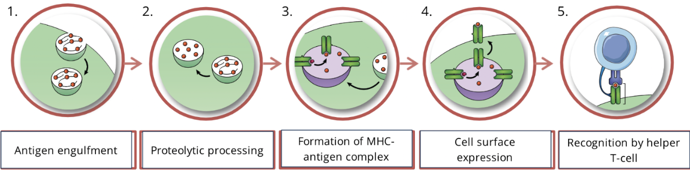
Exogenous pathway
Forms peptide:MHC class II complex (recognized by CD4+ helper T-cells)
Steps of exogenous pathway
Antigen engulfment: Foregin antigen recognized by PRRs is engulfed by an APC by endocytosis forming an endosome.
Proteolytic processing: Foreign antigens inside the endosome are broken down into fragments
Formation of antigen:MHC complex: vesicle containing the foreign fragments fuses with vesicle containing MHC molecules (originating from the ER via the Golgi), forming MHC-antigen complexes
Cell-surface expression: MHC-antigen complex is transported to the plasma membrane, for display on the surface of the cell
Recognition by helper T-cell: T-cell receptor on the surface of a helper T-cell binds to MHC-antigen complex on the surface of the APC, which will initiate adaptive immunity
Proteolytic processing
proteases cleave one or more bonds in a target protein to modify its activity (activation, inhibition, or destruction of activity)
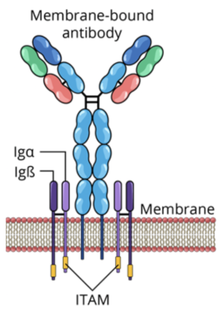
B-cell receptor (BCR)
composed of a membrane-bound antibody and signal transduction molecules (ITAMs).
BCRs recognize and bind to extracellular pathogens or toxins directly
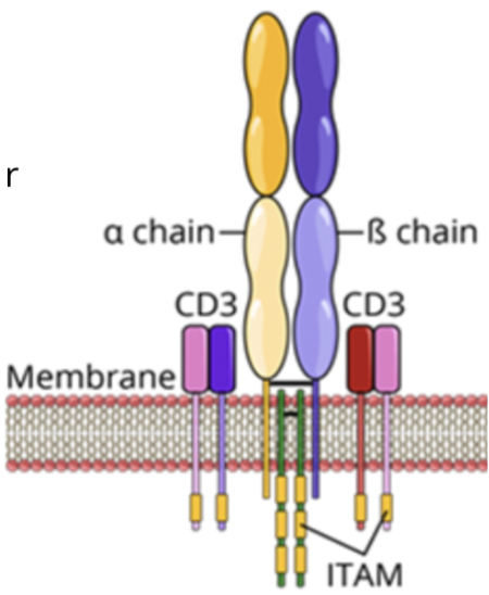
T-cell receptor (TCR)
formed of a membrane-bound antigen-specific molecule and signal transduction molecules (CD3 and ITAMs).
TCRs in association with a co-receptor (CD4 or CD8) recognize and bind to peptide:MHC complex
CD4 vs CD8 and MHC Classes
CD4 co-receptors are expressed on helper T-cells, which recognize peptide: MHC class II complex.
CD8 co-receptors are expressed on cytotoxic T-cells, which recognize peptide: MHC class I complex
How does the immune system communicate?
Components of the immune system communicate between themselves using specific cell surface receptors found on immune cells and a cytokine network
Lymphocyte activation involves many interactions with other immune cells
Cytokine networks
coordinate appropriate immune responses and modulate the balance between humoral and cell-mediated immunity
T-cell dependent activation of B-cells
interaction between a T-cell and a B-cell that involves the exchange of activation signals to produce plasmocytes and memory B-cells for humoral immunity
Thymus-independent antigens
antigens that can induce the production of antibodies by B-cells without the presence of a T-cell (but the majority are thymus dependent and do require a T-cell)
What are the steps of the signalling pathway for T-cell-dependent activation of B-cells?
Peptide MHC-Class II complex
Signal 1: TCR - Peptide:MHC class II complex
Expression of co-stimulatory molecules
Signal 2: co-stimulation (B7:CD28 and CD40:CD40L)
Signal 3: cytokines
Outcome of the 3 signals
Step 1 of signaling: peptide:MHC Class II complex
when an antigen binding to the BCR does not produce enough signal to activate the cell, the antigen is internalized by receptor mediated endocytosis and displayed as an MHC on the membrane

Step 2 of signaling: signal 1 TCR - Peptide:MHC class II complex:
The specific TCR complex and CD4 co-receptor on the T-cell recognizes and binds to the peptide:MHC class II complex

Step 3 of signaling: expression of co-stimulatory molecules
Signal 1 (TCR- peptide:MHC complex II) induces the expression of CD40L on the cell surface of the helper T-cell.

Step 4 of signaling: Signal 2: co-stimulation (B7:CD28 and CD40:CD40L)
CD40L and CD28 expressed on the T-cell, bind to CD40 and B7 (respectively) expressed on the B-cell, inducing a costimulatory signal in both cells.

Step 5 of signaling: Signal 3: cytokines
The activated helper T-cell secretes cytokines which bind to their associated cytokine receptor located on both cells (a paracrine and an autocrine effect).

Step 6 of signaling: outcome of the 3 signals
The combination of these 3 signals stimulate the proliferation and differentiation of B-cells into plasma cells and memory B-cells, forming the humoral immune response against the specific antigen

Immune synapse
structure formed by the interaction between a T-cell and an APC, consisting of signal molecules and adhesions proteins
Often arranged into SMACs

Supra molecular activating clusters (SMACs)
‘bullseye’ of three rings depicting three different cell clusters with similar functions
Includes cSMAC, pSMAC, and dSMAC
central SMAC/c SMAC
Signal molecules: contains the molecules responsible for signaling between the two cells (TCR and peptide:MHC molecules)
peripheral SMAC/p SMAC
Adhesion molecules: contains adhesion proteins (integrins and cytoskeletal linker proteins) responsible for keeping cells in contact long enough for signals to propagate
distal SMAC/d SMAC
Signal regulation molecules: consists of proteins with large extracellular domains that are responsible for helping regulate signal transduction
Function of immune synapse
Primary goal of the immune synapse is the effective activation of the T-cell
The synapse holds signal proteins together to form stronger connections, which give enough time for the right amount of signals to be produced.
The synapse leads to the reorganization of structures inside the T-cell, directing the release of cytokines close to the target cell.
The synapse regulates lymphocyte activation
Cytokines
chemical messengers secreted by immune cells that play a key role in cell-to-cell communication
What are the types of cytokines?
Chemokines
Interleukins (IL)
Interferons (IFN)
Tumor necrosis factor (TNF)
Growth factors
Chemokines
Induce chemotaxis
Call in cells to the region of infection or injury
Play a key role in:
Inflammation; wound healing
Cell-mediated and humoral immune responses
Hematopoiesis
Interleukins
Contain over ten subfamilies (IL-1, IL-2, IL-4, IL-12, etc.)
Regulate immune and inflammatory responses
Primarily affect the proliferation and differentiation of various hematopoietic and immune cells
Interferons (IFN)
The most common and well-known interferon molecules are IFN-α, IFN-β, and IFN-γ
Induce an antiviral state - inhibit the replication process of viruses
Help regulate immune responses
Tumor necrosis factor (TNF)
The most common and well-known are TNF-α and TNF-β
Involved in systemic inflammation (septic shock)
Involved in tumor regression
Can cause apoptosis (cell death)
Growth factors
Stimulate growth, proliferation, healing, cellular differentiation
Regulate a variety of cellular processes such as immune response
Chemotaxis
the movement of an immune cell in direction of an elevated concentration of chemoattractant molecules, such as chemokines.
Septic shock
A serious medical condition associated with a significant drop in blood pressure that can lead to respiratory or heart failure, and death
Immunological memory
ability of lymphocytes to respond more efficiently with a greater and faster immune response to reinfection by a previously encountered antigen
occurs when there is a second encounter with an antigen, that induces a heightened state of immune reactivity
mediated by memory B-cells

Memory B-cells vs Naive B-cells
Memory B-cells differentiate from naive B-cells and display the same antibody as their parent cell
Memory B-cells have a longer lifespan than naive B-cells
Memory B-cells can differentiate immediately into plasma cells, making the immune response more efficient than naive B-cells

Natural passive immunity
Acquired by the fetus or newborn from the mother
Placental transfer of antibodies during pregnancy or breastfeeding
Short-lived immunity (~6 months)
No immunological memory for the recipient
Artificial passive immunity
Acquired by injection of serum containing antibodies
Immunity is temporary
No immunological memory for the recipient
Natural active immunity
Acquired through infection by a pathogen, possibly leading to symptoms/a disease state
Development of innate and adaptive immune responses
Immunological memory has a significant chance of being developed
Artificial active immunity
Acquired through vaccination
Development of innate and adaptive immune responses
Normally, no symptoms/disease states are present
Immunological memory has a significant chance of being developed
Serum
Fluid portion of the blood, which is free of cells and clotting factors
Serology
serological analysis involves the diagnostic evaluation of blood serum to test for the presence of a host antibody response against a foreign particle/pathogen
Chicken pox/shingles case study
95% of the population shows serological evidence of having chicken pox due to the varicella-zoster virus (VZV) vaccine
Even if you did not actually have chicken-pox, the VZV vaccine produces antibodies in our systems
Chicken pox remains in the immune system in latency (inactive state) after infection
Shingles is the reactivation of the latent virus in the immune system causing painful rashes
There is a vaccine for shingles (can reduce risk up to 90%)