MLS 2211 Exam 6
 Call Kai
 Learn
Learn Practice Test
 Spaced Repetition
 Match
Match Flashcards
 Knowt Play
1/131

There's no tags or description
Looks like no tags are added yet.
Name | Mastery | Learn | Test | Matching | Spaced | Call with Kai | Chat |
|---|
No analytics yet
Send a link to your students to track their progress
132 Terms
Explain the general principle of Ion-Selective Electrodes (ISE) for measuring electrolytes
The activity (concentration) of electrolyte on one side of an electrode membrane, as compared to a fixed concentration of that electrolyte on the other side of the membrane, causes an electrical potential to be built-up across the membrane.
The electrical potential is proportional to the electrolyte concentration of the test solution.
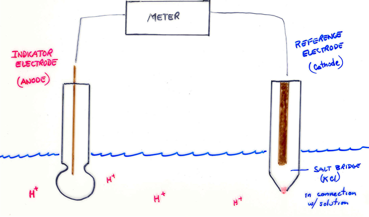
What is the selectivity of a pH ISE?
pH spec. due to H+ ions
What is the filler in a pH ISE
HCl
What is the selectivity for a sodium ISE
Na+
What is the filler for a sodium ISE
NaCl
What is the selectivity for a potassium ISE
Coated w/ Valinomycin
What is the filler for a potassium ISE
KCl
What is the selectivity for a chloride ISE
AgCl crystals
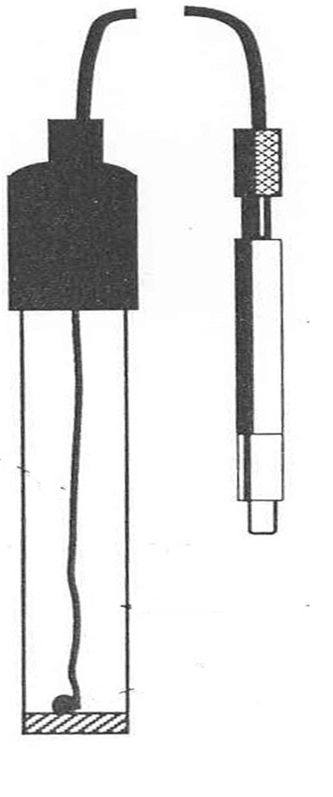
What is the filler for a chloride ISE
Theres no filler
What is the selectivity for a CO2 ISE
Sample reacts with weak acid
What is another name for a CO2 ISE
Severinghaus electrode
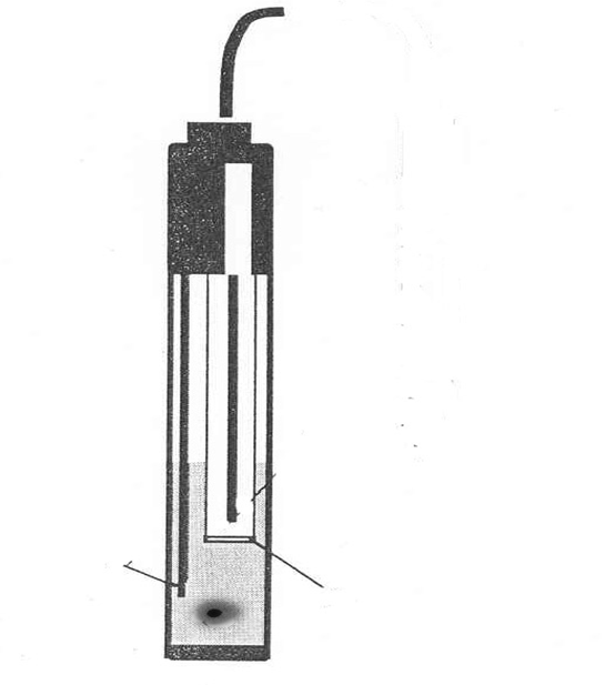
What is the filler for a severinghaus electrode
No filler
For coulometric chloridometers such as the Buchler-Cotlove chloridometer, what is a common source of error
Generator electrode gets dirty and pitted – irratic response and premature assay termination.
Other electrodes get dirty – must be cleaned often.
Cannot distinguish between Chloride and other Halogens or radicals that act like halogens (perchlorates, thiocyanates, cyanates, etc.)
What are the two most abundant electrolytes (2) in Blood Plasma:
Sodium and chloride
What are the two most abundant electrolytes (2) in RBCs (intracellular):
Potassium and magnesium
What electrolytes that would be falsely elevated on hemolyzed specimen:
K+
Mg++
Phosphates (Inorganic Phosphorous)
Iron
What electrolytes that are impacted by incorrect anticoagulants and their corresponding values
EDTA or Oxalate/Fluoride plasma will have:
K+ = Stupid High (9.0 – 25.0 mmol/L)
Ca++ = Stupid Low (0.0 – 2.2 mg/dL)
Mg++ = Stupid Low (0.0 – 1.0 mg/dL)
What is the function of sodium in the body
Main electrolyte controlling osmotic pressure of blood and body fluid.
All parts of renal tubules can secrete Na into the glomerular filtrate, but is regulated by Aldosterone in the Distal Tubules. Aldosterone causes retention of Na, with a corresponding loss of K (by exchange).
What happens in hypernatremia and what can cause it
Hypertension, strokes, renal failure, and heart failure due to over-exertion.
Conn’s Disease (excessive Aldosterone) High dietary intake, Cushing’s Syndrome, dehydration.
What happens in hyponatremia and what can cause it
Hypotension, edema, muscle weakness, neurological failure (seizures, etc.)
Adrenal insufficiency (Addison’s) Low Dietary intake, diarrhea, vomiting, burns, ATN, hypothyroidism.
What is the refrence range for sodium
136-146 mMol/L
What does potassium do in the body
Very critical to Cardiac Nodes and myocardial muscle.
Very critical to other Muscle function (both striated and smooth)
All parts of nephron tubules can secrete K+, but in Distal Tubule elimination is regulated by Aldosterone, in an exchange with Na+. Aldosterone causes excretion of K+.
What happens in hyperkalemia
Bradycardia, flat-line EKG. Heart fails to pump blood
Muscle weakness, poor muscle coordination.
What happens in hypokalemia
Super-excitability of nodes of heart and myocardium. Arrhythmias, finally ending with Ventricular Fibrillation.
Muscle cramping, spasms, seizures.
Potassium Shift: Shifts______ in Acidosis, shifts______ with Alkalosis. Each _____ pH increase, causes a K+ decrease of _______. The inverse is also true.
Shifts out in Acidosis, shifts in with Alkalosis. Each 0.1 pH increase causes a K+ decrease of 0.6. The inverse is also true.
If a pH of 7.4 shifts to 7.3 and the K was 5.0 before, what will the K be after the pH shifts?
5.6
If a pH of 7.4 shifts to 7.5, and the K was 5.0 what would the K be after the pH shift?
4.4
What is the reference range for potassium
3.5-5.1 mMol/L
What is the function of chloride
Tends to follow what Na+ does.
We measure it as part of the Electrolyte panel, but not because it (itself) does anything. We measure it to see if another electrolyte is there in elevated amounts, or decreased amounts
No critical values for Chloride. If it is low, it means that some other electrolyte is high.
If it is high, it means that something else is low.
What happens in high chloride
It means that some other electrolyte is low
What happens in low chloride
It means that some other electrolyte is high
What is the function of CO2 (Bicarb)
The Base (salt) component of the major blood buffer system (Carbonic Acid/Bicarb). At a pH of 7.40, the ratio of base to acid is 20 (20 to 1).
Low levels = Metabolic Acidosis
High levels = Metabolic Alkalosis
What happens in metabolic acidosis
Theres a low level of bicarb
Not eating
Barfing (anorexia + bulimia)
Gets consumed when buffering acids
Losing it in the urine (renal disease)
What happens in metabolic alkalosis
Theres a high level of bicarb
Iatrogenically caused - Excessive intake (Tums, Rolaids)
But not always -
Compensating for chronic Respiratory Acidosis.
What is the function of calcium
Ca++ has very pronounced effects on smooth muscle and skeletal muscle.
Low levels causes muscle spasms, charley-horses, cramps, etc.
High levels cause muscle weakness (flacid paralysis), but also cause calcification of soft tissues and the formation of Ca++Phosphate crystals (kidney stones and arthritis). High Calcium is being scrutinized as a MI (heart attack) risk factor.
What happens in the body with low calcium levels
Low levels causes muscle spasms, charley-horses, cramps, etc.
What happens in the body with high calcium
High levels cause muscle weakness (flacid paralysis), but also cause calcification of soft tissues and the formation of Ca++Phosphate crystals (kidney stones and arthritis). High Calcium is being scrutinized as a MI (heart attack) risk factor.
What happens to Ca in acidosis
In acidosis, the Ionized Ca++ increases (shifts off of proteins). No change in Total Calcium.
What determines potassium blood levels
Dietary Intake
Renal Excretion
All parts of nephron tubules can secrete K+, but in Distal Tubule elimination is regulated by Aldosterone, in an exchange with Na+. Aldosterone causes excretion of K+.
Shifts
Moves in and out of the Cellular compartments vs. pH changes.
What happens to calcium in alkalosis
In alkalosis, the Ionized Ca++ decreases (shifts back onto proteins). No change in Total Calcium.
What are the three forms of Ca in a total calcium
Ionized (free) Ca++ (50%)
Non-ionized (unionized) Ca++ which is bound to protein. Mainly Albumin, but can bind to globulins as well. (49.5%)
Ca-Carbonates and Phosphates (less than 0.5%)
What determines calcium blood levels
Dietary intake
Renal excretion
Renal disease favors Ca++ loss in urine.
Bone metabolism Bone breakdown elevates Ca++
What is the function of magnesium in the body
Metabolism of Magnesium is essentially the same as for Calcium, except that all cells (including RBCs) contain a lot of Magnesium.
Diet, renal function, and bone metabolism affect the blood levels. Chemically works the same way as Calcium does.
Dietary deficiency results in “Calcium-Resistant Ricketts.” Renal disease favors losing Mg++ in the urine.
What happens in the body with low magnesium
In alkalosis, the Ionized Mg++ decreases (shifts back onto proteins). No change in Total Magnesium.
What are the three forms of magnesium
Ionized (free) Mg++ (50%)
Non-ionized (unionized) Mg++ which is bound to protein. Mainly Albumin, cut can bind to globulins as well. (49.5%)
Mg-Carbonates and Phosphates (less than 0.5%)
What happens in the body with high magnesium
In acidosis, the Ionized Mg++ increases (shifts off of proteins). No change in Total Magnesium.
Magnesium Shift: Shifts _____ in acidosis, shifts ______ with alkalosis.
Shifts off of proteins in acidosis, shifts back onto proteins with alkalosis.
What do phosphates do in the body
An anion that normally shows an inverse relationship with Calcium.
Bone salts are the bodies’ stores of Phosphorous. It, along with Calcium are important bone metabolism markers.
A component of the Phosphate Buffer system of the blood.
Describe the "Chloride Shift" and identify the two anions participating in the shift.
A shift between Chloride and Bicarbonate.
Shift between Intracellular compartments and the Extracellular compartments
Explain what is meant by the Anion Gap
Normal Anion Gap means there is NOT a significant amount of abnormal, ugly, or perverse anions present (ketones, lactates, salicylates, formates, etc.)
If the Chloride Shift is not apparent, then you got wierdos and pervo anions!
explain the physiological significance of an abnormal anion gap.
If the gap is high (greater than 16) it means you are not seeing the expected Chloride Shift, and suggests that there is a significant amount of some other anion present (Dang weirdo, pervo, or unnatural anions like Salicylates, Drugs, Lactate, Oxalates, etc.).
Addison’s Disease is about the only time you expect to get Negative Gaps. Usually a Low or Negative Gap means an error in the assay (low Na+ or high Chlorides).
What is the formula for anion gap
Na - (Bicarb + Chloride)
What is a normal anion gap?
7-16
What test are in an electrolyte panel
pH, Total CO2, Na, K, and Cl
What would you expect an electrolyte panel to look like for a patient with Addisons disease
Low sodium
high potassium
normal chloride
normal bicarb
low glucose
normal BUN
normal creatinine
normal calcium
Abnormal anion gap
What would you expect an electrolyte panel to look like for a patient with Cushing’s disease
High sodium
low potassium
normal chloride
normal bicarb
high glucose
normal BUN
normal creatinine
normal calcium
normal anion gap
What would you expect an electrolyte panel to look like for a patient with early renal failure
low sodium
high potassium
normal chloride
normal bicarb
normal glucose
high BUN
high Creatinine
low calcium
normal anion gap
What would you expect an electrolyte panel to look like for a patient with late renal failure
Low sodium
High potassium
low chloride
low bicarb
normal glucose
High BUN
high creatinine
normal Ca
abnormal anion gap
What would you expect an electrolyte panel to look like for a patient with type II DM with ketosis
low sodium
high potassium
low chloride
low bicarb
high glucose
normal BUN
high creatinine
normal calcium
abnormal anion gap
What would you expect an electrolyte panel to look like for a patient with type I DM with ketosis
low sodium
high potassium
low chloride
low bicarb
high glucose
normal BUN
high creatinine
normal calcium
abnormal anion gap
What would you expect an electrolyte panel to look like for a patient with metabolic acidosis (salicylate overdose)
Normal sodium
high potassium
low chloride
low bicarb
normal glucose
normal BUN
normal creatinine
normal calcium
What would you expect an electrolyte panel to look like for a patient with lactic acidosis
Low sodium
high potassium
low chloride
low bicarb
low glucose
high BUN
high creatinine
low ca
abnormal anion gap
What would you expect an electrolyte panel to look like for a patient with Anorexia Nervosa - Bulimia
low sodium
high potassium
low chloride
low bicarb
low glucose
Low BUN
high creatinine
normal ca
normal anion gap
Explain the clinical rationale of performing CSF Chloride and CSF Lactate assays, and how these tests distinguish between bacterial and viral meningitis.
Bacteria ferment glucose into Lactic Acid, thus CSF Lactate will be elevated.
Viruses don’t cause CSF Lactate to increase.
Normal CSF Lactate rules-out Bacterial Meningitis, and is consistent with Viral Meningitis.
Bacteria ferment glucose into Lactic Acid, thus CSF Lactate will be elevated.
As Lactic Acid increases, CSF Chloride must decrease (anion gap thing in CSF).
Note: Normal CSF Chloride is118 –132 mmol/L.
Viruses don’t affect CSF Chloride levels.
Explain the relationship of sweat chloride with Cystic Fibrosis
Cystic Fibrosis also involves defective distal tubules of the sweat glands. This causes decreased electrolyte reabsorption.
Affects Chloride, Na+, and K+, and results in high levels of these electrolytes.
explain how a sweat specimen is collected using Pilocarpine Iontophoresis.
PCN must be iontophoresed into the sweat gland by an electrical current.
Clean appropriate area of skin.
Apply PCN, place positive electrode over it. Negative electrode is placed on skin nearby.
What is the clinical significance of a high iron level
Hemolytic Anemia (Fe from Hemoglobin)
Megaloblastic Anemia (Fe – cells dying)
Heme Synthesis blockages (heavy metals)
Severe Hepatitis (Iron stores released)
Iron Intoxication (biggie for children)
If Serum Iron is high, there is probably a hemolytic condition (free Fe++ from hemoglobin), or Ferritin and Hemosiderin spilling from the liver into the blood, or an increased intake of Iron
What is the clinical significance of a low iron level
Dietary Iron Deficiency (least common)
Chronic blood loss (most common)
Pregnancy (increased demand for Fe)
Malabsorption
If Serum Iron is low, there is an Iron deficiency from something
What is the clinical significance of a high TIBC level
Pregnancy (compensation)
Iron Deficiency (compensation)
Chronic hemorrhage (compensation)
Hepatic disease (not compensation – due to release of Hemosiderin and Ferratin).
Big-time Iron Poisoning. (not compensation, you’re measuring free Iron, Dude!).
What is the clinical significance of a low TIBC level
Chronic Infections (decreased production of Transferrin)
Chronic Inflammation (decreased production of Transferrin)
Renal Disease (lose proteins in urine)
Nephrotic Syndrome (lose proteins in urine)
Hepatic Cirrhosis (not making Transferrin)
Mild Hepatic Disease (not making Transferrin)
What is UIBC
The results you get on the second assay is called the Total Iron-Binding Capacity (TIBC - how much Iron Transferrin can bind). Tells the total number of Iron seats on the bus! (Serum Iron + the UIBC)
The difference between the first and second serum Iron assays is called the Unbound Iron-Binding Capacity (UIBC).
Tells how many empty Iron seats were on the bus!
How do TIBC and iron relate to each other
Low Iron = High TIBC
High Iron = Low TIBC
What would you expect the iron and TIBC to be in Hemolytic anemia due to transfusion reaction
High iron and high TIBC
What would you expect the iron and TIBC to be in hemochromatosis (iron poisoning)
High iron and low TIBC
What would you expect the iron and TIBC to be in IDA or chronic blood loss
Low iron and high TIBC
What would you expect the iron and TIBC to be in anemia of chronic infections or inflammation
Low iron and low TIBC
What would you expect the iron and TIBC to be in hepatic necrosis
High iron and high TIBC
Explain the relationship of serum copper, ceruloplasmin, and Wilson's Disease
Wilson’s Disease is the most common cause of Copper Overload. In this disease, the liver cannot make Ceruloplasmin. Lacking Ceruloplasmin, copper gets deposited in the tissues.
Wilson’s Disease = Hemochromatosis with Copper “halo” in Iris of eyes, accompanied by low Ceruloplasmin levels.
What methods are used to assay sodium, potassium and lithium
Ion-Selective Electrode
What methods are used to assay chloride
Coulometric (Buchler-Cotlove)
Mercuric Thiocyanate
Ion-Selective Electrode
What methods are used to assay total CO1
pCO2 Severinghaus Electrode
PEPC Enzymatic method
What methods are used to assay calcium
Atomic Absorption
OCPC (o-Cresolphthalein Complexone)
Ion-Selective Electrode
What methods are used to assay inorganic phosphate
Molybdenum Blue (Fiske-Subbarow)
What methods are used to assay magnesium
Titan Yellow Dye or Methylthymol Blue Dye
Calmagite Dye
What methods are used to assay Iron and TIBC
Ferrozine
Bathophenanthroline
The liver is pretty dang cool for many reasons. One in particular is when circulating iron levels become too high, leading to the risk of iron deposition in tissues, the liver compensates by decreasing the production of__________ to reduce iron transport in the bloodstream.
Transferrin
In a type I Diabetes Mellitus patient, what type of pattern would we expect to see on a BMP?
Low Na+, Cl-, and Bicarb, high K+
I am also a highly clinically significant electrolyte that is routinely ordered for nearly every patient. In MLS 2211/2213, my reference range is 3.5 mMol/L to 5.1 mMol/L. Which electrolyte am I?
Potassium
Magnesium can be measured using which method?
Buchler-Cotlove
Fiske-Subbarow
Ferrozine
Methylthymol Blue Dye
Methylthymol Blue Dye
Inorganic Phosphorus can be measured using which method?
OCPC
Fiske-Subbarow
Buchler-Cotlove
Methylthymol Blue Dye
Fiske-Subbarow
Iron can be measured using which method?
Ferrozine
Buchler-Cotlove
Methylthymol Blue Dye
Mercuric Thiocyanate
Ferrozine
Are we all totally on board to never ever draw, or knowingly run, a BMP on a EDTA (purple top) or Oxalate (grey top)?
Yes, I promise
In the CSF of a patient with bacterial meningitis, we would expect to see an increased CSF protein, decreased CSF glucose, and an increased CSF lactic acid. But what about CSF chloride??
We would expect to see a decrease in CSF Chloride
What condition is associated with excessive Aldosterone and hypernatremia?
Conn’s Disease
I am an ISE method who’s filler substance is KCl. Which ISE method am I?
Potassium
Which of the following ions shift into the cell in alkalosis to replace H+ ions moving from the cell into the plasma? Fun fact, magnesium does this too!
Potassium
A symptom of hemochromatosis is what?
bronzing of the skin
Chloride can be measured using which method?
Ferrozine
Mercuric Thiocyanate
Methylthymol Blue Dye
Buchler-Cotlove
Mercuric Thiocyanate
What is a common error for ISE membranes that causes irreproducible results.. aka test results that are inconsistent and cannot be reliably duplicated under the same conditions.
protein build-up
What should we be looking for results wise if a patient BMP is run on a purple or grey top?
Stupid High K+, Stupid Low Ca