Articulations
1/58
There's no tags or description
Looks like no tags are added yet.
Name | Mastery | Learn | Test | Matching | Spaced | Call with Kai | Chat |
|---|
No analytics yet
Send a link to your students to track their progress
59 Terms
Synarthrosis
immovable joint
Amphiarthrosis
slightly movable joint
Diarthosis
freely movable joint
Bony joint
- Synostosis
-an immovable joint formed when the gap between two bones ossifies and the bones become, in effect a single bone
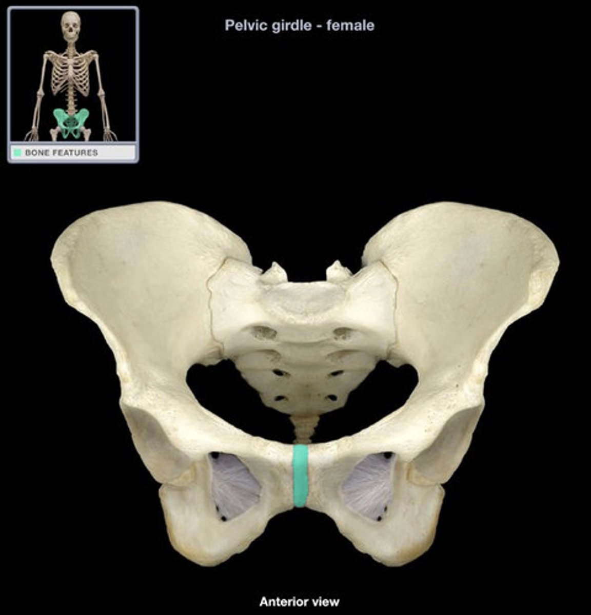
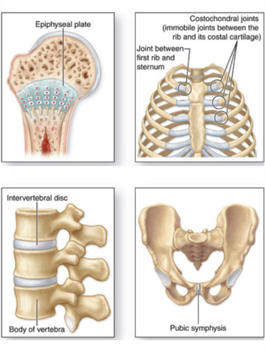
Cartilaginous joint
adjoining bones united by cartilage; no joint cavity
1) Synchondrosis
2) Symphasis
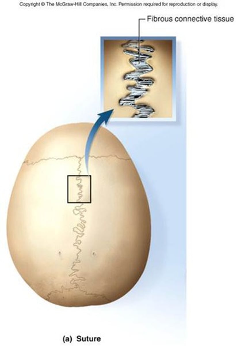
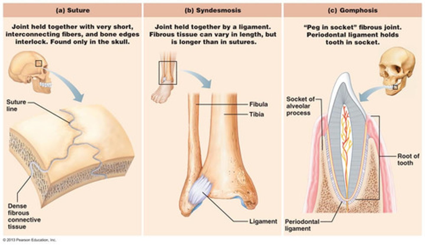
Fibrous joint
consists of inflexible layers of dense connective tissue, holds the bones tightly together
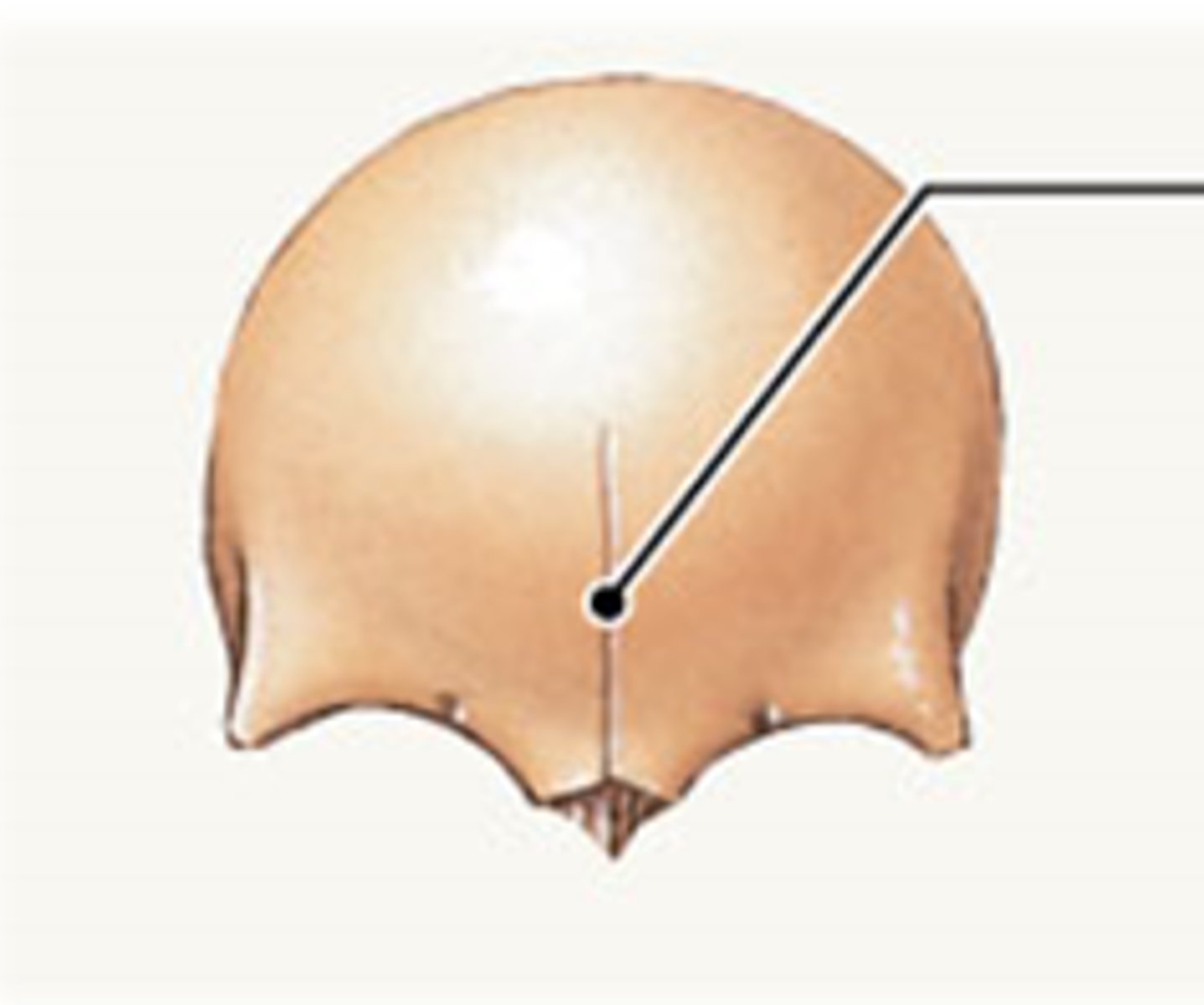
1) suture
2) gomphosis
3) syndesmosis
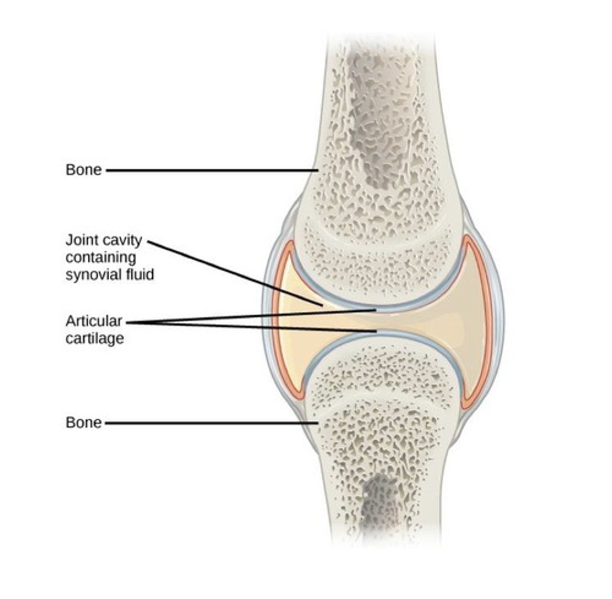
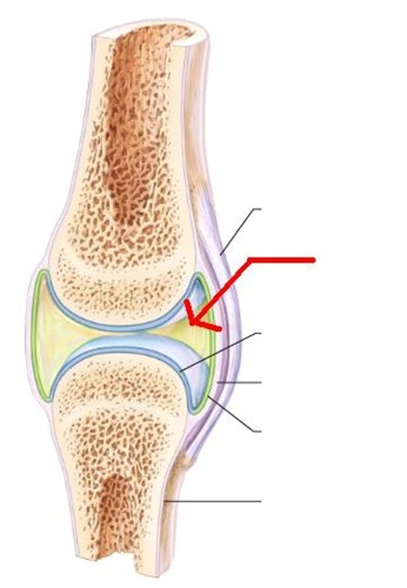
synovial joints
freely movable joints that have a joint cavity filled with synovial fluid
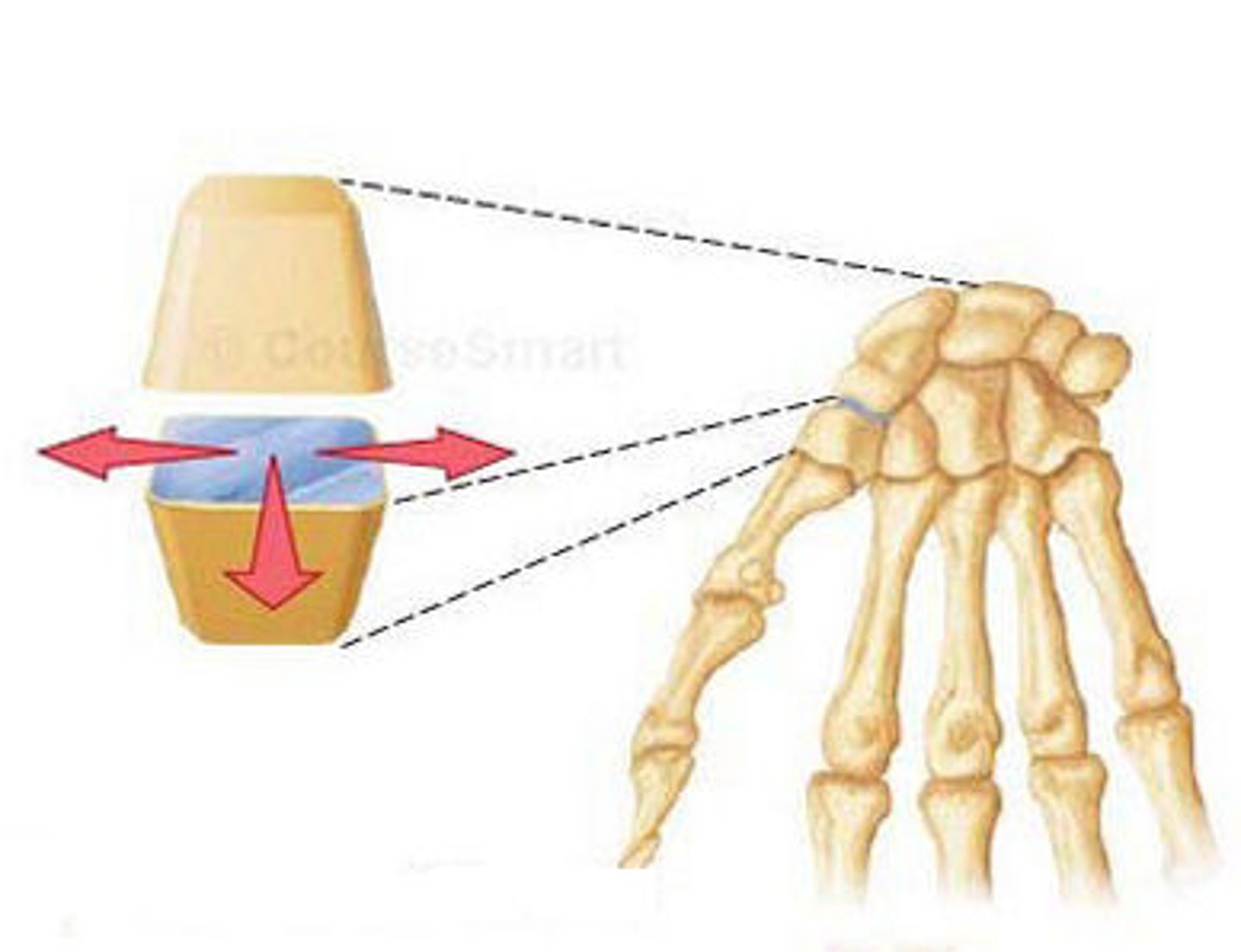
gliding joint
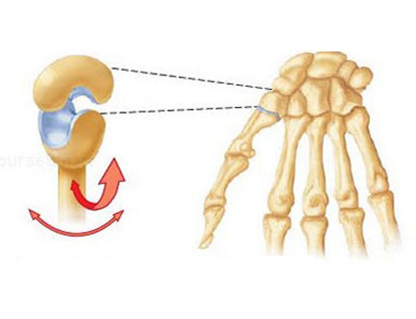
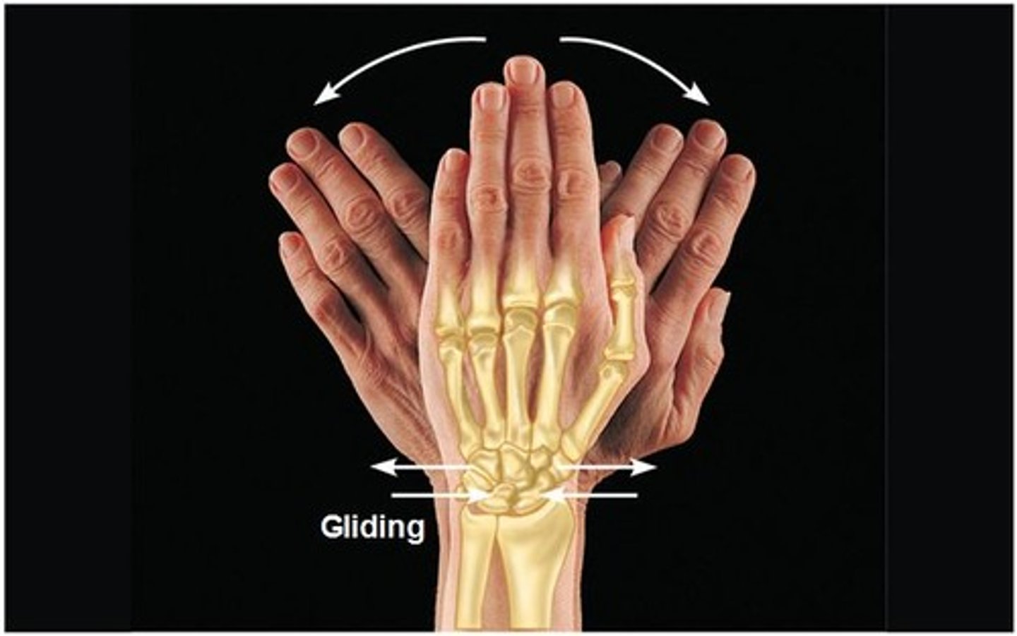
type of synovial joint: allows one bone to slide over another; found in wrist and ankles
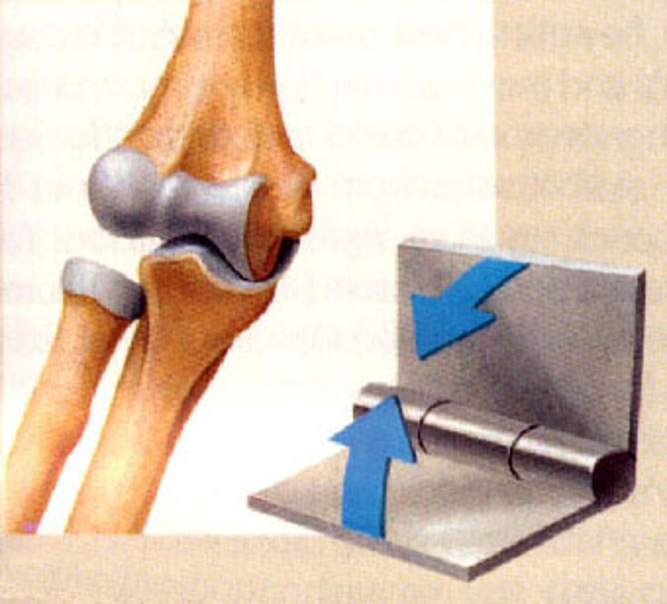
hinge joint
type of synovial joint which is formed between two or more bones where the bones can only move along the axis to flex or extend
(i.e. elbow and knee)
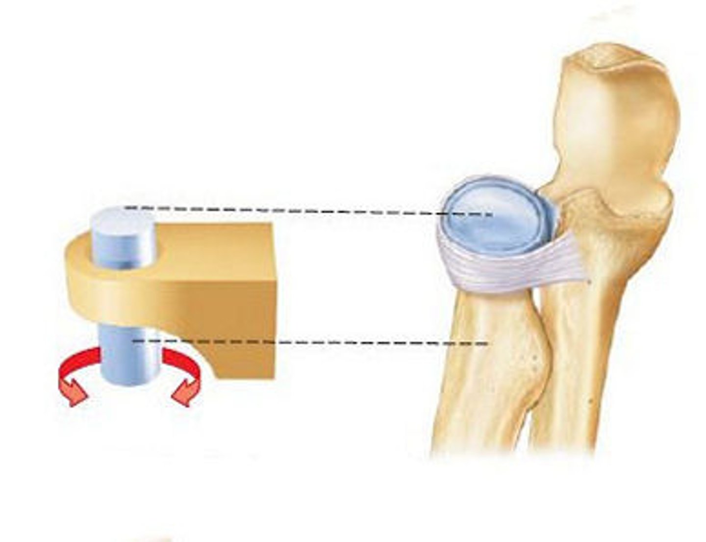
pivot joint
type of synovial joint: rotating bone turns around an axis; i.e. connection between radius/ulna
condylar joint
type of synovial joint:
convex oval surface articulates with concave oval
surface; biaxial (2 axes) rotation (wrist, metacarpophalangeal 2-5 joints)
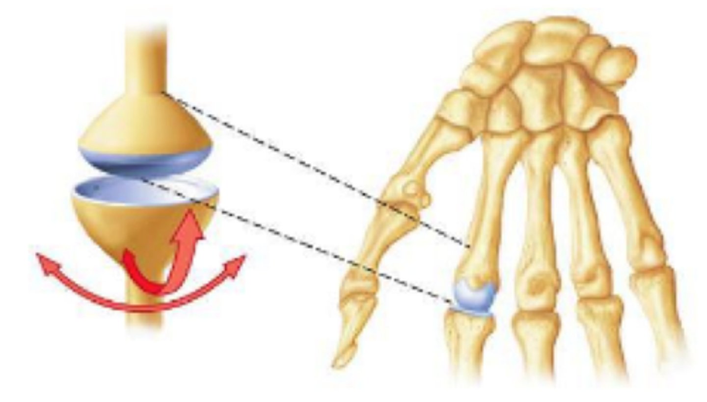
saddle joint
type of synovial joint: found at the base of each thumb; allows grasping and rotation
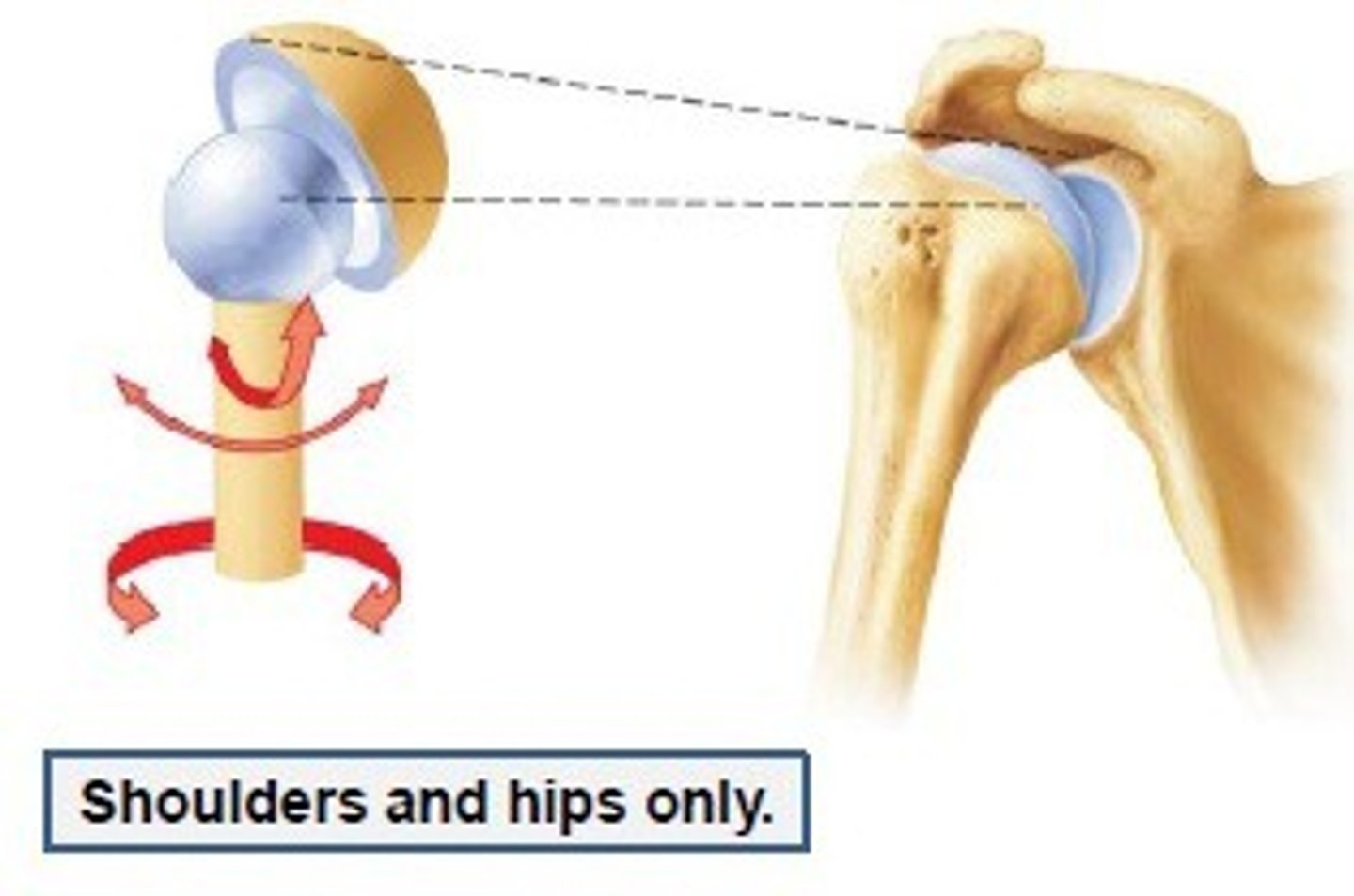
ball and socket joint
type of synovial joint: allows movement in all 3 planes (i.e. hip and shoulder)
gliding
Two surfaces slide past each other
Between carpal or tarsal bones
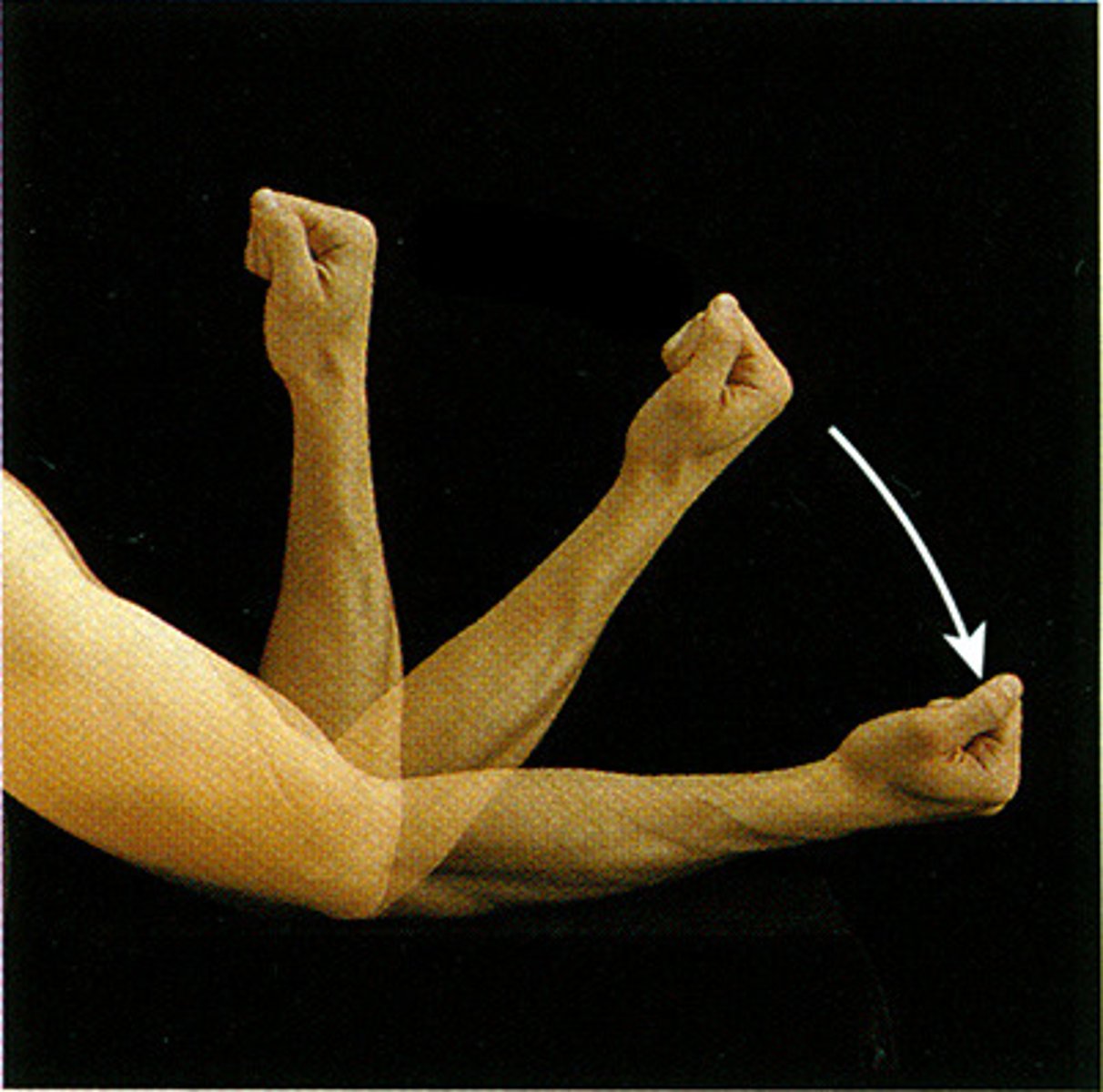
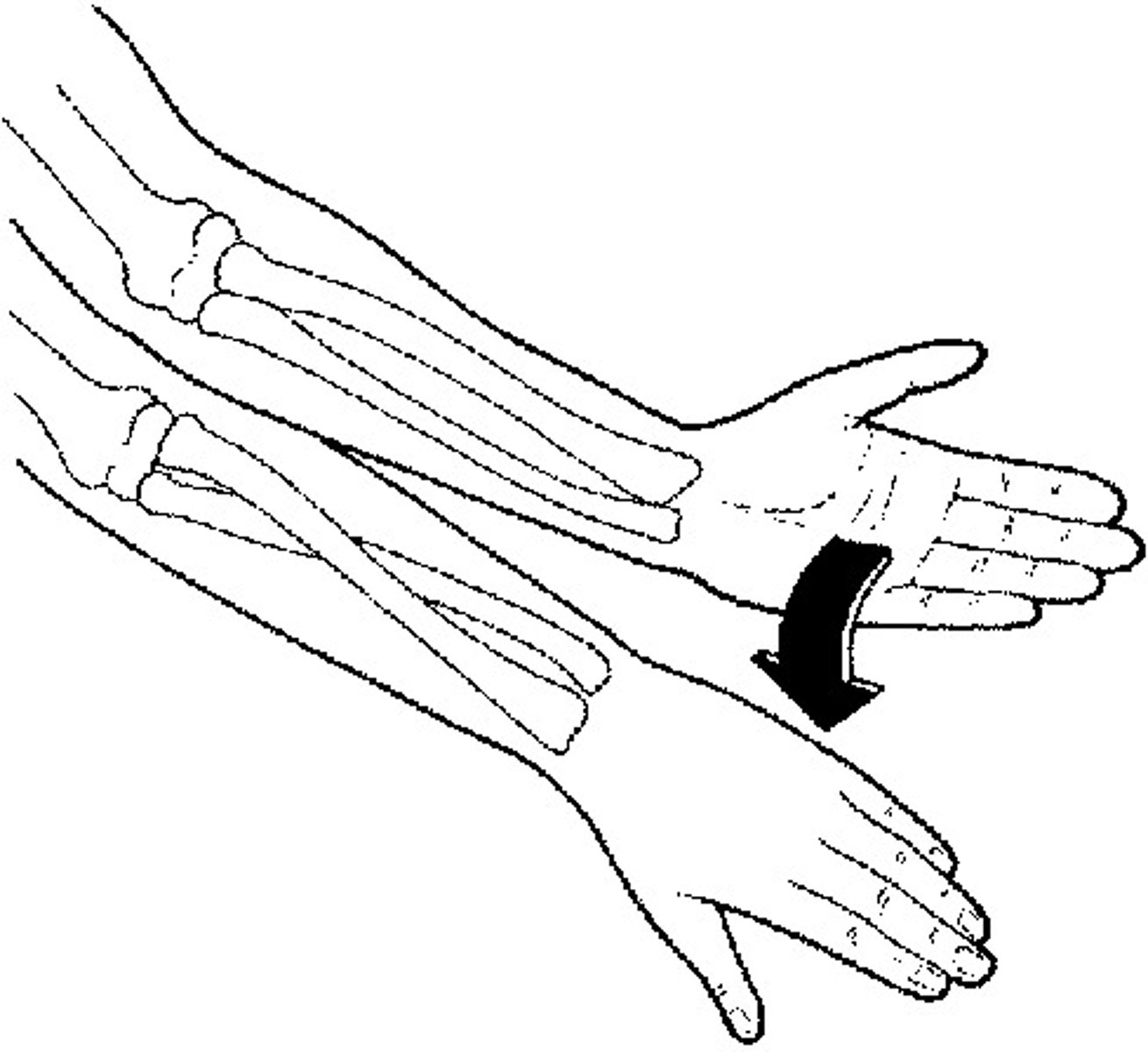
flexion
Decreases the angle of a joint
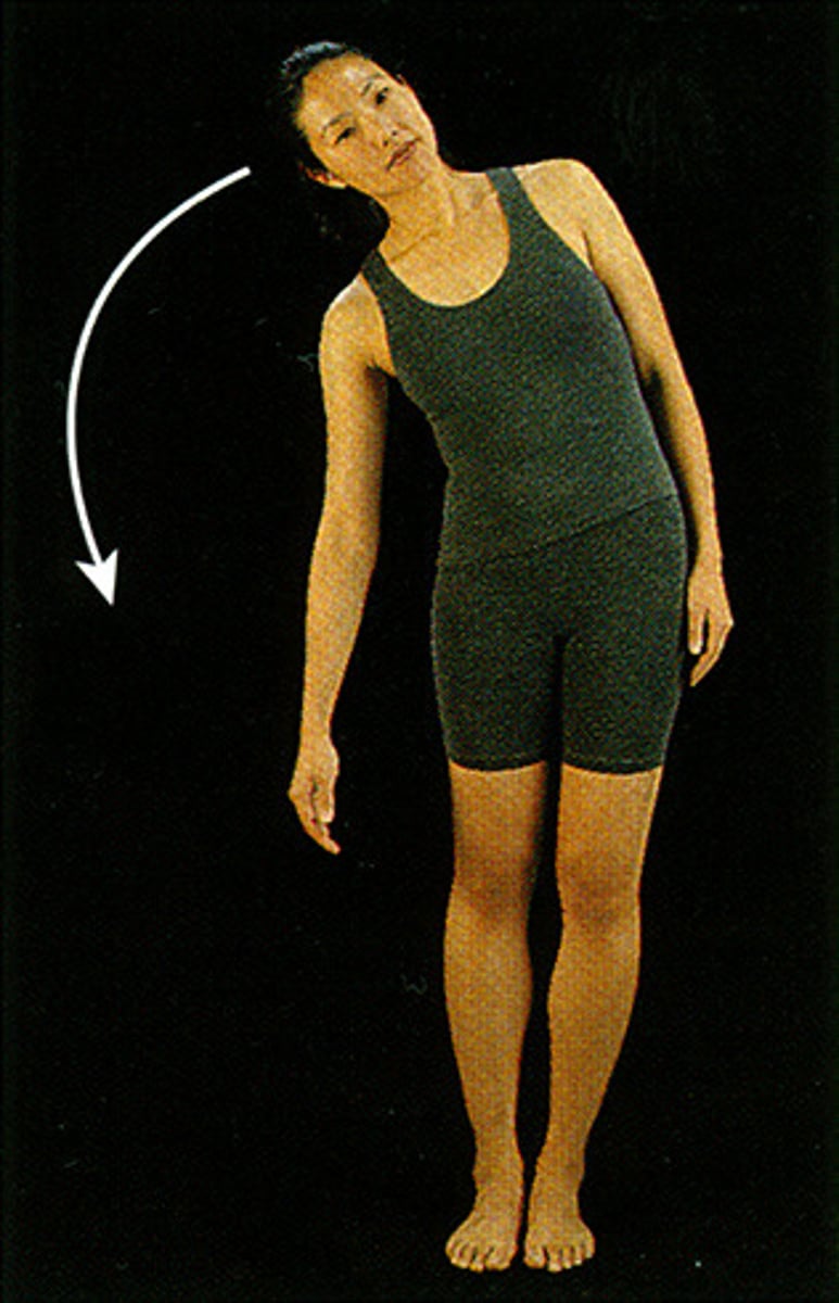
lateral flexion
movement of trunk in frontal plane
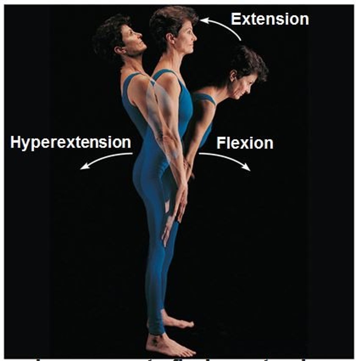
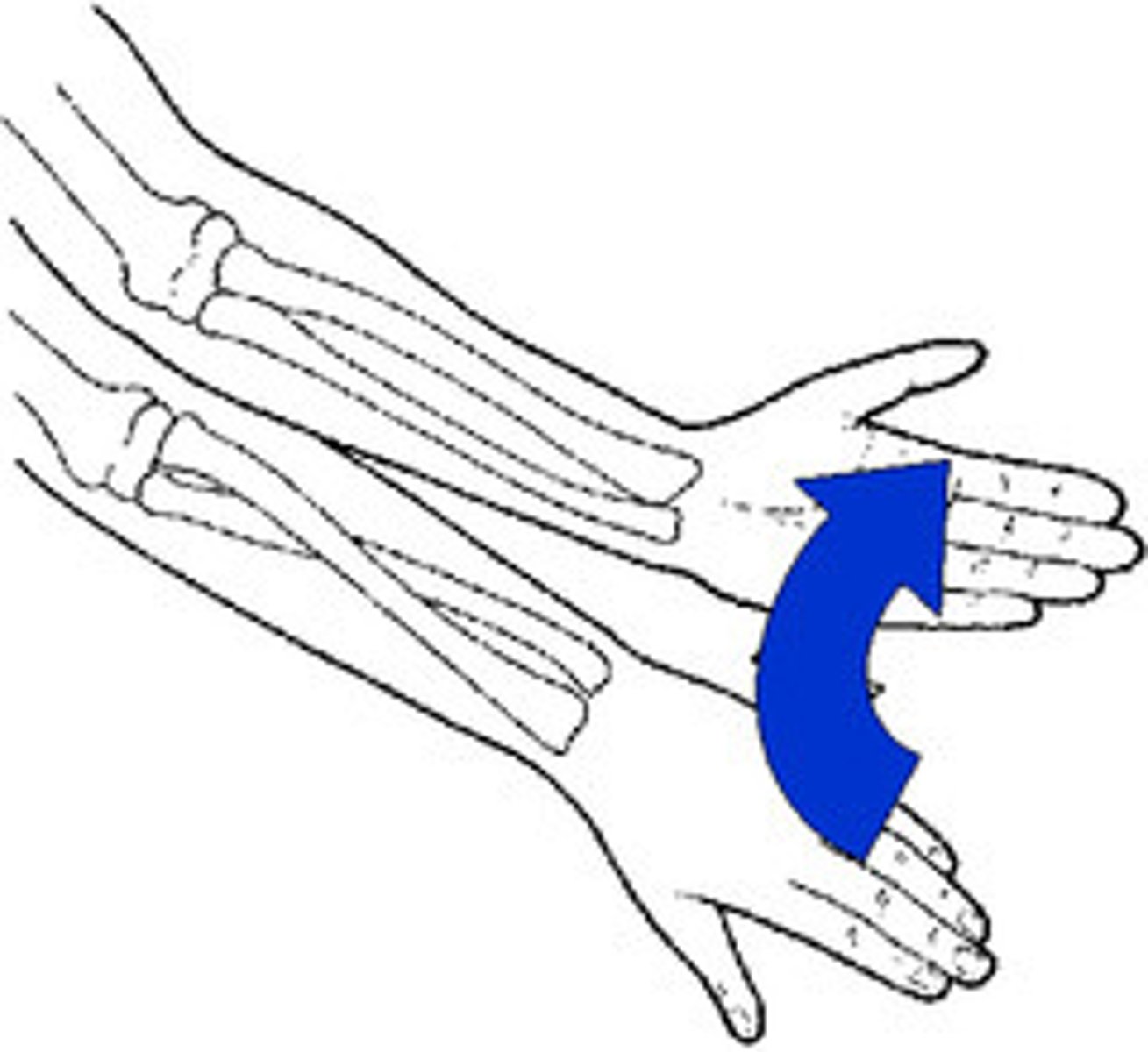
extension
increase angle between bones
hyperextension
extension beyond anatomical position
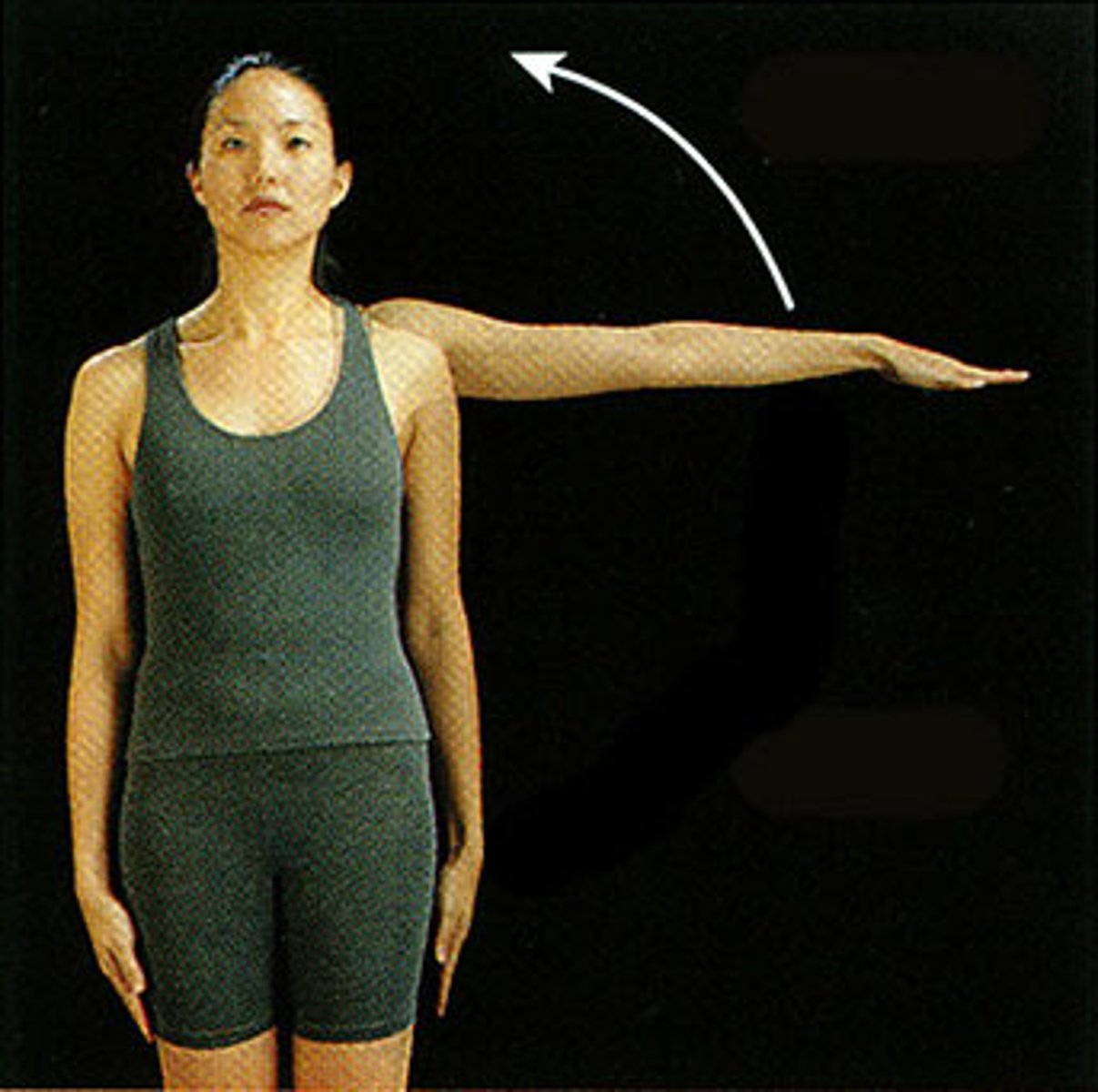
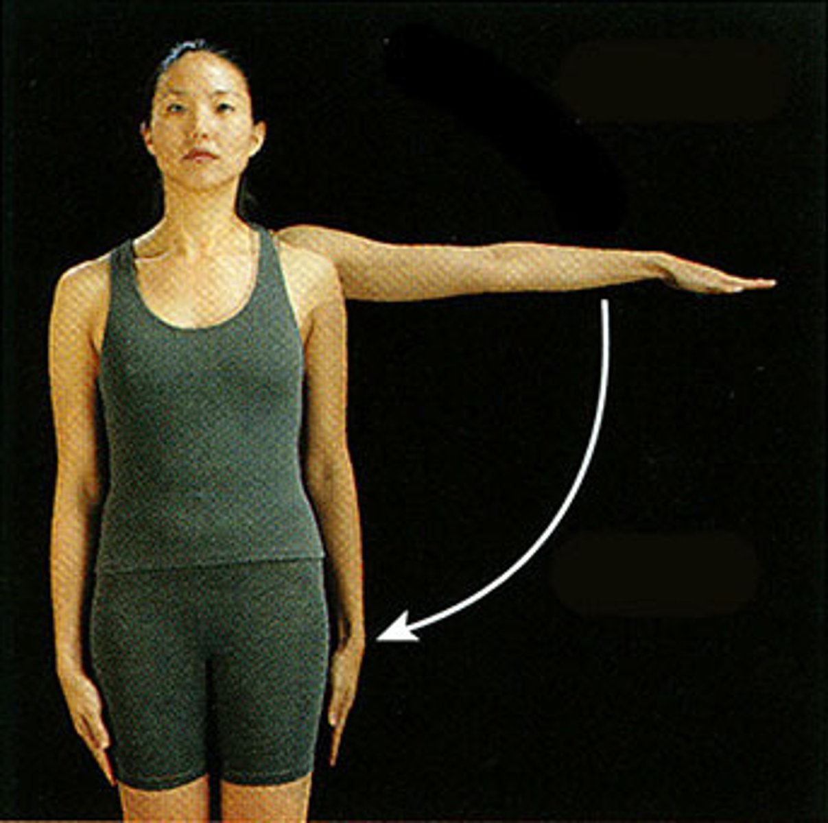
abduction
movement away from the midline
adduction
movement toward the body midline
medial rotation
rotational movement towards the midline

lateral rotation
rotation away from the midline
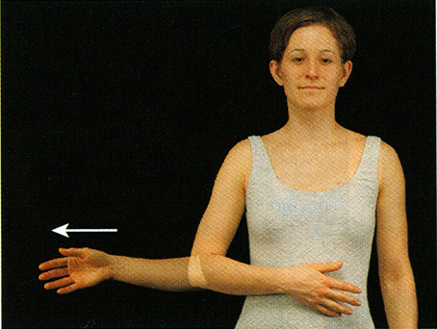
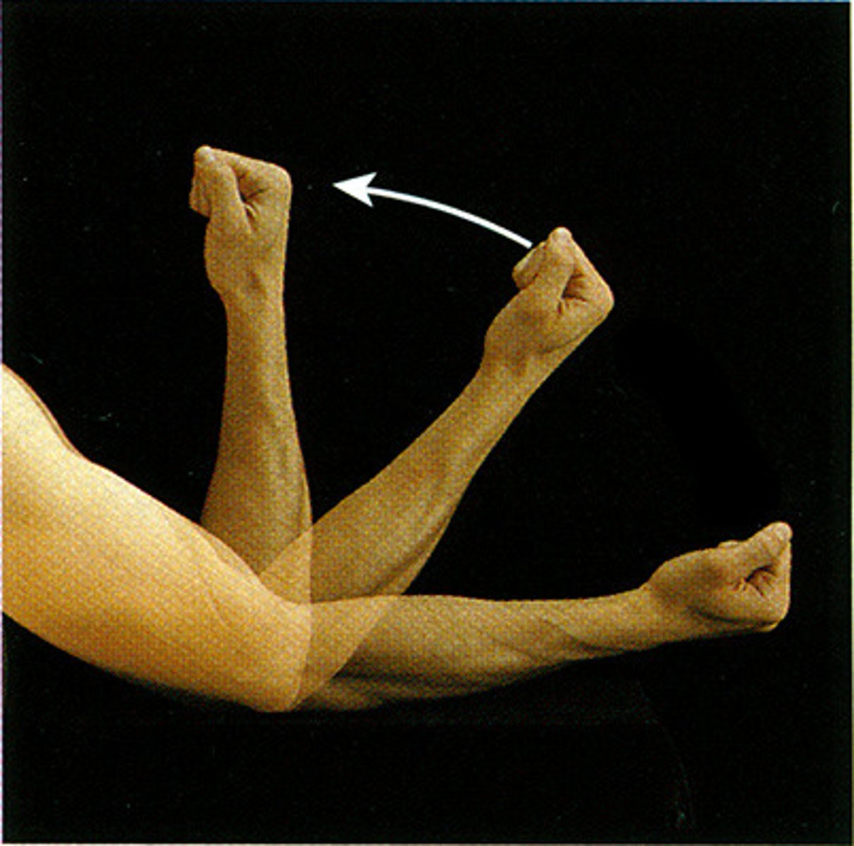
pronation
movement that turns the palm down
supination
movement that turns the palm up
inversion
Turning the sole of the foot inward
eversion
turning the sole of the foot outward
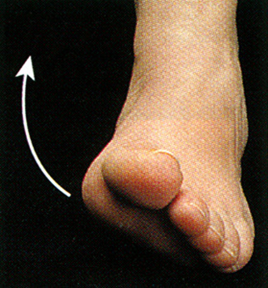
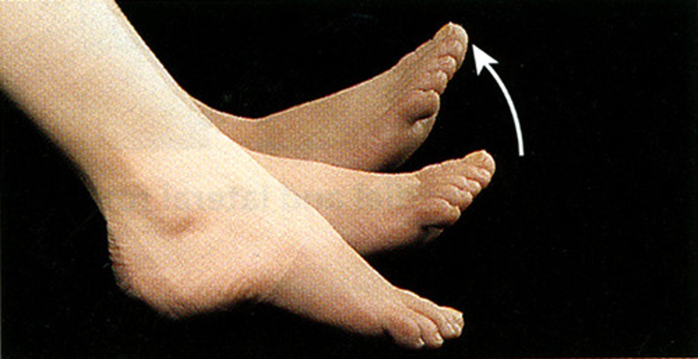
dorsiflexion
bending of the foot or the toes upward
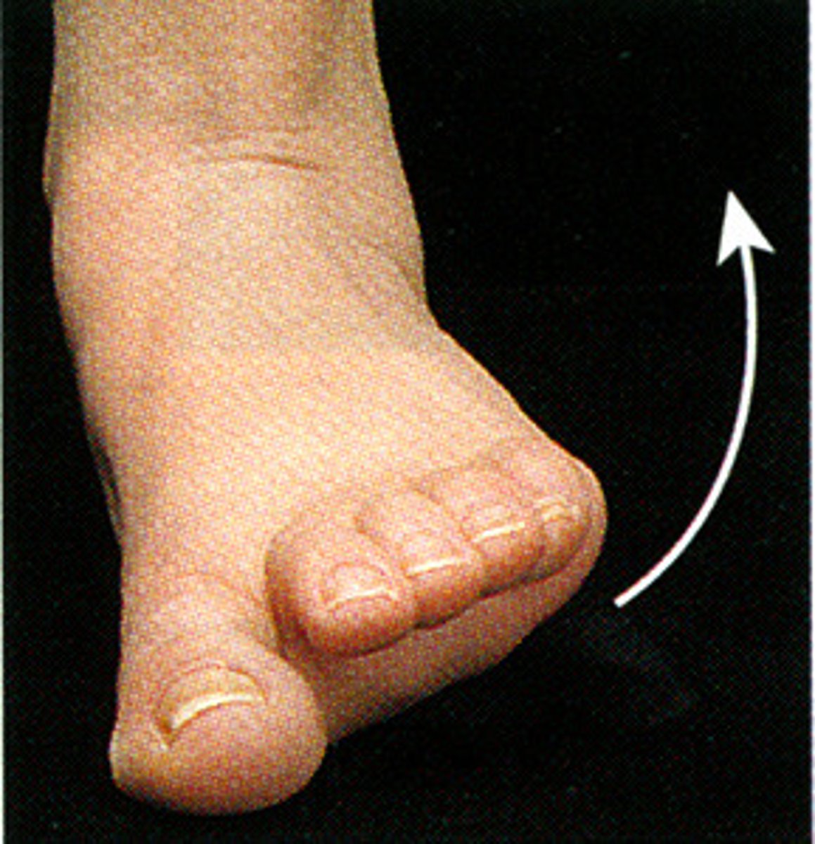
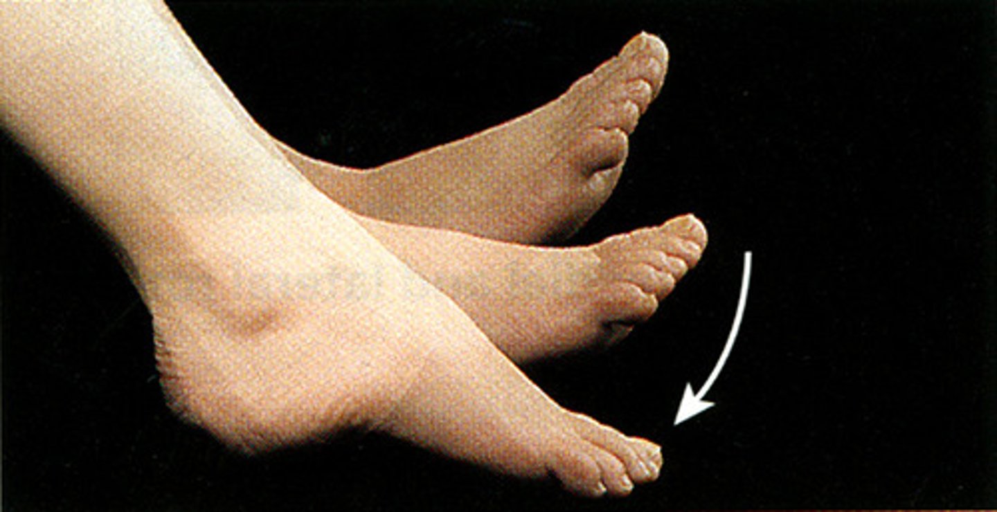
plantar flexion
bends the foot downward at the ankle
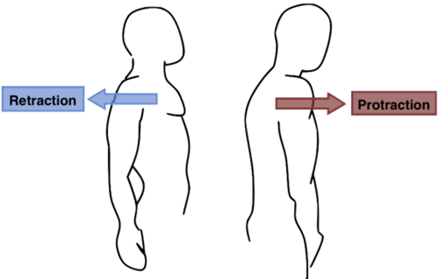
protraction
moving a body part forward

retraction
moving a body part backward
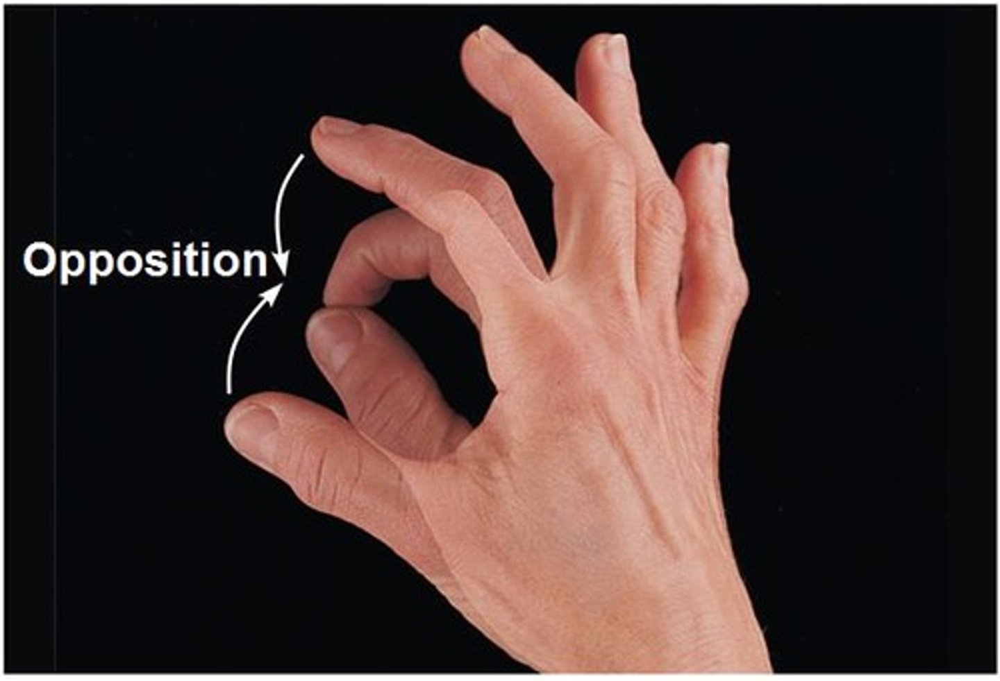
opposition
Movement of the thumb to touch the fingertips
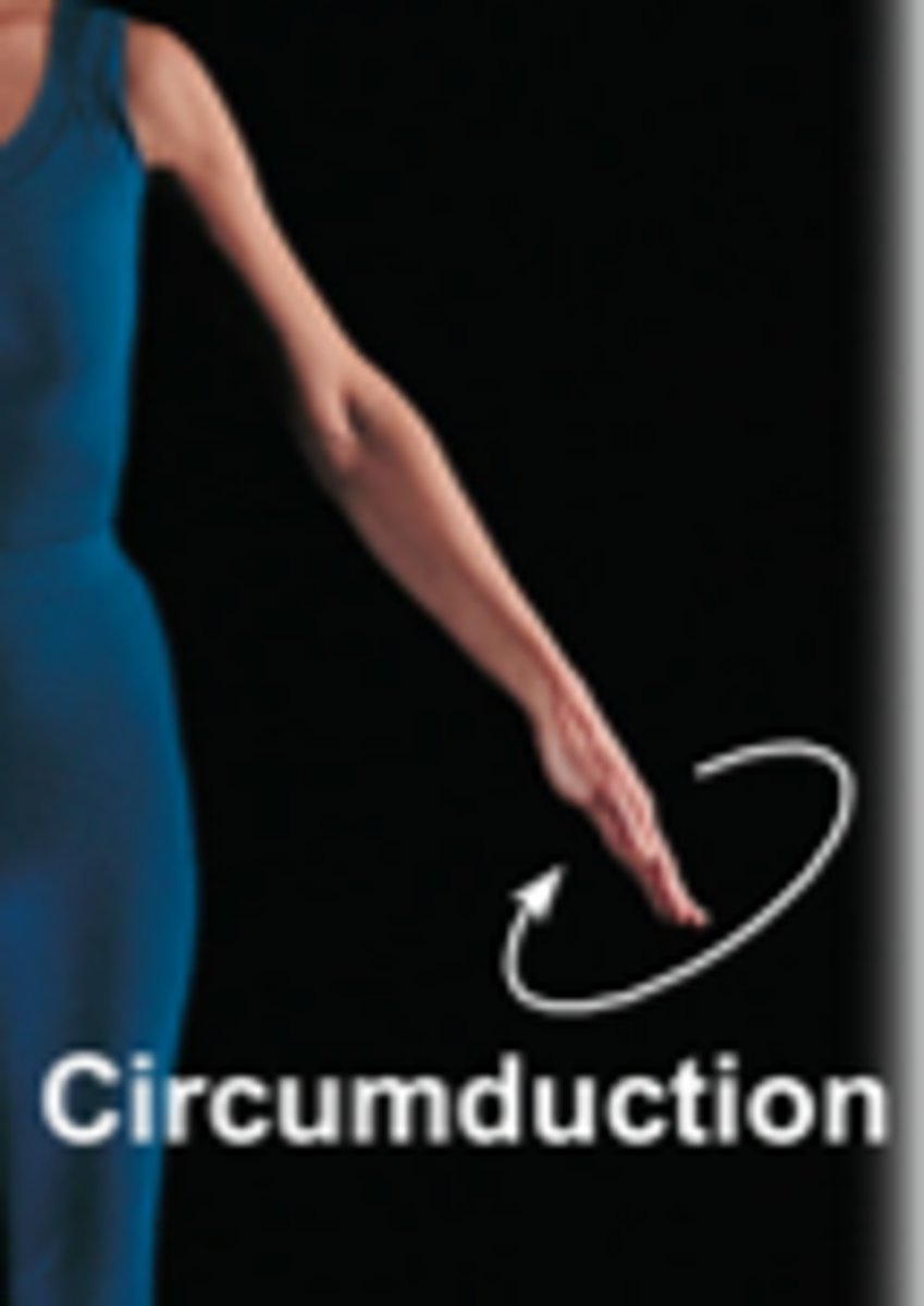
circumduction
circular movement of a limb at the far end, combination of flexion, extension, abduction and adduction
elevation
raising a body part
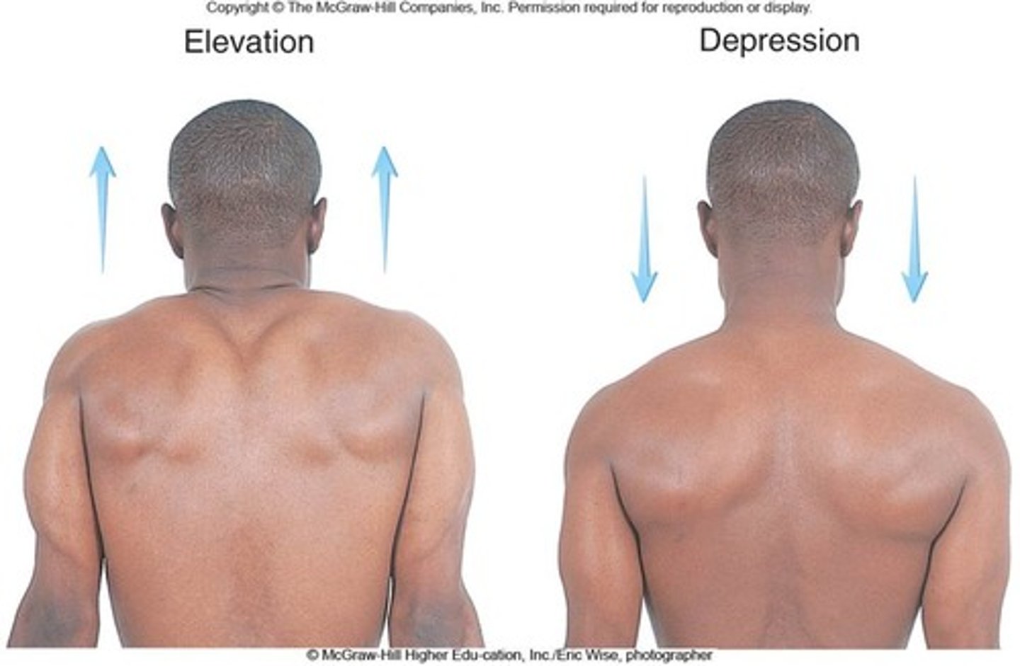
depression
lowering a body part

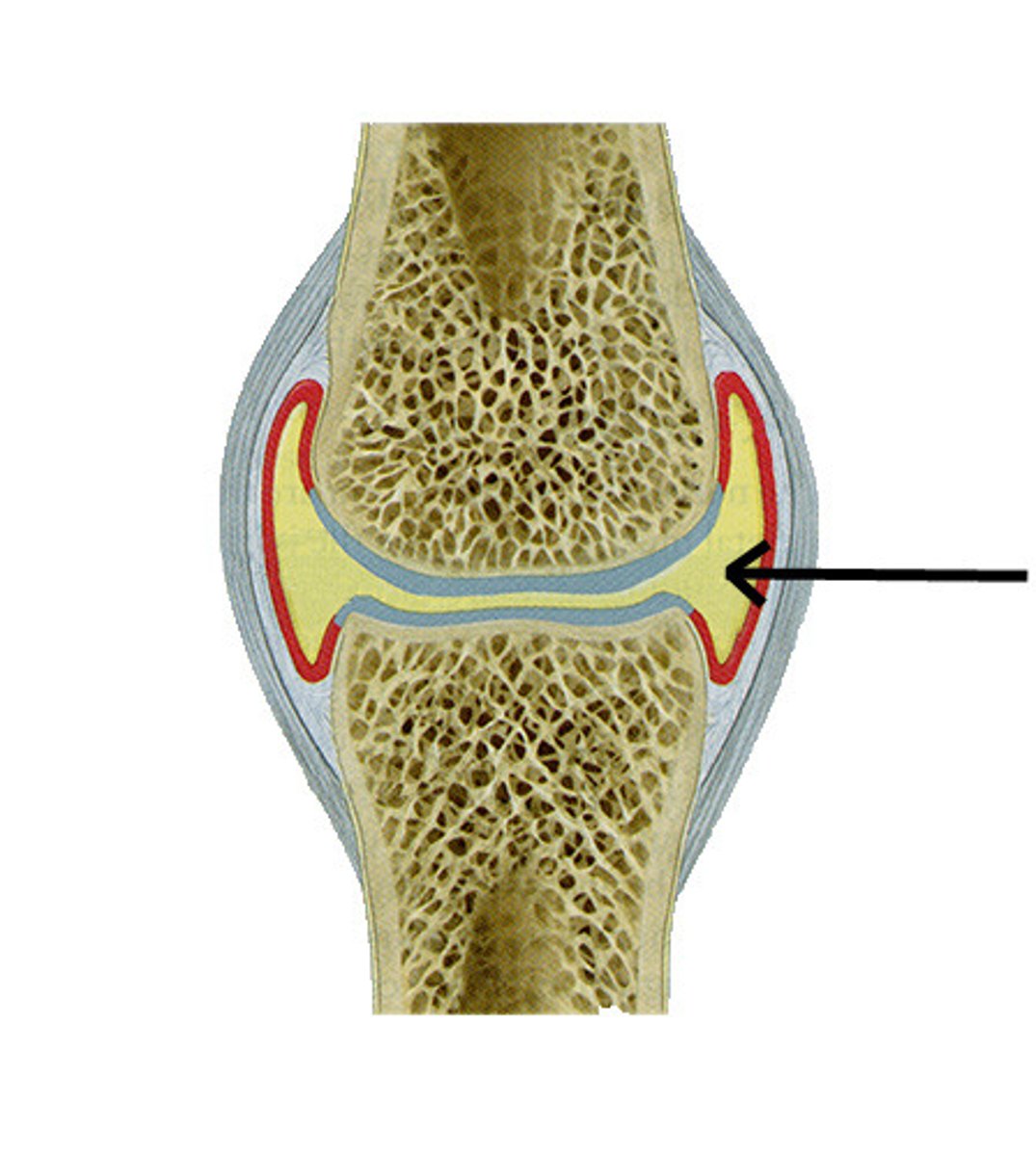
articular capsule
Fibrous envelope that encloses a synovial joint
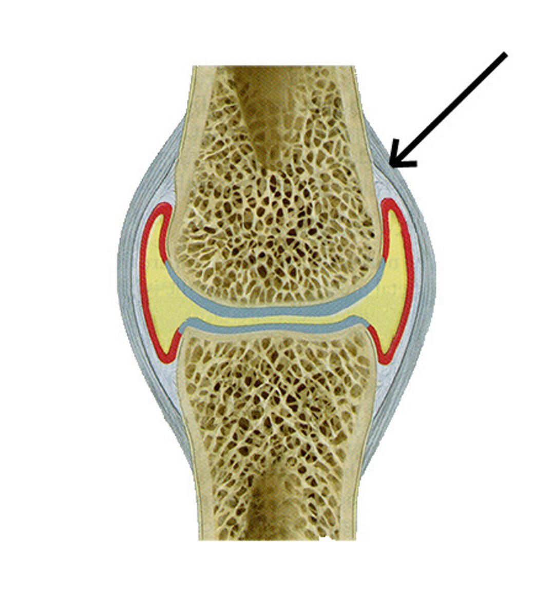
articular cartilage
covers the surfaces of bones where they come together to form joints
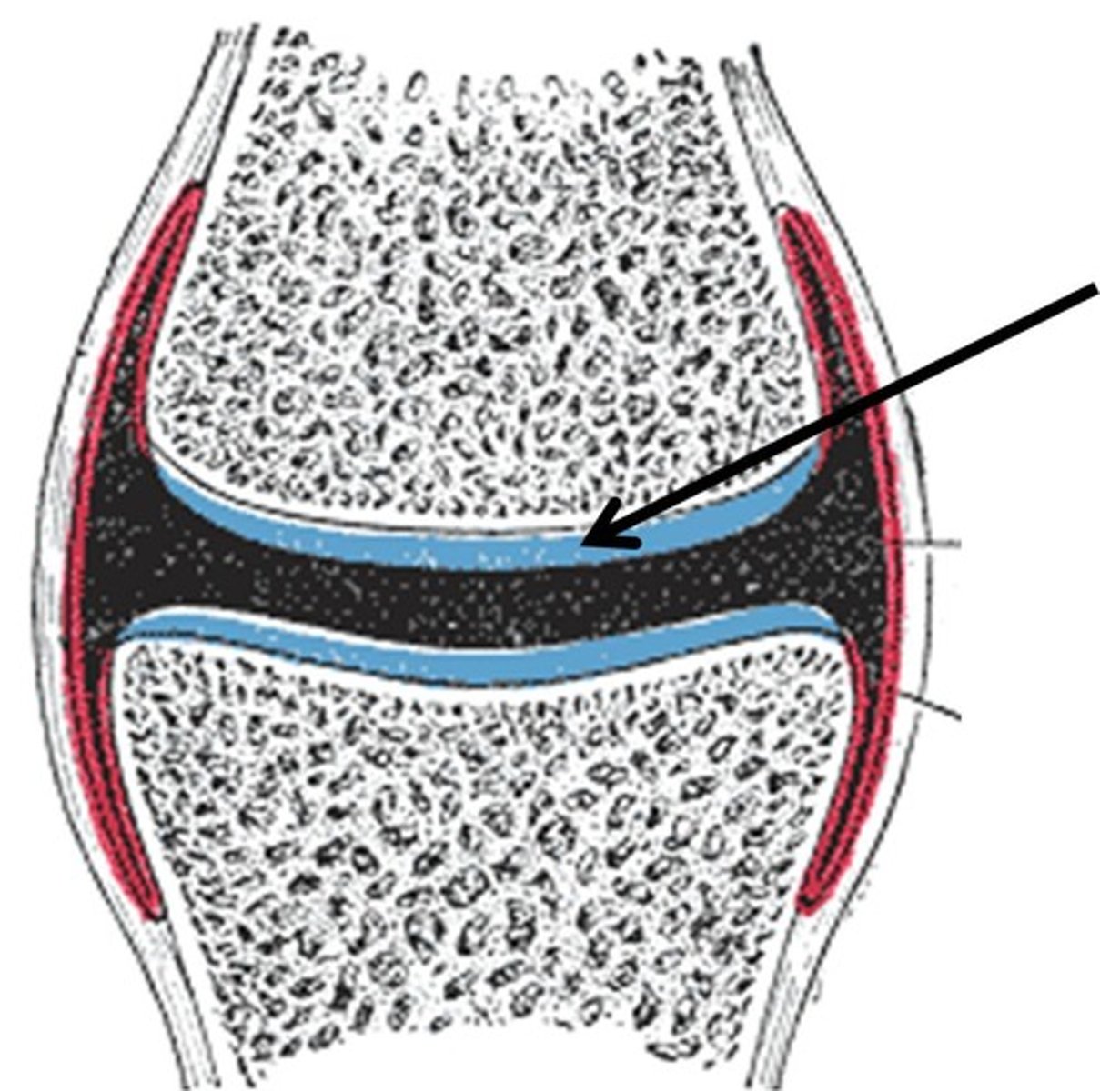
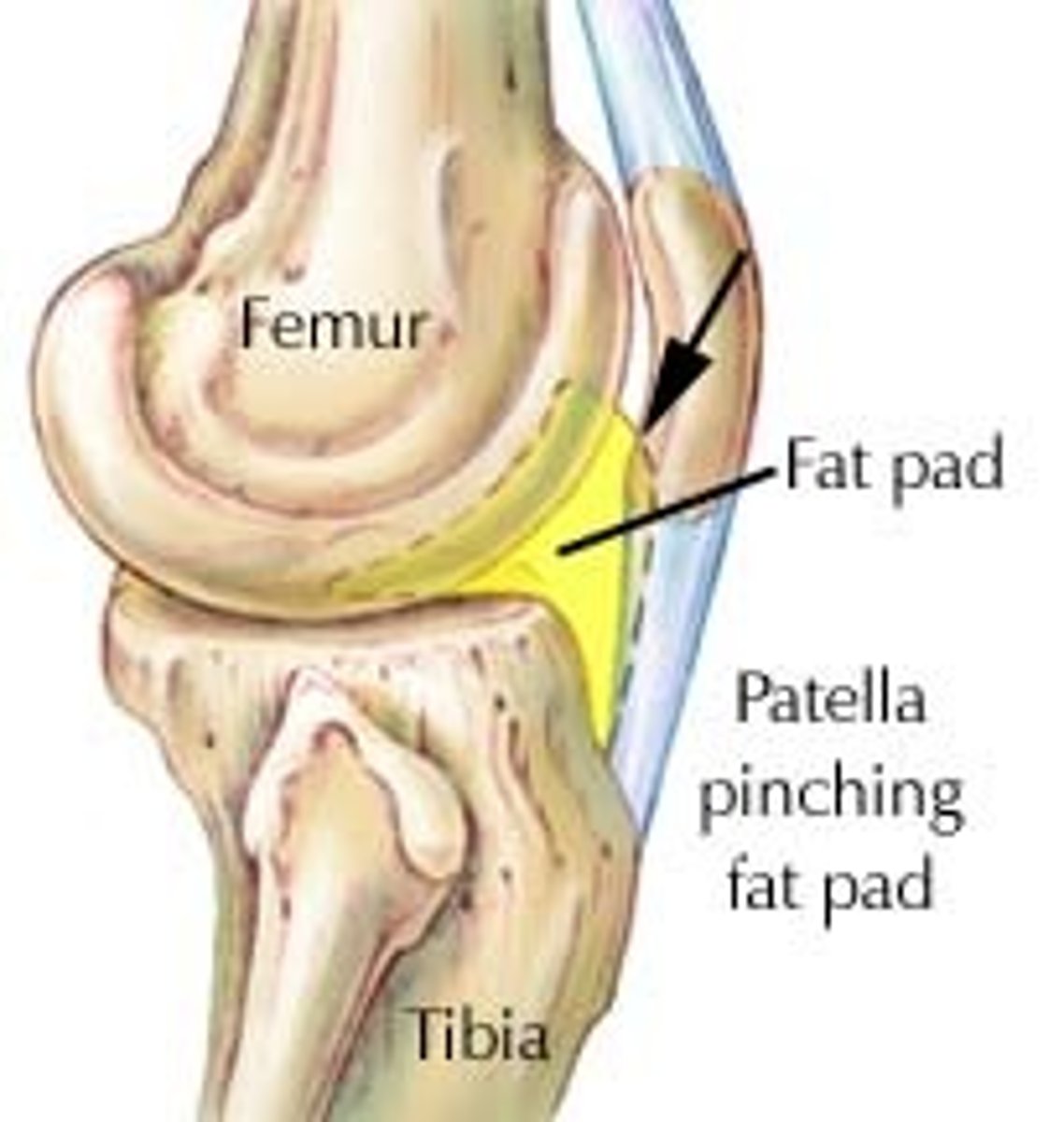
fat pads
localized masses of adipose tissue covered by a layer of synovial membrane
capsular ligament
ligaments within the wall of the capsule; thickening in the capsule wall
intracapsular ligaments
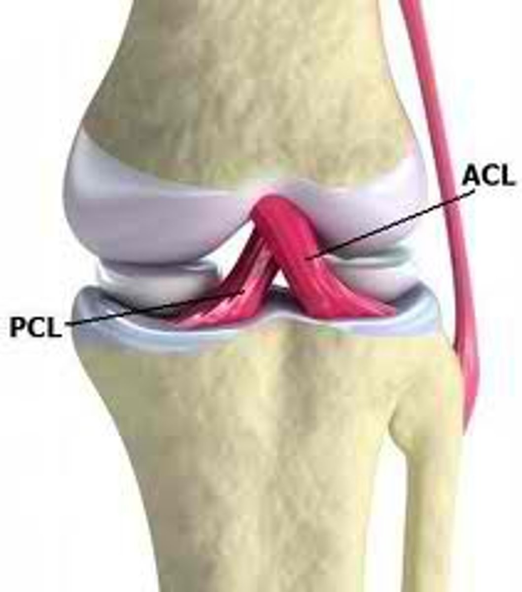
stabilizing ligaments located inside joint capsule (ie ACL, PCL)
extracapsular ligaments
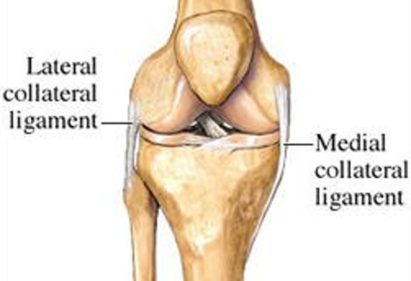
stabilizing ligaments located outside joint capsule (ie LCL, MCL)
synovial membrane and fluid
membrane lines inside of joint capsule except at actual articulation of articular cartilages. Secretes fluid to lubricate joints
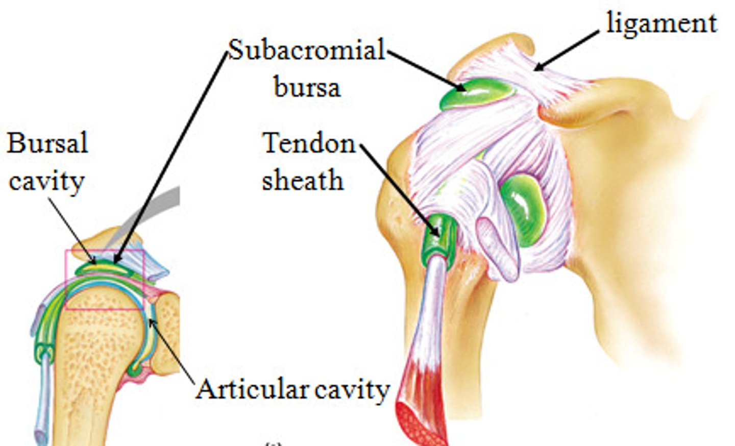
bursae and tendon sheaths
friction-reducing structures commonly associated with synovial joints
fibrocartilage pads
Articular discs may be present within capsule. (ie meniscus in knee)
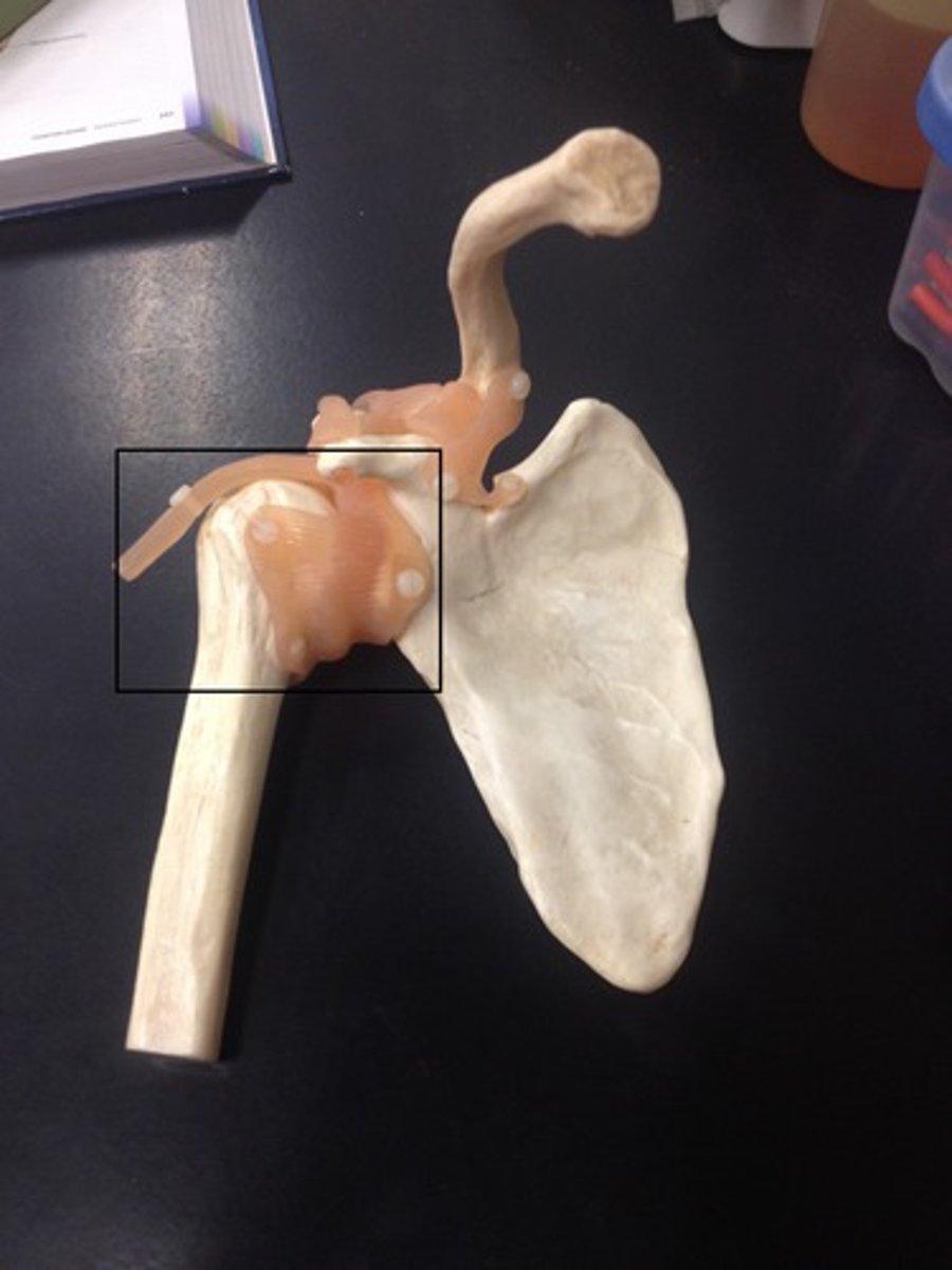
acromioclavicular ligament
fx: connects the clavicle to the acromion
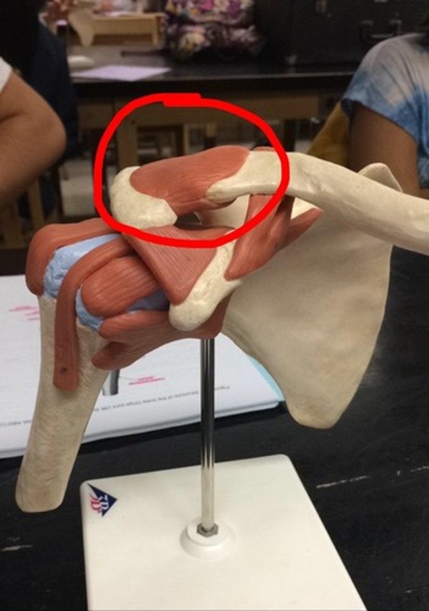
coracoclavicular ligament
Fx: connects the clavicle to the coracoid process
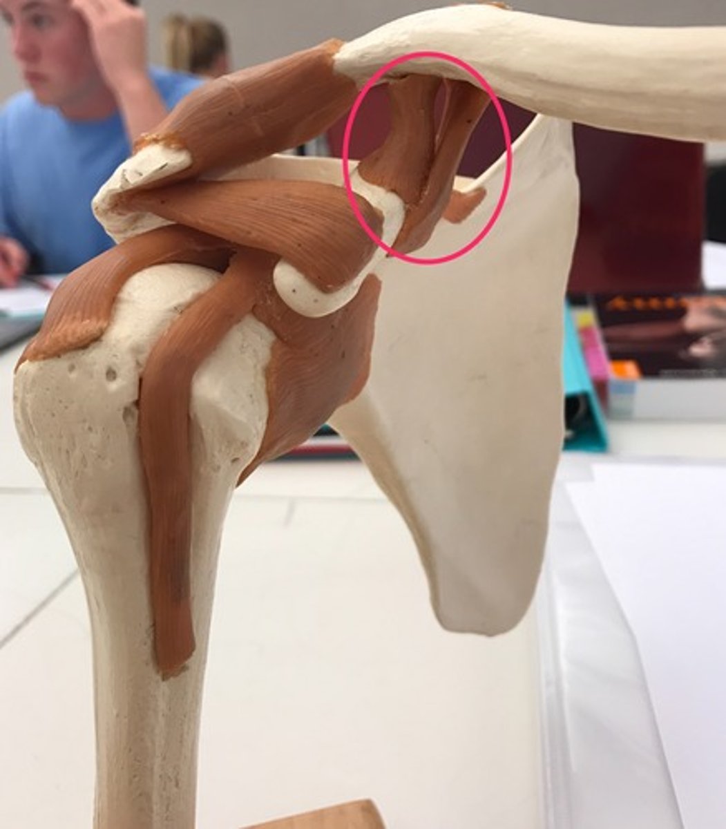
coracoacromial ligament
Fx: connection between the coracoid process and the acromion

glenohumeral ligament
Fx: Connects humerus to the glenoid cavity
iliofemoral ligament
Fx: connects ilium and femur
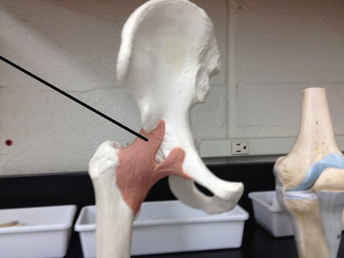
ischiofemoral ligament
Fx: connects ischium to femur
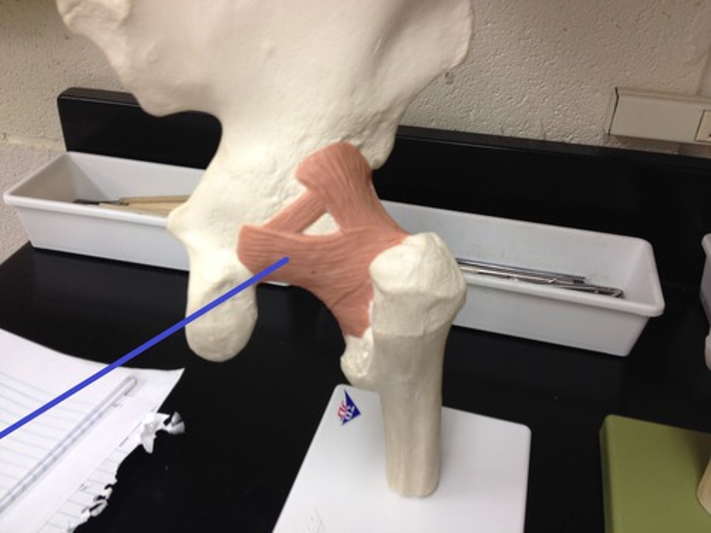
pubofemoral ligament
Fx: connects pubis to femur
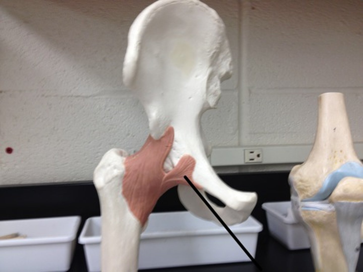
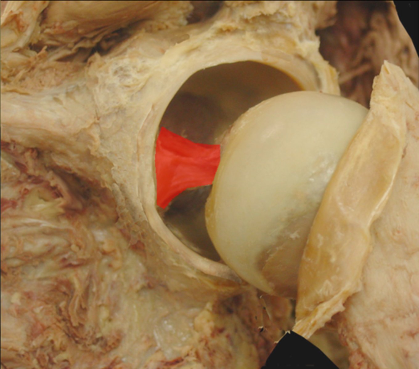
ligamentum teres
Fx: connects the acetabulum to favea capitis
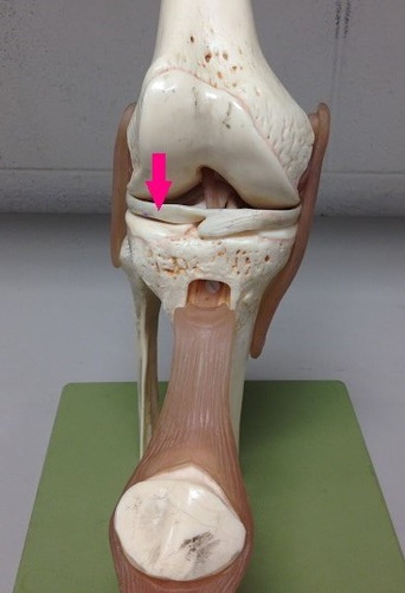
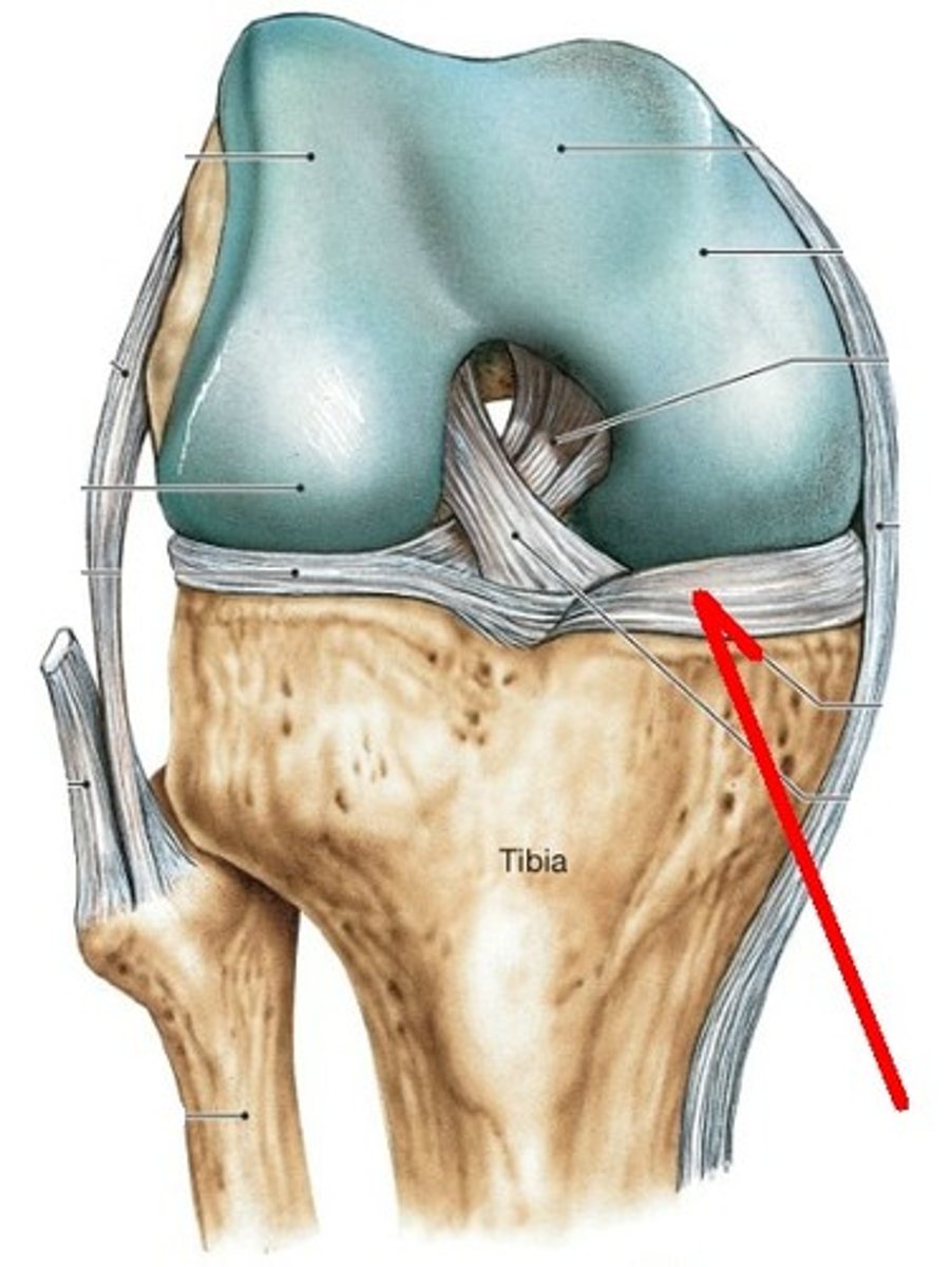
anterior cruciate ligament
Fx: prevent anterior movement of the tibia on the femur
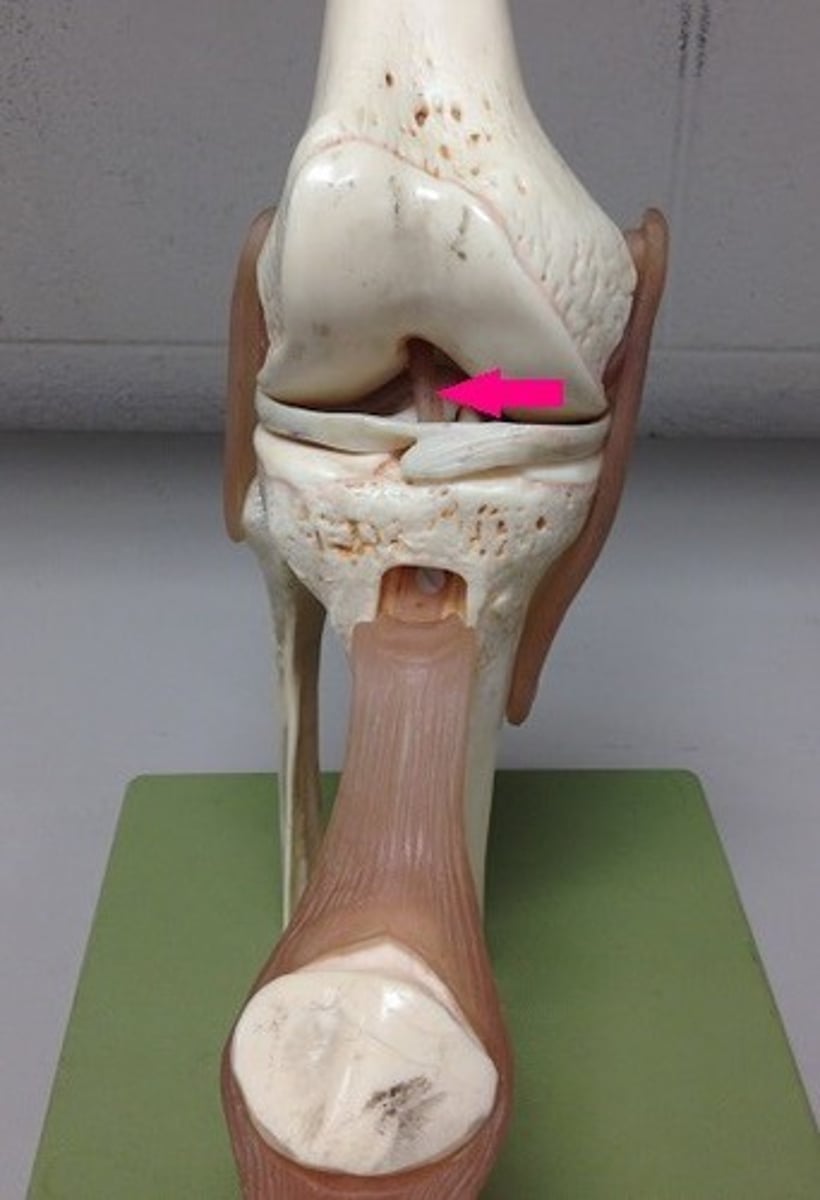
posterior cruciate ligament
Fx: prevents posterior displacement of tibia

tibial collateral ligament
Fx: connects the medial epicondyle of the femur to the tibia (MCL)
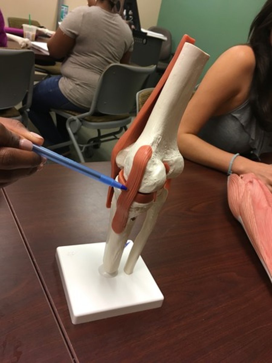
fibular collateral ligament
Fx: connects the lateral epicondyle of the femur to the fibula (LCL)
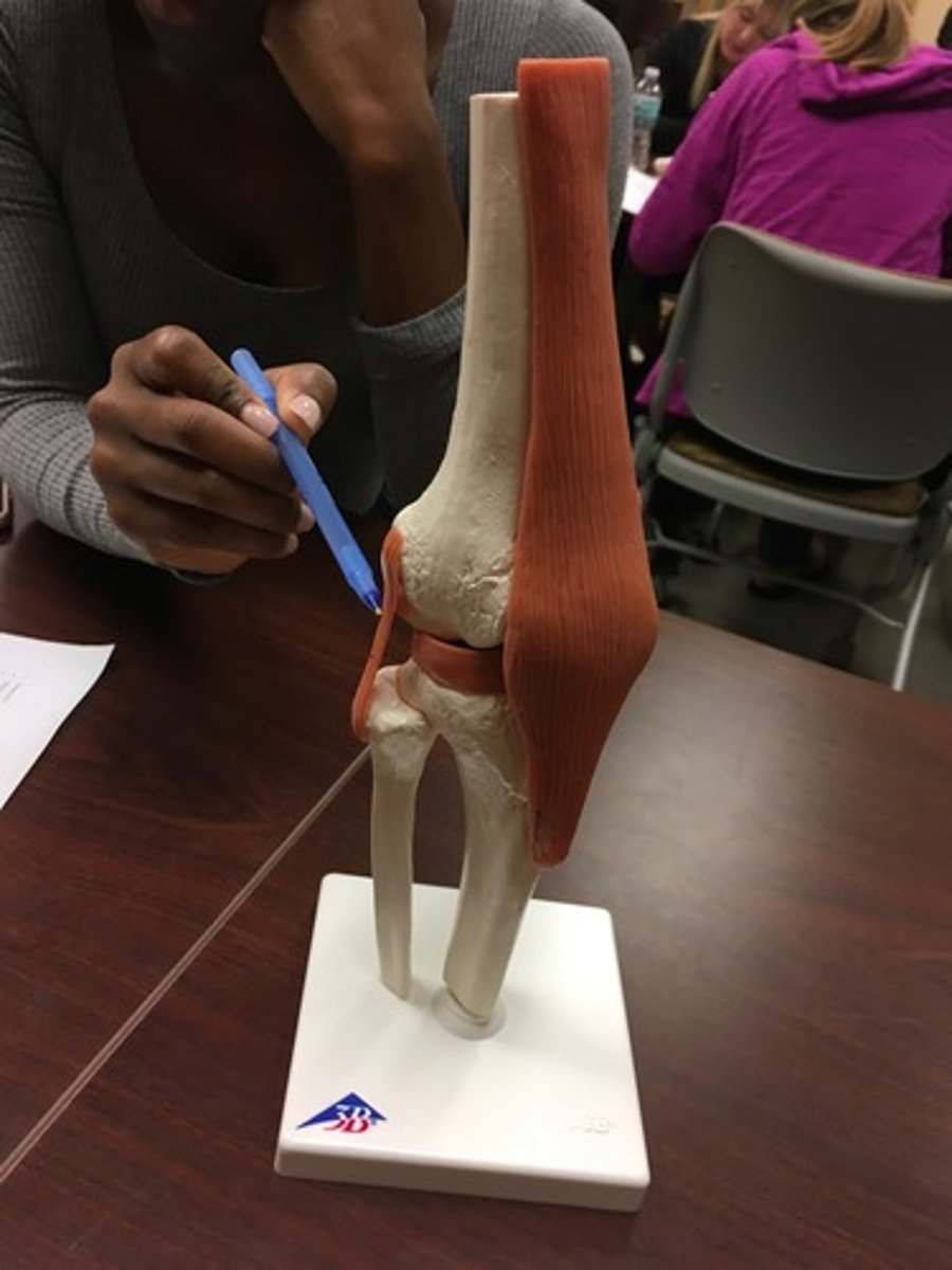
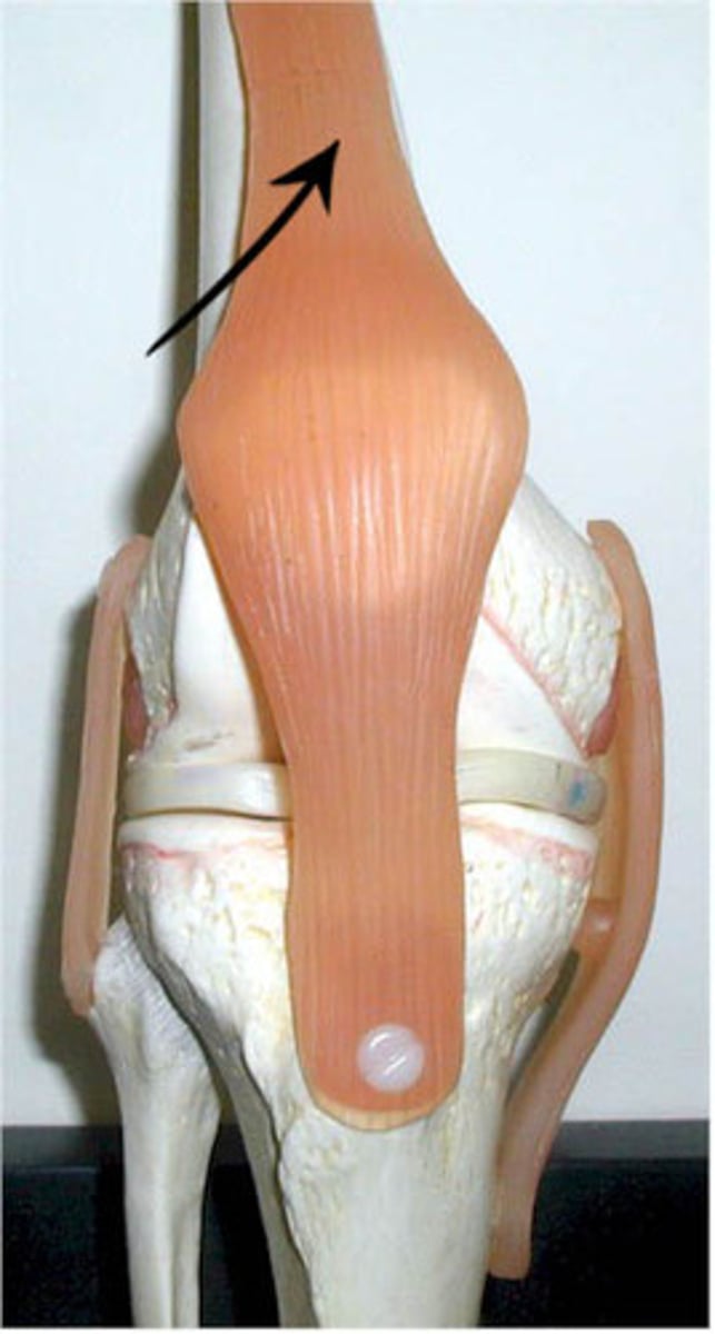
patellar ligament
Fx: connects patella to tibial tuberosity
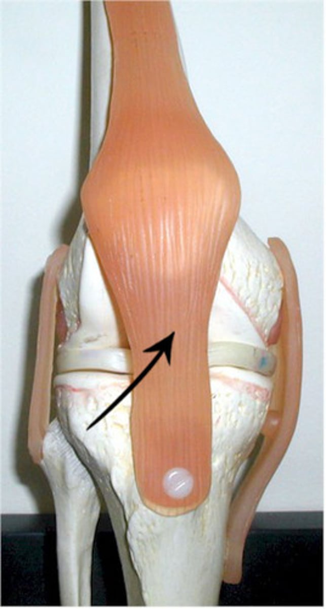
quadriceps tendon
common tendon for quadriceps group, attaches to patella
medial meniscus
Cartilage in the knee between the femoral condyle and the medial tibial plateau

lateral meniscus
cartilage in the knee between the lateral femoral condyle and the lateral tibial plateau