electron microscopy - part 2 (electron optics)
1/18
There's no tags or description
Looks like no tags are added yet.
Name | Mastery | Learn | Test | Matching | Spaced | Call with Kai |
|---|
No analytics yet
Send a link to your students to track their progress
19 Terms

SEM operation
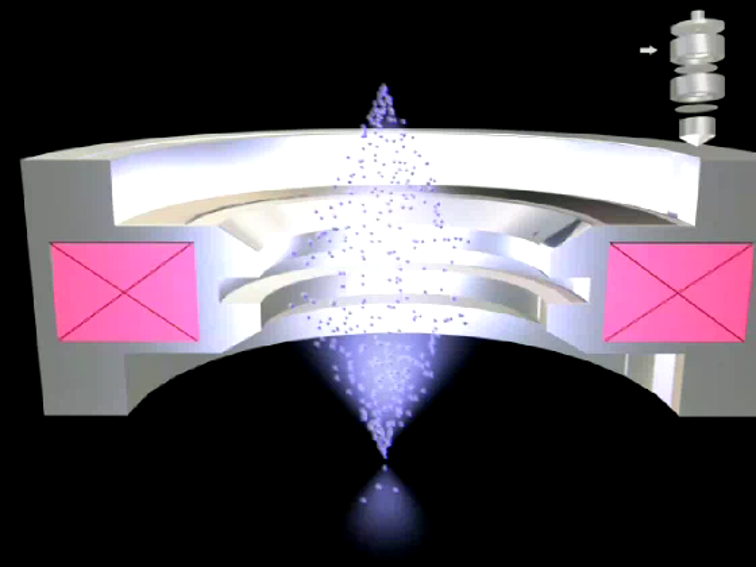
Magnetic Electron Lenses
The first magnetic electron lenses were developed by M. Knoll and E. Ruska in Germany in 1932.
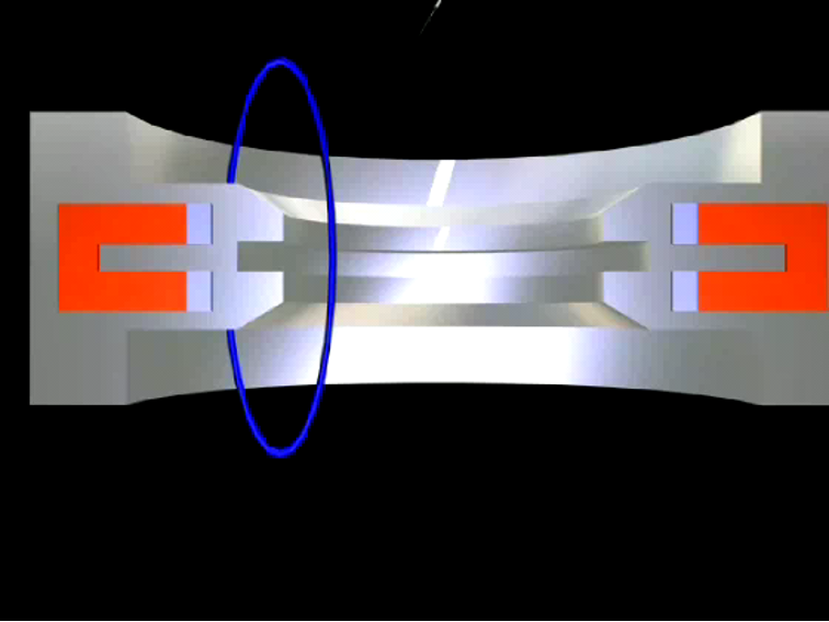
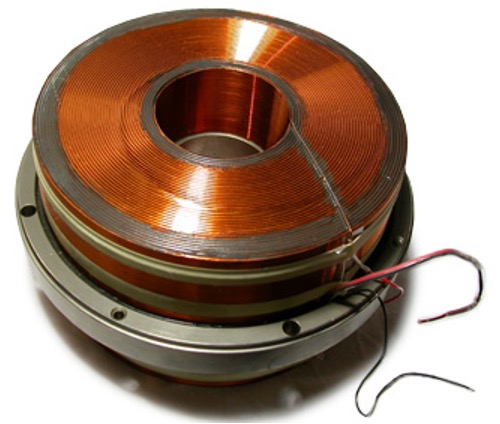
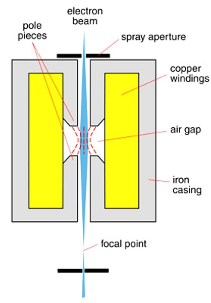
Magnetic lens consist of two circularly symmetric iron pole-pieces with copper windings with a hole in centre through which beam passes.
The two pole pieces are separated by "air-gap" where focusing actually takes place.
Electrons have a charge and their direction of travel can be altered by an electromagnetic field.
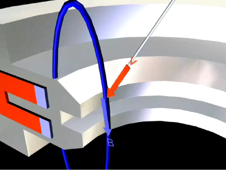
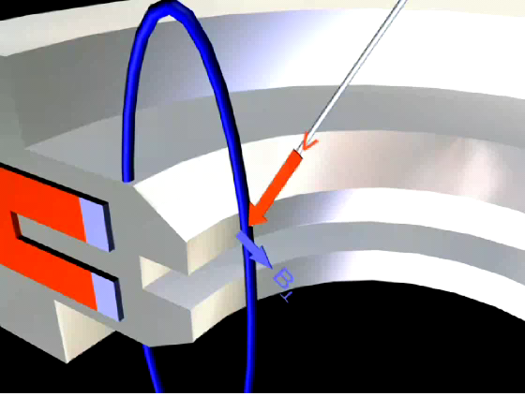
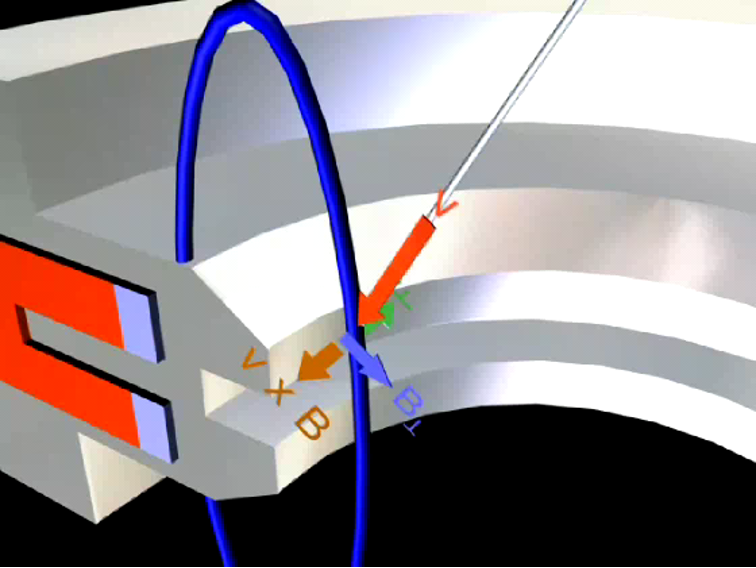
An electron traveling in off-axis to a uniform magnetic field follows a helical path.
Electrons can be brought to focus by engineering the electrostatic and/or magnetic fields.
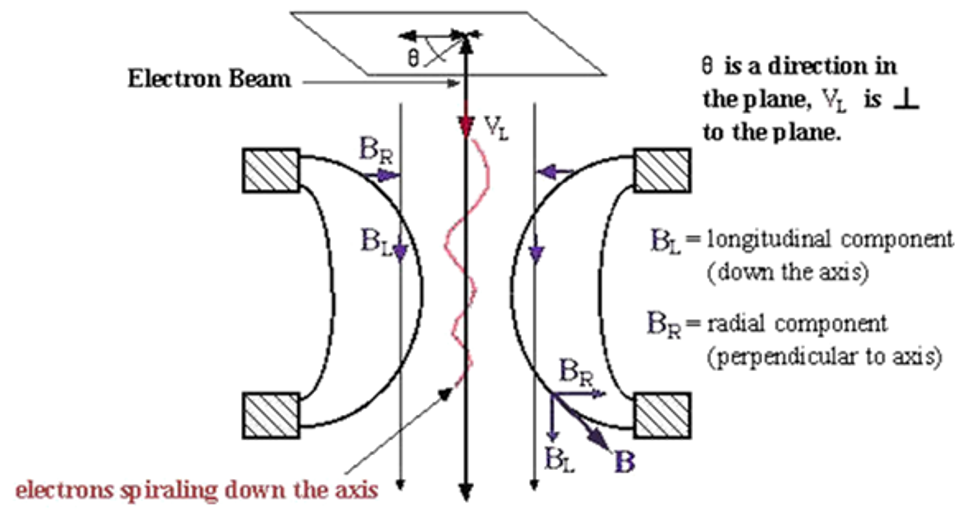

Magnetic Electron Lenses
An off-axis electron is acted on by a magnetic force:
where,
v = electron velocity and
B = the magnetic field.
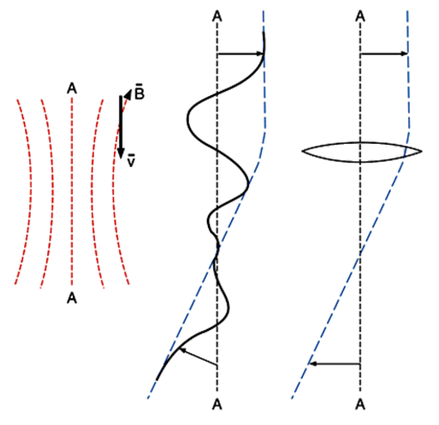
Magnetic Electron Lenses
The focal length (f) of the electromagnetic lens is:
f = K (V / I2)
where:
K = constant based on the number of turns in the magnet coil and geometry of lens
V = accelerating voltage
I = current
SEM operation
Magnetic Electron Lenses
Electron lenses are not as good as optical lenses in terms of defects of focus, called aberrations.
Spherical aberrations are minimized by placing a spray aperture in front of the magnetic lens, confining electrons to the center. This results in greatly reduced beam currents.

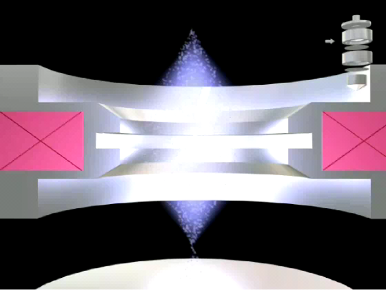
magnetic electron lenses
The condenser lens controls the amount of current that passes down the column.
This is accomplished by focusing the electron beam to variable degrees onto a lower aperture.
The sharper the focus, the less of the beam intercepted by the aperture and the higher the current.
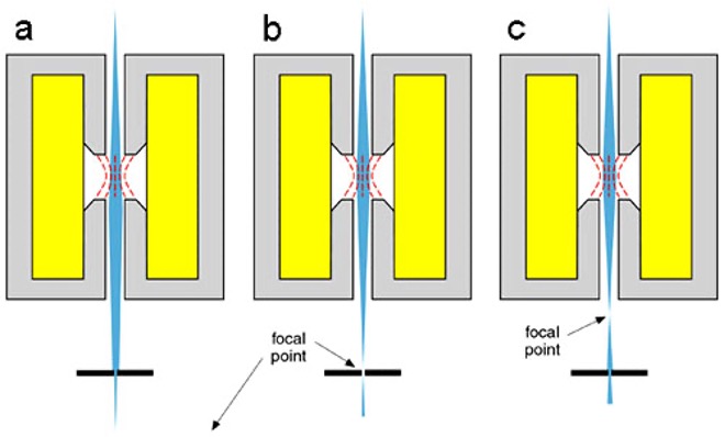
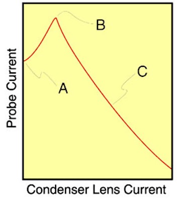
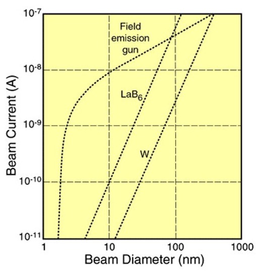
The ultimate electron beam spot size depends on the beam current, which is controlled by the condenser lens, and the type of filament in use.
Objective Lens
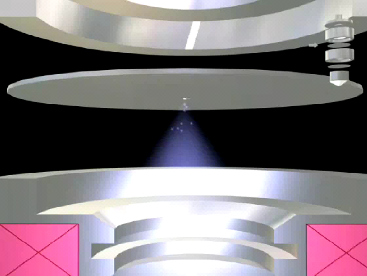
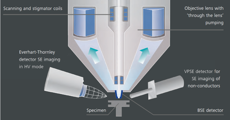
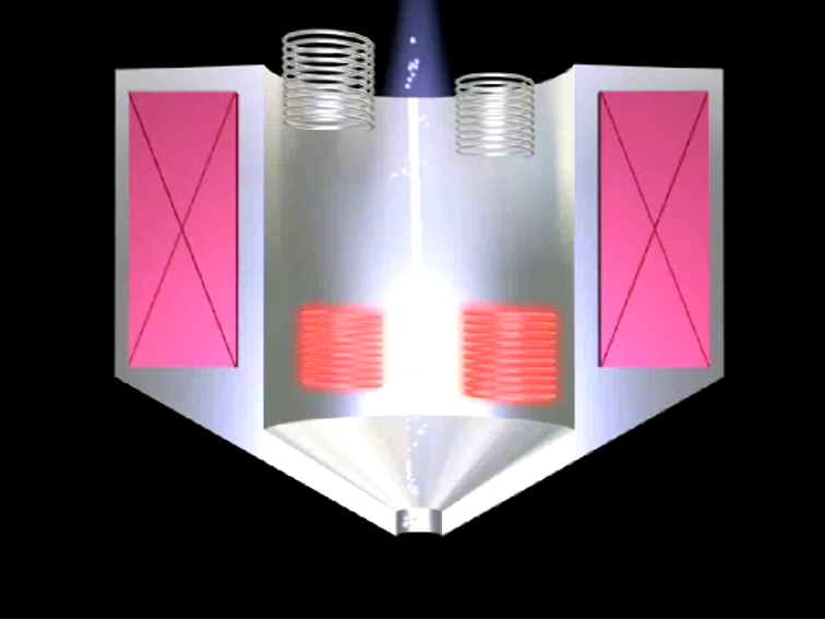
The lens nearest to the specimen and used to focus the electron beam onto the specimen surface.
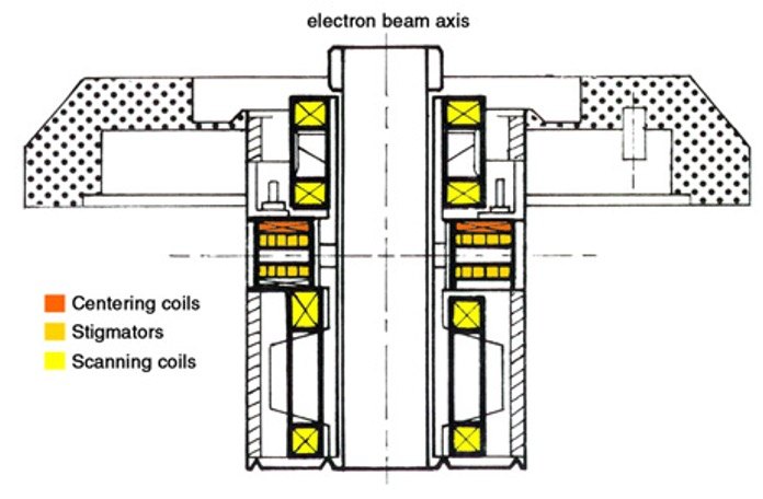
Stigmator/scanning coils also housed in objective.
The objective lens also influences the spot size.
Objective Lens: Scanning Coils
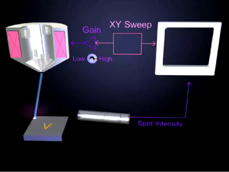
Scanning coils raster the beam across the sample.
The coils consist of four magnets.
The beam is rastered by varying the current through these magnets.
Deflecting the beam off-axis introduces aberrations.
Objective lens: Astigmatism
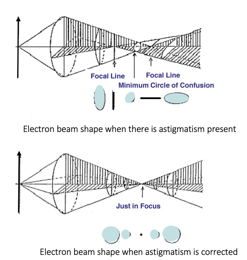
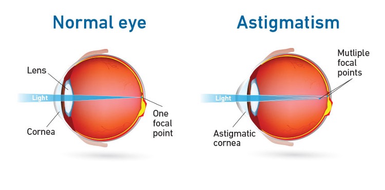
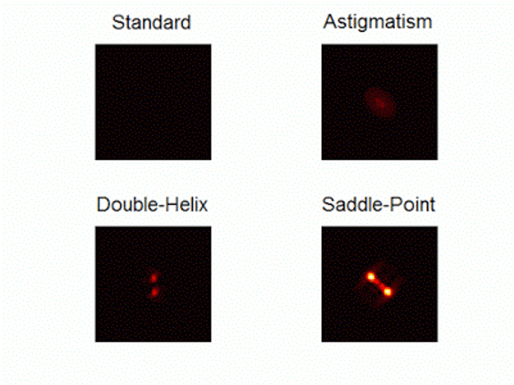
Magnetic lenses do not have perfect symmetry and the electron beam may be elliptical.
Astigmatism produces a reduction in resolution.
This astigmatism can be removed by adjusting the X and Y correction controls of the stigmator coils.

The Vacuum
When a SEM is used, the column must always be at a vacuum. There are many reasons for this:
If the sample is in a gas filled environment, an electron beam cannot be generated or maintained.
Gases could react with the electron source, causing it to burn out, or cause ionization.
Cont’d
The transmission of the beam through the electron optic column would also be hindered by the presence of other molecules.
Other molecules could form compounds and condense on the sample. This would lower the contrast and obscure detail in the image.